Abstract
Objective
To evaluate trunk and knee muscle strength in patients with chronic sciatica.
Patients and method
Twenty-eight patients with right-side chronic sciatica (group 1, G1) and 40 healthy controls (group 2, G2) were evaluated using an isokinetic dynamometer (Cybex Norm II). Quadriceps and hamstring muscle strength were evaluated at concentric velocities of 60 and 120°/s and the trunk muscles were evaluated at concentric velocities of 60 and 90°/s.
Results
G1 comprised 15 women and 13 men (mean ± SD age: 34.787 ± 6.06; weight: 65.85 ± 5.33 kg; height: 165 ± 6.92 cm). G2 comprised 20 women and 20 men (mean ± SD age: 35.92 ± 6.66; weight: 67.07 ± 6.12 kg; height: 165.82 ± 7.65 cm. There were no significant inter-group differences concerning these parameters. In G1, the peak torque values for the trunk extensors were 123.71 ± 32.45 and 108.85 ± 32.07 Newton metres (Nm) at angular velocities of 60 and 90°/s, respectively. In G2, the values were 192.73 ± 64.24 and 168.65 ± 53.96 Nm, respectively. In G1, the peak torque values for the trunk flexors were 134.32 ± 30.77 and 124.39 ± 32.59 Nm at angular velocities of 60 and 90°/s, respectively. In G2, they were 177.44 ± 44.1 and 157.81 ± 39.01 Nm, respectively. The difference between G1 and G2 was statistically significant. The peak torque for the right quadriceps in G1 was 100.03 ± 24.45 and 78.71 ± 22.92 Nm at angular velocities of 60 and 120°/s, respectively. In G2, these values were 160.5 ± 36.34 and 131.05 ± 33.42 Nm. The peak torque for the hamstrings in G1 was 56.42 ± 13.02 and 50 ± 13.55 Nm at angular velocities of 60 and 120°/s, respectively. In G2, these values were 97.77 ± 33.32 and 84.72 ± 31.41 Nm. Again the difference between G1 and G2 was statistically significant. We also noted a statistically significant difference between G1 and G2 in terms of the peak quadriceps and hamstring torque values on the left side. In G1, the quadriceps and hamstrings were significantly weaker on the sciatica side than on the unaffected side.
Conclusion
The present study demonstrated trunk and knee muscle weakness in patients with chronic sciatica, when compared with healthy subjects. This weakness was predominant on the sciatica side. Consequently, the management of these patients should include a knee muscle reinforcement programme.
Résumé
Objectif
Évaluer la force musculaire du tronc et des genoux chez des adultes ayant une lombosciatique chronique.
Patients et méthodes
Vingt-huit patients ayant une lombosciatique chronique droite (G1) et 40 sujets sains (G2) ont bénéficié d’une évaluation isocinétique en concentrique de la force du quadriceps et des ischio-jambiers des genoux aux vitesses angulaires 60 et 120°/s (s : seconde) et des muscles extenseurs et fléchisseurs du rachis lombaire aux vitesses angulaires 60 et 90°/s à l’aide d’un dynamomètre isocinétique (Cybex Norm II ). L’évaluation isocinétique de ces muscles a été réalisée le même jour à un intervalle d’une heure pour chacun de nos patients.
Résultats
Le premier groupe G1 a comporté 15 femmes et 13 hommes d’âge, de poids et de taille moyens respectivement : 34,787 ± 6,06 ans ; 65,85 ± 5,33 kg et 165 ± 6,92 cm. Le deuxième groupe G2 a comporté 20 femmes et 20 hommes d’âge, de poids et de taille moyens respectivement : 35,92 ± 6,66 ans ; 67,07 ± 6,12 kg et 165,82 ± 7,65 cm. Les deux groupes ont été jugés comparables selon ces différents paramètres. Pour les muscles du tronc, les moyennes des moments de force maximaux des extenseurs du tronc des patients du G1 étaient respectivement de 123,71 ± 32,45 et 108,85 ± 32,07 Nm aux vitesses 60 et 90°/s. Celles du G2 étaient respectivement de 192,73 ± 64,24 et 168,65 ± 53,96 Nm. Les moyennes des moments de force maximaux des fléchisseurs du tronc du G1 étaient respectivement de 134,32 ± 30,77 et 124,39 ± 32,59 Nm aux vitesses 60 et 90°/s. Celles du G2 étaient respectivement de 177,44 ± 44,1 et 157,81 ± 39,01 Nm. Les différences de ces moyennes entre les deux groupes étaient statistiquement significatives. Les moyennes des moments de force maximaux du quadriceps du côté droit du G1 étaient de 100,03 ± 24,45 et 78,71 ± 22,92 Nm aux vitesses 60 et 120°/s. Celles du G2 étaient respectivement de 160,5 ± 36,34 et 131,05 ± 33,42 Nm. Les moyennes des moments de force maximaux des ischio-jambiers du G1 étaient de 56,42 ± 13,02 et 50 ± 13,55 Nm aux vitesses 60 et 120°/s. Celles du G2 étaient respectivement de 97,77 ± 33,32 et 84,72 ± 31,41 Nm. Les différences de ces moyennes entre les deux groupes étaient statistiquement significatives. Nous avons constaté également des différences significatives entre les moyennes des moments de force maximaux des muscles quadriceps et ischio-jambiers du côté gauche du G1 par rapport à celles du G2. En comparant le côté sciatique au côté controlatéral, nous avons noté une faiblesse du quadriceps et des ischio-jambiers du côté de la sciatique.
Conclusion
Cette étude montre une faiblesse des muscles du tronc et des genoux chez les patients ayant une lombosciatique chronique comparativement aux sujets normaux. Cette faiblesse est plus marquée du côté de la sciatique. Par conséquent, la prise en charge en rééducation des patients ayant une lombosciatique chronique doit inclure un programme de renforcement des muscles du genou.
1
English version
1.1
Introduction
Functional retraining is becoming increasingly important in the therapeutic management of chronic sciatica. The objective is to improve the patient’s physical, psychosocial and socio-economic situation by using a physical reactivation approach. This multidisciplinary programme should include trunk and leg muscle toning to improve the patient’s overall physical condition.
However, the initiation of a strength training programme and the assessment of the latter’s efficacy must be based on a standardized, sensitive, reproducible evaluation of muscle function.
The clinical analysis of trunk and leg muscle strength is based not only on muscle testing but also on specific tests, such as the Shirado test for the abdominal muscles and the Sorensen test for spinal muscles. These tests constitute a simple, cheap way of assessing trunk muscle strength and endurance and enable a patient’s progress to be monitored during the retraining programme.
However, these isometric tests provide limited information on capacities in lower back pain or sciatica patients because they are too far removed from the trunk muscles’ normal conditions of use.
The use of an isokinetic dynamometer is an alternative method for measuring muscle strength . Several studies have demonstrated the reproducibility and sensitivity of this technique . In addition to its value as a measurement tool, isokinetic movement could also have diagnostic and even therapeutic value in certain situations .
The objective of the present study was to evaluate the isokinetic strength of the trunk and knee extensor and flexor muscles in adults with chronic sciatica, in order to optimize a muscle toning programme in a rehabilitation setting.
1.2
Patients and methods
1.2.1
Patients
Sixty-eight subjects were included in our study: 28 right-handed patients with right-side chronic sciatica (group G1) and 40 healthy subjects (group G2).
In a patient interview: we noted the patient’s age, gender and the characteristics of his/her sciatica: the side (right or left), the path (L5 or S1) and the intensity of the lumbar and radicular pain evaluated on a 0 to 10 visual analogue scale (VAS).
The patients in G1 had right side, L5- or S1-dominant chronic sciatica related to lateralized and non-foraminal L4L5 spinal disc herniation or non-operated L5S1 spinal disc herniation (evidenced by computed tomography or magnetic resonance imaging) in which a L5 or S1 disc-nerve root conflict had always been present. The mean time since onset of sciatica was 11.71 ± 4.72 months [range: 6–24 months] and the duration of the last painful episode was at least 3 months. Sixteen patients (57.14%) in G1 had L5 sciatica and the other twelve (42.85%) had S1 sciatica. Isokinetic muscle strength evaluation in all patients was made possible by the low intensity of the lumbar and radicular pain (rated as no higher than 3 out of 10 on the VAS). In G1, the mean lumbar and radicular pain scores on the VAS at the time of isokinetic muscle strength testing were 2.37 ± 0.42 and 2.58 ± 0.38, respectively. The lumbar and radicular pains were sequelae and corresponded to a stable, non-progressing state (close to full recovery) in our population of sciatica sufferers.
The clinical examination recorded each subject’s weight, height and spinal mobility (as the Schöber index) and the presence or absence of Lasegue’s sign. The neurological examination included manual strength testing of the main leg muscles (the tibialis anterior, peroneus, extensor digitorum longus , extensor hallucis longus , gluteus medius , triceps surae , hamstrings and quadriceps), an evaluation of skin sensitivity and the presence or absence of patellar and Achilles tendon reflexes.
Manual muscle strength testing was based on the United Kingdom Medical Research Council system . The mean Schöber index was 14.2 cm and Lasegue’s sign was absent in all our patients. In G1, all the muscle strength tests (and notably those on the quadriceps and the hamstrings) were normal and there were no sensory disorders. Patellar and Achilles tendon reflexes were present on both body sides.
The control group G2 comprised 40 healthy, right-handed, volunteer subjects with moderate levels of physical activity.
After the patient interview and the clinical examination, G1 and G2 were compared in terms of the main factors that can influence the muscle strength (i.e. age, gender, height and weight); there were no significant inter-group differences other than sciatica status. The groups’ respective anthropometric characteristics (age, gender, weight and height) and the statistical significance of the inter-group comparison are summarized in Table 1 .
Furthermore, we exclude patients with secondary sciatica, significant spinal stiffness (a Schöber index below 13 cm), restricted knee movement (less than 90° of flexion or extension below 0°), a chronic, progressing disease contra-indicating effort or a psychiatric condition.
1.2.2
Methods
Each subject underwent isokinetic evaluation of the trunk, quadriceps and hamstring muscles. We used the Cybex Norm II Medimex system, together with its Trunk Extension/Flexion (TEF) unit.
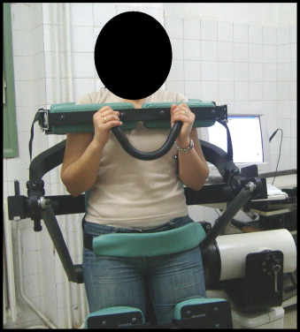
For the trunk muscle evaluation, the subject was placed upright in the TEF unit. After adjusting for height and the anteroposterior position, the dynamometer’s axis of rotation is situated at the L5-S1 level (i.e. close to the anterior superior iliac spine). The knees were positioned with around 15° of flexion. The upper part of the trunk was supported by an adjustable cushion placed near the spine of the scapula. The subject was secured to the unit:
- •
at the top of the thorax with a shoulder bar (fitted with a front handle, which the subject grasps);
- •
by a pelvic belt that ensures that the pelvis does not move during the test;
- •
at the legs by two anterior cushions (one femoral and the other tibial).
These straps held the legs and the upper part of the trunk firmly, in order to prevent any compensatory muscle activity (notably that of the hip flexors and extensors). The amplitude of the movement was limited by electronic stops at 70° of flexion (the starting marker is set to 0° of flexion). A screen on the left side of the apparatus enabled the subject to visualise the exercise curves ( Fig. 1 ). The concentric-mode evaluation was performed at two velocities: 60 and 90°/s. A series of five repetitions was performed at each speed, with a one-minute rest interval between the two series.
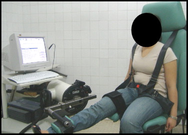
For the evaluation of quadriceps and hamstring muscle strength, the subject sat in the dynamometer with the chair back inclined at an angle of 10° to the vertical. The knees were flexed at 90°. Thorax, pelvis and thigh straps held the subject firmly to the apparatus and thus reduced compensatory movements as much as possible. An adjustable fitting was used to attach the lever arm to the lower third of the leg to be tested. The contralateral leg was immobilized at the ankle ( Fig. 2 ). The concentric-mode evaluation was performed at two velocities: 60 and 120°/s. Again a series of five repetitions was performed at each speed, with a one-minute rest interval between series.
Each patient was informed about how the apparatus worked and the various phases of the isokinetic evaluation. All subjects were instructed to exert maximum effort throughout the amplitude of the movement for each repetition, whether in flexion or extension (i.e. to push or pull as hard and fast as possible). Each session was preceded by a 10-minute warm-up session on a cycle ergometer at a resistance of 30 watts. Before each series, the subject performed three practice movements, in order to accustom themselves with the corresponding velocity. During each series, the subject was verbally encouraged by the examiner to develop maximum effort. The effect of gravity was systematically taken into account before each test.
The knee and trunk muscle strength evaluations were performed on the same day by an examiner trained in the use of this technique. To avoid muscle fatigue, each subject was allowed an hour’s rest between the tests. For the selected muscle groups, we measured the peak torque (in Newton metres, Nm) and the agonist-antagonist ratios (mean values for the subjects). Each subject was informed of the study’s objective and procedures and gave his/her consent to participation.
1.2.3
Statistics
Data entry and analysis were performed using SPSS 11.0 software. The results were expressed as the mean ± standard deviation. Student’s t test was used to compare mean values and the Chi 2 test was used to compare frequencies (used for the gender factor). The statistical significance threshold was set to p < 0.05.
1.3
Results
All subjects performed the isokinetic tests without any incidents or complaints. There were no adverse effects and, in particular, pain did not recur during or after the tests.
The mean peak torque values for the trunk flexor and extensor muscles were significantly lower in G1 than in G2. Furthermore, the flexor/extensor ratio was reversed in G1 (i.e. greater than 1), since it is usually below 1 in healthy subjects (as we also found in the present study). These results were confirmed at both velocities used for trunk muscle evaluation (60 and 90°/s) and are summarized in Table 2 .
| Mean ± SD peak torque (Nm) | G1 ( n = 28) | G2 ( n = 40) | p |
|---|---|---|---|
| Trunk flexors at 60°/s | 134.32 ± 30.77 | 177.44 ± 44.1 | 0.002 |
| Trunk flexors at 90°/s | 124.39 ± 32.59 | 157.81 ± 39.01 | 0.01 |
| Trunk extensors at 60°/s | 123.71 ± 32.45 | 192.73 ± 64.24 | < 0.001 |
| Trunk extensors at 90°/s | 108.85 ± 32.07 | 168.65 ± 53.96 | 0.001 |
| Flexors/extensor ratio at 60°/s | 1.08 | 0.85 | < 0.001 |
| Flexor/extensor ratio at 90°/s | 1.14 | 0.85 | < 0.001 |
For the right-side quadriceps and hamstring muscles in the two groups, we observed significantly lower mean peak torque values in G1 than in G2. The right knee’s flexor/extensor ratio was also lower in G1 than in G2. These results were observed at both velocities (60 and 120°/s) and are summarized in Table 3 . Similarly, we evidenced lower mean peak torque values on the left side in G1, when compared with G2. These results are summarised in Table 4 .
| Mean ± SD peak torque (Nm) | G1 ( n = 28) | G2 ( n = 40) | p |
|---|---|---|---|
| Quadriceps at 60°/s | 100.03 ± 24.45 | 160.5 ± 36.34 | < 0.001 |
| Quadriceps at 120°/s | 78.71 ± 22.92 | 131.05 ± 33.42 | < 0.001 |
| Hamstrings at 60°/s | 56.42 ± 13.02 | 97.77 ± 33.32 | < 0.001 |
| Hamstrings at 120°/s | 50 ± 13.55 | 84.72 ± 31.41 | < 0.001 |
| Hamstrings/Quadriceps at 60°/s | 0.55 | 0.6 | 0.004 |
| Hamstrings/Quadriceps at 120°/s | 0.59 | 0.65 | 0.002 |
| Mean ± SD peak torque (Nm) | G1 ( n = 28) | G2 ( n = 40) | p |
|---|---|---|---|
| Quadriceps at 60°/s | 131.07 ± 24.19 | 146.07 ± 33.96 | 0.004 |
| Quadriceps at 120°/s | 105.785 ± 22.84 | 115.45 ± 28.02 | 0.005 |
| Hamstrings at 60°/s | 73.03 ± 16.52 | 89.02 ± 30.88 | < 0.001 |
| Hamstrings at 120°/s | 62.57 ± 9.44 | 72.05 ± 26.8 | 0.001 |
| Hamstrings/quadriceps at 60°/s | 0.58 | 0.62 | 0.004 |
| Hamstrings/quadriceps at 120°/s | 0.56 | 0.61 | 0.001 |
The mean peak torque values for the quadriceps and hamstring muscles on the right (sciatica) side were significantly lower than those for the contralateral limb in the patients in G1. These results are presented in Table 5 .
| Mean ± SD peak torque (Nm) | G1 | ||
|---|---|---|---|
| Affected side (right) | Healthy side (left) | p | |
| Quadriceps at 60°/s | 100.03 ± 24.45 | 131.07 ± 24.19 | < 0.001 |
| Quadriceps at 120°/s | 78.71 ± 22.92 | 105.785 ± 22.84 | < 0.001 |
| Hamstrings at 60°/s | 56.42 ± 13.02 | 73.03 ± 16.52 | < 0.001 |
| Hamstrings at 120°/s | 50 ± 13.55 | 62.57 ± 9.44 | < 0.001 |
1.4
Discussion
Sciatica commonly results from disc-nerve root conflict after disc herniation. In many cases, the sciatica becomes chronic. At this stage, functional rehabilitation is very important, so that sciatica patients can normalize their physical, psychosocial and economic situation. This rehabilitation includes the treatment of impairments and, in particular, muscle weakness (via a toning programme in a rehabilitation setting). The training programme is based on an accurate evaluation of muscle strength, with particular emphasis on the trunk (abdominal and spinal muscles) and the legs (the quadriceps and hamstrings). Impairments in these muscles have been studied widely in lower back pain but rarely in sciatica.
Isokinetic movement enables quantified, accurate evaluation that is not possible with other methods . As a benchmark for maximum trunk muscle strength, the technique’s reliability, reproducibility and sensitivity have been clearly demonstrated .
A number of authors have agreed that the isokinetic method is more sensitive than manual testing , which significantly under-estimates the severity of muscle weakness. Isokinetic testing can detect improvements, which go unobserved by manual testing (particularly for scores above 3). In the present study, isokinetic measurements of quadriceps and hamstring muscle strength revealed impairments which had not be identified by manual testing during the clinical examination; this should enable better follow-up.
However, given the non-negligible cardiovascular impact of isokinetic efforts, it appears advisable to perform an effort test before the isokinetic evaluation, so as not to overlook a cardiovascular condition (especially a coronary problem) that could reveal itself during the testing. The risk of cardiovascular problems may restrict the use of this technique in the investigation of trunk and leg muscle strength in patients with chronic sciatica. Likewise, limited spinal mobility in the lower back (notably flexion below 70°) and the existence of intense pain will prevent this isokinetic technique from being employed.
In the present study, all our patients completed the tests successfully and no incidents or complaints were noted; this confirms the good tolerability of isokinetic evaluation once patients with intense pain and/or a spinal syndrome have been eliminated. However, practical reasons prevented us from performing an effort test here.
A review of the literature reveals that the velocity chosen for evaluating spinal muscle strength varies between 30 and 180°/s. However, it is not possible to affirm that one particular evaluation velocity is more reliable than another . The two most frequently used velocities are 60 and 120°/s. The isokinetic strength measurement protocols vary from one group of researchers to another; some authors use only two velocities, whereas others employ multiple tests with increasing velocities. In the present study, we adopted a muscle protocol based on two increasing velocities for evaluation of the trunk muscles (60 and then 90°/s) and the knee muscles (60 and 120°/s). Moreover, there is no consensus regarding the number of repetitions. Since the value is most commonly between 3 and 5, we decided to use five repetitions for each series of contractions.
According to Genty and Schmidt, the deconditioning syndrome in chronic lower back pain includes the loss of trunk muscle function . Vancelcenaher et al. have described specific impairments of lumbopelvic extensors and rotators and increased fatigability . Roques et al. have shown that lower back pain sufferers display lower peak torque values for the trunk flexors and extensors (and especially for the spinal extensors) . Likewise, Lee et al.’s 5-year prospective study of 67 initially pain-free subjects showed significantly lower peak torque values in the 18 subjects who developed lower back pain and a statistically significant difference in the trunk flexor/extensor ratio between the healthy subjects and the lower back pain sufferers . Furthermore, several studies have shown that the flexor/extensor ratio is inverted in lower back pain sufferers . The ratio is between 0.7 and 0.8 in healthy male subjects, closer to 1 in women and over 1 in lower back pain sufferers .
To the best of our knowledge, only one study has evaluated the trunk muscle strength in patients with disc herniation . A statistically significant reduction in trunk flexor and extensor muscle strength was demonstrated at velocities of 60°/s and 120°/s. However, the sample size was very low.
In the present study, we found a statistically significant decrease in the peak torque values for the spinal flexors and extensors in sciatica patients, when compared with healthy subjects. This difference was seen at all velocities used. It is noteworthy that we observed an inverted flexor/extensor strength ratio (i.e. greater than 1) in the sciatica patients.
The synergies between trunk muscles and leg muscles when maintaining an erect stance and during trunk movements emphasize the importance of evaluating knee muscle strength in patients with lower back pain or chronic sciatica.
Duvallet evidenced a reduction in the strength and fatigue resistance of the quadriceps and especially the hamstring muscles in chronic lower back pain sufferers, when compared with healthy subjects . Work by Bibré et al. gave the same results .
This reduction in quadriceps and hamstring strength was also found in chronic sciatica patients by Poiraudeau et al., who also observed a low flexor/extensor ratio . These results were confirmed by Ho et al. in a study comparing 22 herniated disc patients with 41 healthy subjects.
In the present study, we found significant lower quadriceps and hamstring strengths on the right side (i.e. the sciatica side) in G1, compared with G2. This deficiency was most prominent for the hamstrings.
Similar results were found by comparing G1 and G2 in terms of the left-side strength values.
When comparing the sciatica side to the contralateral side, Ho et al. did not find any statistically significant difference between the quadriceps and hamstring muscle strengths . In contrast, Poiraudeau et al. found a right vs. left muscle imbalance in patients with unilateral sciatica . Hence, our results confirm those of Poiraudeau et al.
We suggest that the overall muscle impairment revealed by isokinetic measurement (and notably trunk and leg muscle weakness on the non-painful side) could be explained by a deconditioning syndrome. A peripheral neurological impairment (caused by an L5 or S1 nerve root problem) cannot be the cause because the knee muscles primarily depend on roots L3 and L4 (for the quadriceps) and L5 and S1 (for the hamstrings). Further, our neurological examination did not reveal impaired sensitivity or tendon reflexes and none of the patients displayed cruralgia symptoms.
In fact, in chronic sciatica patients, a physical deconditioning syndrome can occur after four to six months of inactivity and is evidenced by a decrease in initial physical capacities, a loss of spinal mobility, muscle-tendon hypoextensibility (particularly for the hamstrings) , decreased performance of the main spinal extensor muscles in the trunk (due to atrophy of the various muscle fibres) , a decrease in the anterior and posterior trunk wall muscles ) and a reduced aerobic capacity.
This physical deconditioning may be accompanied by psychosocial factors and thus worsened anxiety and depression scores. However, a systematic literature review has cast doubt on the existence of a physical deconditioning syndrome in chronic sciatica .
In contrast, regular physical exercise or an exercise retraining programme significantly limits this phenomenon.
Moreover, the sciatic pain causes muscle inhibition, which in turn aggravates atrophy through lack of use . Lumbar syndrome probably aggravates the loss of lumbar muscle strength and may explain the observed trunk muscle imbalance .
1.5
Conclusion
Functional rehabilitation in chronic sciatica must include a trunk and leg muscle toning programme.
The nature of this muscle strengthening programme depends on the extent of the impairment and the affected muscles. Accurate muscle strength evaluation thus enables the programme to be fine-tuned.
Isokinetic movement is one of the best ways to perform this evaluation. It enables the knee and trunk muscle weakness to be accurately quantified and can be used to identify extensor/flexor imbalance, if present. The technique is a reliable, sensitive means of evaluating and monitoring muscle weakness.
Our study showed a reduction in trunk and knee muscle strength in patients with chronic, unilateral sciatica, when compared with healthy subjects. This impairment was most prominent for the trunk extensor muscles and the knee flexor muscles. For the knee flexor and extensor muscles, the impairment was more marked on the sciatica side.
Hence, the rehabilitational management of chronic sciatica patients should feature knee and trunk muscle strengthening, with special emphasis on the trunk extensors and the hamstrings on the affected side.
2
Version française
2.1
Introduction
La restauration fonctionnelle des patients souffrants d’une lombosciatalgie chronique occupe une place de plus en plus importante dans la prise en charge thérapeutique de ces patients. L’objectif de cette restauration est de récupérer la situation physique, psychosociale et socio-économique de ces patients en utilisant une démarche active de ceux-ci basée sur une réactivation physique. Ce programme pluridisciplinaire comporte une tonification des muscles du tronc et des membres inférieurs afin d’améliorer la forme physique générale.
Cependant la mise en place de ce programme de tonification musculaire et l’estimation de leur efficacité doivent pouvoir reposer sur une évaluation de la fonction musculaire qui soit standardisée, sensible et reproductible.
L’étude clinique de la force musculaire du tronc et des membres inférieurs repose essentiellement sur le testing musculaire mais aussi sur des tests spécifiques tels que le test de Schirado pour les muscles abdominaux et le test de Sorensen pour les muscles spinaux. Ces tests constituent une approche simple, peu coûteuse, de la force et de l’endurance des muscles du tronc permettant un bon suivi des progrès réalisés en rééducation.
Cependant, ces tests isométriques restent limités dans les renseignements qu’ils fournissent sur les capacités musculaires des patients lombalgiques et lomboradiculalgiques car trop éloignés des conditions d’utilisation physiologique de la musculature du tronc.
L’utilisation des dynamomètres isocinétiques offre une alternative dans l’évaluation de ces forces musculaires . Diverses études témoignent de la reproductibilité et de la sensibilité de cette technique . En plus de sa valeur d’outil d’évaluation, l’isocinétisme pourrait également dans certaines situations avoir un intérêt diagnostic mais surtout thérapeutique .
L’objectif de notre étude est d’évaluer les forces musculaires isocinétiques du tronc et des deux genoux chez des adultes ayant une lombosciatique chronique afin de mieux cibler le programme de tonification musculaire en milieu de rééducation.
2.2
Patients et méthodes
2.2.1
Patients
Soixante-huit sujets, ont été inclus dans notre étude : 28 patients droitiers ayant une lombosciatique chronique droite (groupe G1) et 40 sujets sains (groupe G2).
L’interrogatoire a comporté essentiellement le recueil de l’âge, du sexe et les caractéristiques de la lombosciatique à savoir le côté (droit ou gauche), le trajet (L5 ou S1) et l’intensité de la douleur lombaire et radiculaire évaluée sur une échelle visuelle analogique de 0 à 10.
Les patients du premier groupe (G1) avaient une lombosciatalgie L5 ou S1 chronique droite (côté dominant), en rapport avec des hernies discales lombaires L4L5 (latéralisées et non foraminales) ou L5S1 non opérées (objectivées par un scanner ou une imagerie par résonance magnétique), dans lesquelles un conflit discoradiculaire L5 ou S1 a été constamment évoqué. La moyenne de la durée d’évolution de ces sciatiques était 11,71 ± 4,72 mois (minimum six mois, maximum 24 mois), Le délai du dernier épisode douloureux était supérieur à trois mois. Seize patients (57,14%) de ce groupe G1 avaient une lombosciatique L5, les 12 autres patients (42,85%) avaient une lombosciatique S1. L’intensité de la douleur lombaire et radiculaire de chaque patient était faible permettant l’évaluation musculaire en isocinétisme. Elle était cotée au maximum à 3/10 sur l’échelle visuelle analogique (EVA). Les moyennes de l’EVA douleur lombaire et radiculaire du groupe G1, au moment de l’évaluation étaient respectivement de 2,37 ± 0,42 et de 2,58 ± 0,38. Ces douleurs lombaires et radiculaires sont des séquelles et correspondent à un état stable et non évolutif proche de la guérison de notre population lombalgique.
L’examen clinique précisait le poids, la taille, la mobilité rachidienne (par la mesure de l’indice de Schöber), la recherche du signe de Lassègue et un examen neurologique comportant un testing musculaire manuel des principaux muscles des membres inférieurs (tibial antérieur, fibulaires, extenseur commun des orteils, extenseur propre du gros orteil, moyen fessier, triceps sural, ischio-jambiers et quadriceps), l’évaluation de la sensibilité superficielle et la recherche des réflexes ostéotendineux (réflexes rotulien et Achilléen).
Le testing musculaire manuel a été basé sur la cotation internationale de British Medical Council (BMC) . L’indice de Shöber moyen était de 14,2 cm et le signe de Lassègue était négatif chez tous nos patients. Le testing des forces musculaires du quadriceps et des ischio-jambiers (ainsi que des autres muscles testés) dans le G1 était normal et il n’y avait pas de trouble sensitif. Les réflexes ostéotendineux étaient présents et symétriques.
Le second groupe (G2) comportait 40 sujets sains volontaires droitiers qui faisaient des activités physiques de loisir.
Au terme de cet interrogatoire et l’examen clinique, les deux groupes G1 et G2 ont été comparés selon les principaux facteurs qui pourraient influencer les forces musculaires des sujets à savoir l’âge, le sexe, la taille et le poids.
Il n’y avait pas de différence significative entre les deux groupes concernant ces différents paramètres. Les deux groupes ont été jugés donc comparables selon les facteurs qui peuvent influencer les résultats des forces musculaires à part le facteur lombosciatique. Les caractéristiques anthropométriques (âge, sexe, poids et taille) des sujets des deux groupes G1 et G2 ainsi que les taux de signification de leur comparaison sont présentés dans le Tableau 1 .
Par ailleurs, ont été exclus de l’étude, les patients qui avaient une lombosciatique secondaire, une raideur rachidienne importante avec un indice de Schöber inférieur à 13 cm ou une limitation de la mobilité des genoux (flexion moins de 90° ou une extension inférieure à 0°). Ont été exclus, de même, les patients ayant une maladie évolutive chronique contre-indiquant les efforts ou une affection psychiatrique.
2.2.2
Méthodes
Chaque sujet a bénéficié d’une évaluation isocinétique des muscles du tronc et ceux des membres inférieurs (quadriceps et ischio-jambiers) : nous avons utilisé l’appareil Cybex Norm II Medimex associé à son module TEF.
Pour l’évaluation des muscles du tronc, le sujet a été installé en position debout dans le module TEF. L’axe de rotation du dynamomètre est au niveau du disque intervertébral L5-S1, soit à la hauteur de l’épine iliaque antéro-supérieure, après réglage en hauteur et en antéropostérieur. Les genoux sont positionnés à environ 15° de flexion. La partie haute du tronc repose sur un coussin scapulaire réglable positionné à la hauteur de l’épine du scapula. Le sujet est sanglé au niveau thoracique par une ceinture scapulaire munie d’une poignée antérieure que le sujet prend dans ses mains, au niveau du bassin par une ceinture pelvienne qui assure le maintien et la fixité du bassin au cours du test et au niveau des genoux par deux coussins antérieurs l’un fémoral et l’autre tibial. Ces sangles maintiennent les membres inférieurs et la partie supérieure du tronc afin d’éviter toute compensation notamment celle des fléchisseurs et extenseurs des hanches. L’amplitude du mouvement est limitée par des butées électroniques à 70° de flexion (le repère de départ est 0° de flexion). Un écran disposé du côté gauche de l’appareil permet au sujet de visualiser les courbes représentant l’exercice effectué ( Fig. 1 ). L’évaluation a été réalisée avec deux vitesses : 60 et 90°/s (s : seconde) sur un mode concentrique. Pour chaque vitesse une série de cinq répétitions a été effectuée. Un intervalle de repos d’une minute entre les séries a été respecté.

Related posts:
 Physical and rehabilitation medicine in Europe, from the White Book to the eBooks
Non-steroidal anti-inflammatory drugs for athletes: An update
Seeking care for lower back pain in the French population aged from 30 to 69: The results of the 2002–2003 Décennale Santésurvey
Assessment of compliance with prescribed activity by hemiplegic stroke patients after an exercise programme and physical activity education
Influence of electrical stimulation frequency on skeletal muscle force and fatigue
Analyse de livre
Physical and rehabilitation medicine in Europe, from the White Book to the eBooks
Non-steroidal anti-inflammatory drugs for athletes: An update
Seeking care for lower back pain in the French population aged from 30 to 69: The results of the 2002–2003 Décennale Santésurvey
Assessment of compliance with prescribed activity by hemiplegic stroke patients after an exercise programme and physical activity education
Influence of electrical stimulation frequency on skeletal muscle force and fatigue
Analyse de livre
Stay updated, free articles. Join our Telegram channel

Full access? Get Clinical Tree


