Color
Turbidity
Viscosity
Cell count/mm3
Others
Normal
Amber
No
High
100
–
Chronic polyarthritis
Yellow brown, grey
Opaque, fluffy
Low
5,000–50,000
Rhagocytes +++
Gout
Yellow
Opaque
Low
10,000–20,000
Leukocytes, urate crystals
Pseudogout
Milky
Opaque
Low
20,000
Leukocytes, calcium pyrophosphate
Bacterial arthritis
Yellow brown, green
Fluffy
Low
>50,000
Leukocytes, bacteria
Tuberculosis
Yellow grey
Opaque
Low
20,000–50,000
Leukocytes, lymphocytes, tubercle bacterium
Trauma
Red
Red
High or low
>2,000
Erythrocytes
A chronic painful knee should be considered infected until otherwise disapproved by clinical and paraclinical test.
41.1.1 Technique for Joint Aspiration
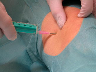
The patient should be placed supine in a comfortable position. Sterile gloves, extensive disinfection, and sterile draping are compulsory. There is no need for local anesthesia prior to aspiration. The superolateral approach, as shown in Fig. 41.1, is most commonly used [3]. An 18-gauge needle (yellow) (Birmingham wire gauge) should be inserted 1 cm proximal and dorsal of the proximal-lateral edge of the patella (Fig. 41.1). Other approaches are the mid-lateral and mid-medial approach with the knee in full extension, the anteromedial and anterolateral approach with the knee in 90° of flexion, and the Waddell’s approach using the anterolateral approach in 30° of knee flexion.


Fig. 41.1
Superolateral approach to the knee joint. The site of injection should be 1 cm proximal and 1 cm dorsal from the superolateral border of the patella (rule of thumb)
The most accurate approach for intra-articular needle placement in the knee joint seems to be the superolateral approach with the knee in extension as reported in a clinical review [9]. This is due to the fact that the suprapatellar pouch is distended by the effusion, which helps to introduce the needle very safely. An 18-gauge needle is recommended for aspiration, since the risk of clogging is very high when smaller gauge needles are used.
If an injection is performed after aspiration, the needle could be left in place and used for both procedures. Aspiration of joint fluid prior to injection is the best evidence for correct intra-articular needle placement.
The comparison between the ultrasound-guided and palpation-guided joint injection between less and more in experienced physicians yielded 100 % success in both groups, when the injection was guided by ultrasound. In contrast, the palpation-guided joint injection showed an accuracy of 55 % in less experienced and 100 % in more experienced physicians [6]. Other studies have also reported superior results when ultrasound-guided knee aspiration was performed [17].
Joint aspiration into the suprapatellar pouch is recommended using an 18-gauge (yellow) needle.
41.2 Knee Joint Injection
The indication for therapeutic joint injections after TKR is rare. It may be considered as a diagnostic tool in patients in whom the cause of pain remains unclear. A steroid injection may help when the patient presents symptoms caused by chronic synovitis, such as pain at night and hyperthermia. However, it cannot be overemphasized that it is mandatory to exclude joint infection first. Corticosteroids such as crystalloid or shorter-acting water-soluble glucocorticoids in combination with local anesthetics might be injected at a volume of 10 ml into the knee joint. The longest anti-inflammatory effect has been shown by the crystalloid triamcinolone hexacetonide [14].
Patient consent is required prior to joint aspiration and injection because both are invasive procedures with associated risks of bleeding or infection. The risk of infection after intra-articular injection ranges from 1 in 3,000 to 1 in 50,000 injections of corticosteroid and 1 in 2,000 arthroscopic procedures [5]. In patients with oral anticoagulants, the indication for injection or aspiration has to be made with all due care.
Subcutaneous injection with local anesthetics in trigger points or tender spots might be considered in chronic myofascial pain using bupivacaine.
A neuroma of the infrapatellar branch of the saphenous nerve causes chronic pain and stiffness after TKR [11]. Repeated subcutaneous injections of anesthetics may help, but surgical revision is required when conservative treatment fails after 12 months. The study by Nahabedian and Johnson showed that only 44 % of the patients reported complete pain relief and 40 % partial pain relief after surgery [12].
Intra-articular injection might be considered for diagnostic purposes or in chronic synovitis. However, the indication is rare.
41.2.1 Technique for Joint Injection
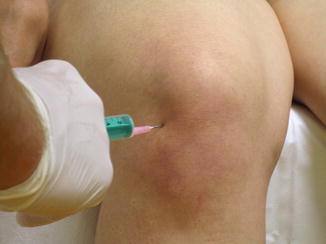
For injection only, the patient might be placed in a sitting position with legs hanging freely in 90° of flexion or supine (Fig. 41.2). The injection site is at the anteromedial or anterolateral region of the knee as used for arthroscopy. The needle should be advanced deep enough to prevent injection into the Hoffa’s fat pad. Scratching on the polyethylene liner or the femoral component needs to be avoided.


Fig. 41.2
Anterolateral approach to the knee joint, showing the soft spot, which is surrounded medially by the patella ligament, laterally by the medial condyle, and inferiorly by the medial tibial plateau
41.3 Arthroscopic Procedure
Arthroscopy has become the most common surgical procedure in orthopedic practice, and the numbers of arthroscopies performed are constantly growing. Along with the increased number in TKR revision, the number of arthroscopic investigations for diagnostic or therapeutic purposes will also increase.
In the situation of a painful patient after TKR, arthroscopy offers the combined benefit of visual assessment of the synovium, the TKR itself, and scar tissue, and biopsies can be easily obtained in different parts of the knee joint. If there is any suspicion of infection, aspiration should be performed routinely, but early biopsy should be considered if needed. The predictive value of preoperative biopsy in revision TKR has been shown to be 94.7 and 100 % [8]. Five samples in different areas of the knee joint should be obtained when infection is considered. This might decrease the chance of false-negative results and reduces the delay of sufficient management. The biopsies should be taken directly after establishment of the approaches prior to starting the flow of arthroscopic fluid. This is due to the fact that the dilution may lead to false-negative results in microbiological testing. The culture bottles for collecting the tissue samples should be placed directly on the operating table in order to reduce the risk of contamination.
Stay updated, free articles. Join our Telegram channel

Full access? Get Clinical Tree


