Lateral Closing Wedge Valgization Osteotomy of the Proximal Femur With Distal and Lateral Advancement of the Greater Trochanter
The greater trochanter and the upper femoral shaft are exposed with the use of the technique described in Procedure 7, steps A to K, on page 22. If the hip adductors are taut, they are released through a separate medial incision.
Operative Technique
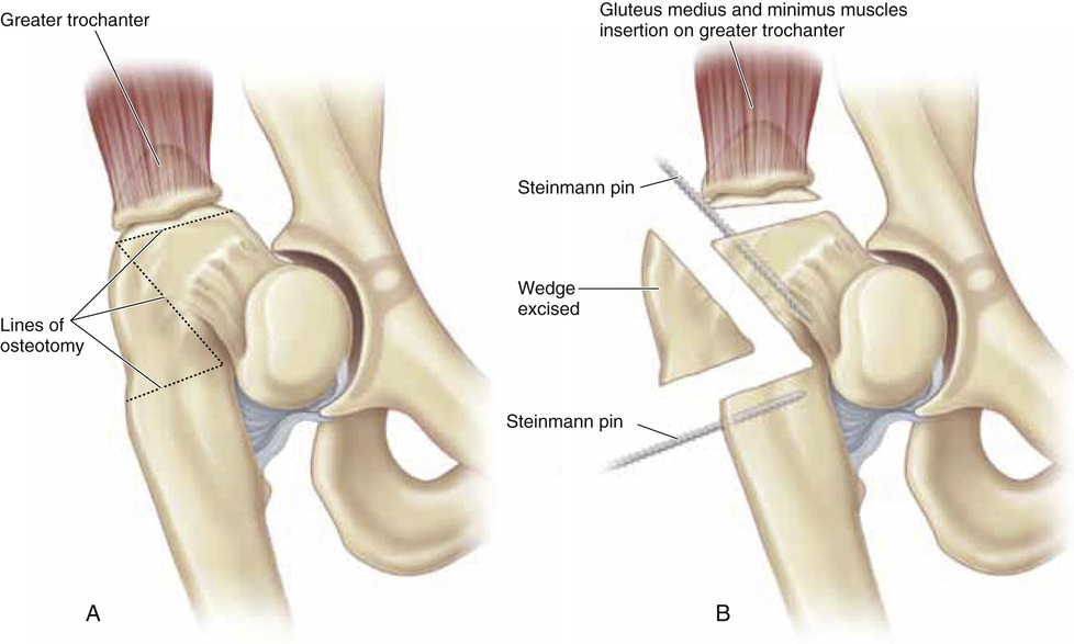
A and B, First, the greater trochanter is osteotomized with the use of the technique described for distal and lateral advancement. Next, two threaded Steinmann pins are inserted to serve as guides for the level and angle of the osteotomy. The apex of the osteotomy stops 1 cm short of the medial cortex. The length of the base of the wedge depends on the degree of correction of the coxa vara that is required. The wedge of bone is resected with an oscillating saw.
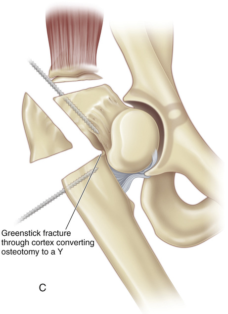
C, With a straight osteotome and leverage from the pins anchored in the femur, a greenstick fracture is produced in the medial cortex, thereby converting the osteotomy to a short-stemmed Y.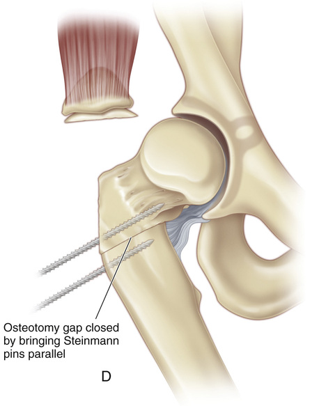
D, The osteotomy gap is closed by bringing the two Steinmann pins together and by aligning the neck, shaft, and the greater trochanter at a preoperatively determined angle.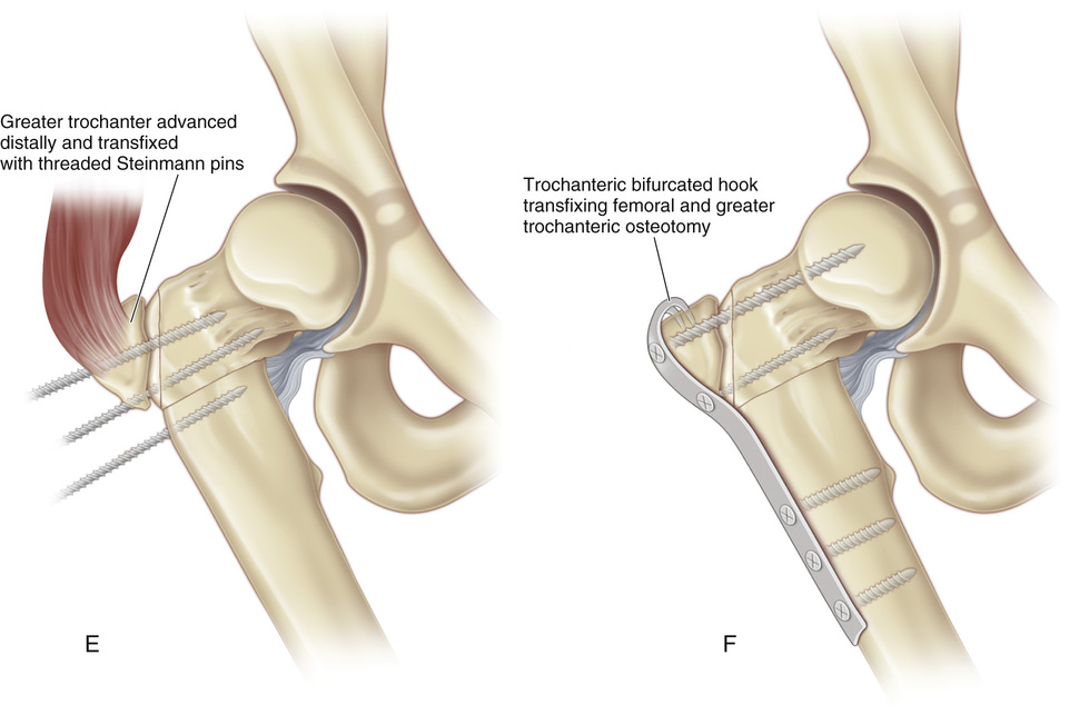
E, The greater trochanter is transfixed with a threaded Steinmann pin that is driven into the neck of the femur.
F, The three fragments are then fixed with a prebent trochanteric hook plate and screws.
Related posts:

Stay updated, free articles. Join our Telegram channel

Full access? Get Clinical Tree


