A, The surgical exposure of the greater trochanter and the upper femoral shaft is similar to that for the distal and lateral transfer of the greater trochanter (see Procedure 7, A to K, on page 22). B, The tip of the greater trochanter is at its normal level, so it is not necessary to advance it distally. It is kept horizontally level with the center of the femoral head, and its position is maintained by two wide-thread positional cancellous screws. The screws are inserted horizontally and perpendicular to the osteotomized lateral surface of the upper femur. The threads of these “positioning” screws grip the trochanter as well as the intertrochanteric region of the femur without compression. The cleft between the greater trochanter and the femur is filled with autogenous cancellous iliac bone, which is taken through a separate incision over the iliac apophysis. C, Internal fixation is augmented by a taut tension band of heavy wire suture that extends from the neck of each trochanteric screw to a small unicortical screw that is anchored 6 D, The detached vastus lateralis is then sutured to the insertion of the gluteus medius. The subcutaneous tissue and the skin are closed in the usual manner.
Lateral Advancement of the Greater Trochanter
Operative Technique
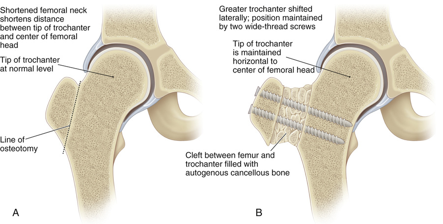
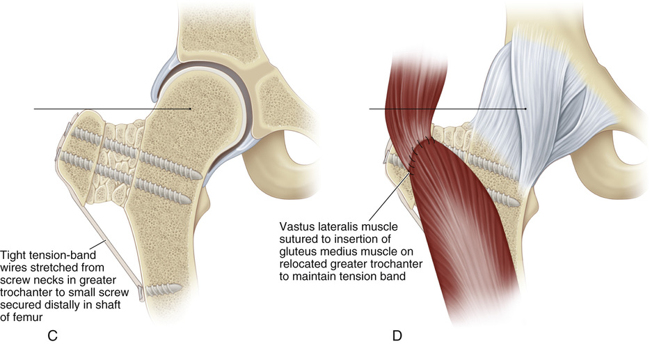 cm distally in the femur. This wire tension band counteracts the pull of the hip abductors.
Related posts:

Stay updated, free articles. Join our Telegram channel

Full access? Get Clinical Tree


