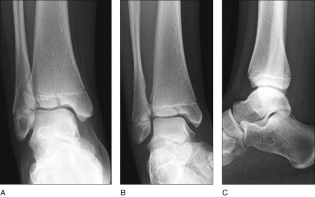
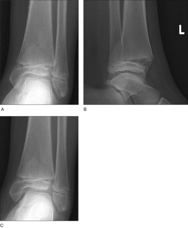
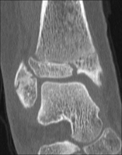
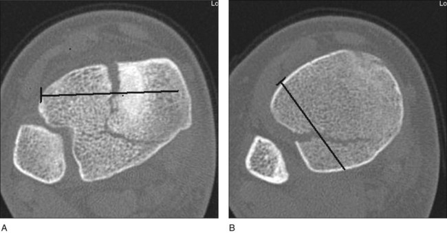
• Figure 1 shows anteroposterior (AP) (Fig. 1A), mortise (Fig. 1B), and lateral (Fig. 1C) radiographs of a three-part triplane fracture. • Figure 2 shows AP (Fig. 2A), mortise (Fig. 2B), and lateral (Fig. 2C) radiographs of an extra-articular medial triplane fracture. • Coronal (Fig. 3) and axial views should be used to accurately measure displacement (stepoff and gap) of the distal tibial articular surface. • Furthermore, coronal, axial, and sagittal CT views enable the surgeon to preoperatively plan the placement of internal fixation (i.e., screw trajectory) based on the fracture pattern, thus allowing for limited exposure of the fracture fragments. Figure 4 illustrates the use of axial CT images in surgical planning of screw placement at the epiphyseal (Fig. 4A) and metaphyseal (Fig. 4B) levels. Pearls
Triplane Fractures
Examination/Imaging
 Intra-articular triplane fractures of the distal tibia are anatomically complex Salter-Harris IV fractures usually consisting of a coronal fracture line in the metaphysis, a transverse fracture line within the physis, and a sagittal, intra-articular fracture through the epiphysis. On the anteroposterior (AP) view, the fracture appears to be a Salter-Harris III fracture; on the lateral view, it appears to be a Salter-Harris II fracture.
 Computed tomography (CT) permits identification of the number and location of fracture fragments.
Positioning
 The patient is placed in the supine position on a radiolucent operating table with a bump under the ipsilateral hip to internally rotate the limb and permit easy medial and lateral access to the ankle (Fig. 6).
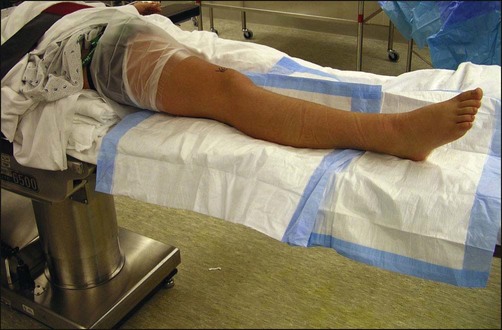
 A nonsterile tourniquet is placed around the ipsilateral thigh.
Related posts:
![]() 41: Operative Treatment of Tillaux Fractures of the Ankle
41: Operative Treatment of Tillaux Fractures of the Ankle
![]() 15: Repair of Proximal Hamstring Avulsion
15: Repair of Proximal Hamstring Avulsion
![]() 28: Femur Fracture: Closed Reduction and Spica Cast
28: Femur Fracture: Closed Reduction and Spica Cast
![]() 18: Percutaneous in situ Cannulated Screw Fixation of Slipped Capital Femoral Epiphysis
18: Percutaneous in situ Cannulated Screw Fixation of Slipped Capital Femoral Epiphysis
![]() 47: Resection of Calcaneonavicular Coalition and Fat Autograft Interposition
47: Resection of Calcaneonavicular Coalition and Fat Autograft Interposition
 60: Thoracoscopic Release and Instrumentation for Scoliosis
![]()
Stay updated, free articles. Join our Telegram channel

Full access? Get Clinical Tree


44: Triplane Fractures