Knee Fusion for Prosthetic Conversion in Proximal Focal Femoral Deficiency
Arthrodesis of the knee converts the proximal focal femoral–deficient limb into a stable limb by removing the intercalated segment. The original technique included a Syme-type ankle disarticulation. Subsequent modifications offer the option of rotationplasty, which retains the foot.
Operative Technique

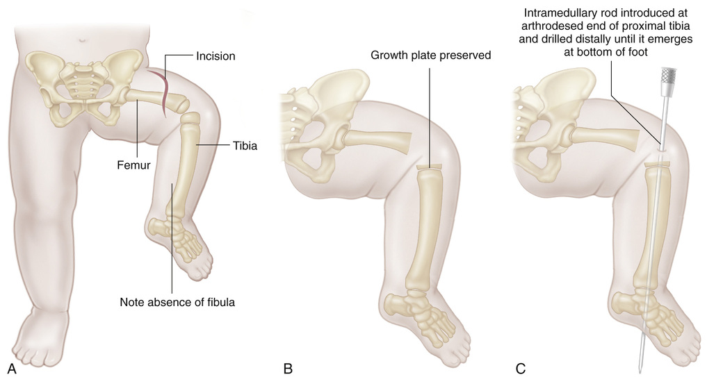
A, With the patient supine, an anterior S-shaped incision is made to expose the anterior aspect of the lower femur and upper tibia. Proximally, the incision is extended laterally to expose the lateral aspect of the upper femur.
B, The capsule and synovium of the knee joint are opened, and the articular cartilage of the upper end of the tibia is excised with an oscillating electric saw until the ossific nucleus of the epiphysis is seen. The distal femoral epiphysis is completely removed.
C, An 8-mm intramedullary nail is inserted. First it is inserted distally into the tibia, and it exits from the sole of the foot.
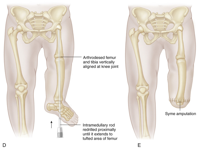
D, The nail is then passed proximally into the femur, thus impacting the lower end of the femur and the upper epiphysis of the tibia in extension. Care is taken to provide proper rotational alignment of the lower limb and ensure that the fused knee is not in flexion. The intramedullary nail should be in the center of the physes of the distal femur and the proximal tibia to avoid growth retardation.
The wound is closed in routine fashion. A one-and-one-half spica cast is applied for immobilization.
E, Syme amputation may be performed at this time or later if desired. The intramedullary nail is removed after 6 weeks.
Stay updated, free articles. Join our Telegram channel

Full access? Get Clinical Tree


