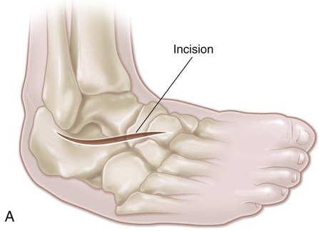
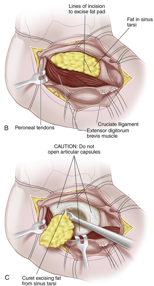
A, A 2-in-long, slightly curved incision is made over the subtalar joint, centered over the sinus tarsi. B, The incision is carried down to the sinus tarsi. The capsules of the posterior and anterior subtalar articulations are identified and left intact. The operation is extraarticular. If the capsule is opened inadvertently, it should be closed with interrupted sutures. The periosteum on the talus corresponding to the lateral margin of the roof of the sinus tarsi is divided and reflected proximally. The fibrofatty tissue in the sinus tarsi along with the periosteum of the calcaneus corresponding to the floor of the sinus tarsi and the tendinous origin of the short toe extensors from the calcaneus are elevated and reflected distally in one mass. C, The remaining fatty and ligamentous tissue from the sinus tarsi is thoroughly removed with a sharp scalpel and curet. D, Next the foot is manipulated into equinus position and inversion and the calcaneus rotated into its normal position beneath the talus to correct the valgus deformity. Broad straight osteotomes of various size (¾ to E, The optimal site of the bone graft bed is marked with the broad osteotome. A thin layer of cortical bone (⅛ to F, A bone graft of appropriate size can be taken from the anteromedial surface of the proximal tibial metaphysis as a single cortical graft, which is then cut into two trapezoidal bone grafts with their cancellous surfaces facing each other. We prefer to use fibular bone grafts with the cortices intact. The corners of the base of the graft are removed with a rongeur so that it is trapezoidal and can be countersunk into cancellous bone and thereby prevent lateral displacement after surgery. The bone graft is placed in the prepared graft bed in the sinus tarsi by holding the foot in varus position. An impactor may be used to fix the cortices of the graft in place. The longitudinal axis of the graft should be parallel to the shaft of the tibia with the ankle in neutral position. With the foot held in the desired position, the distal soft tissue pedicle of fibrofatty tissue of the sinus tarsi, the calcaneal periosteum, and the tendinous origin of the short toe extensors are sutured to the reflected periosteum from the talus. The subcutaneous tissue and skin are closed with interrupted sutures, and an above-knee cast is applied.
Extraarticular Arthrodesis of the Subtalar Joint (Grice Procedure)
Operative Technique
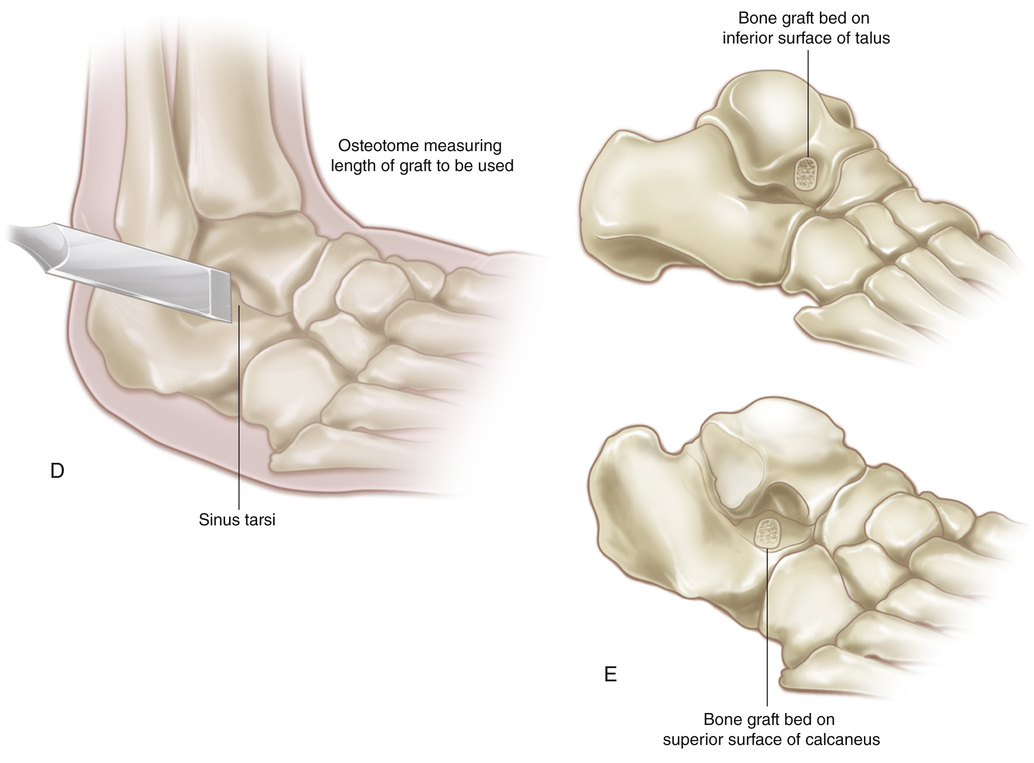
 in or more) are inserted into the sinus tarsi to block the subtalar joint and determine the length and optimum position of the bone graft and the stability that it will provide. The long axis of the graft should be parallel to that of the leg when the ankle is dorsiflexed into neutral position, and the hindfoot should be in 5 degrees valgus or neutral, but never varus. Even a slight degree of varus deformity of the heel seems to increase with growth.
 in) is removed with a dental osteotome from the inferior surface of the talus (the roof of the sinus tarsi) and the superior surface of the calcaneus (the floor of the sinus tarsi) at the site marked for the bone graft. It is best to preserve the most lateral cortical margin of the graft bed to support the bone block and prevent it from sinking into soft cancellous bone.
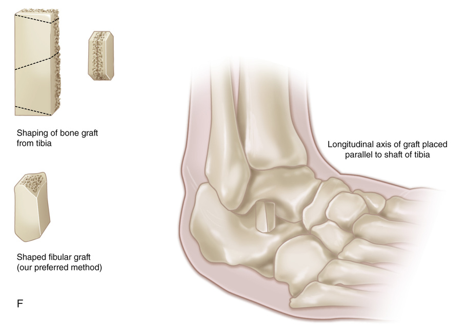
Related posts:
 23 Hamstring Lengthening
 29 Achilles Tendon—Distal Fibular Tenodesis for Mild Ankle Valgus in Skeletally Immature Patients
 10 Pemberton Osteotomy
 33 Triple Arthrodesis
 38 Exposure of the Spine for Posterior Instrumentation and Fusion
 42 Anterior Instrumentation of the Spine for Thoracolumbar or Lumbar Scoliosis
Stay updated, free articles. Join our Telegram channel

Full access? Get Clinical Tree


