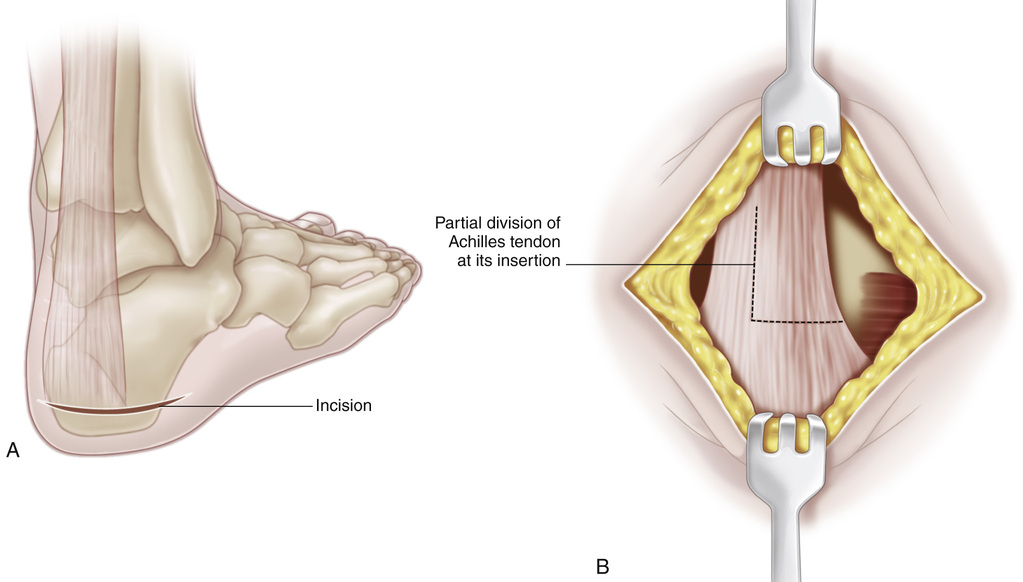
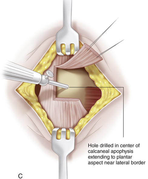
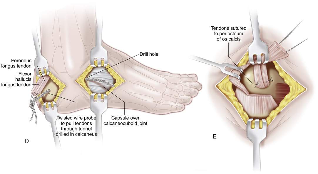
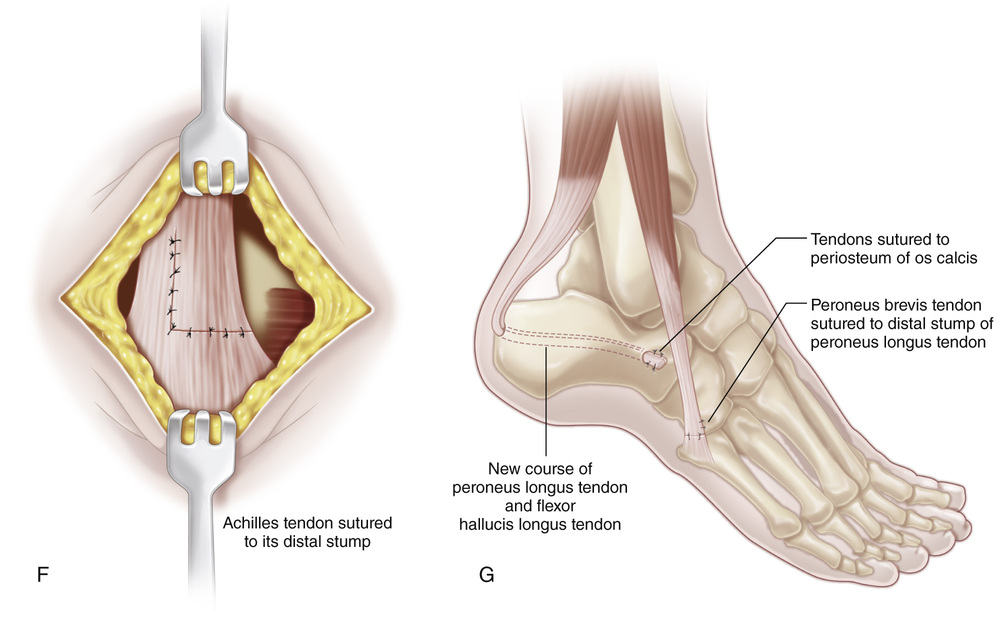
It is best to place the patient in the prone position to facilitate surgical exposure of the heel. The posterior tibial and peroneus longus and brevis tendons are divided distally at their insertion and delivered into the proximal wound. When the flexor hallucis longus tendon is to be transferred, its distal portion is sutured to the flexor hallucis brevis muscle. The anterior tibial tendon is delivered into the calf and heel through the interosseous route. A, A 5-cm-long posterior transverse incision is made around the heel along one of the skin creases in the part that neither presses the shoe nor touches the ground. B, The skin and subcutaneous flaps are undercut and reflected to expose the os calcis and the insertion of the Achilles tendon. An L-shaped cut is made in the lateral two thirds of the insertion of the Achilles tendon. The divided portion is reflected proximally to expose the apophysis of the os calcis. C, Next, with a D, Through a lateral incision, the intermuscular septum is widely divided between the lateral and posterior compartments. An Ober tendon passer is inserted through the wound and directed anterior to the Achilles tendon into the transverse incision over the os calcis. The threads of the whip sutures at the ends of the peroneal tendons are passed through the hole in the tendon passer and the tendons are delivered at the heel. The posterior tibial tendon is delivered at the heel by a similar route, through an incision in the intermuscular septum between the medial and posterior compartments and anterior to the Achilles tendon. Next, with a twisted wire probe, the tendons are inserted into the hole and pulled through the tunnel in the calcaneus. E, At their point of exit on the lateral aspect of the calcaneus the tendons are sutured to the periosteum and ligamentous tissues. The tendons are sutured under enough tension to hold the foot in 15 degrees of equinus when the remaining ankle dorsiflexors are fair in motor strength and in 30 degrees of equinus if they are good or normal. The tendons are sutured to each other and to the periosteum of the apophysis of the calcaneus at the posterior end of the tunnel. F and G, The divided portion of the Achilles tendon is resutured in its original position posterior to the transferred tendons. The wounds are closed and a long-leg cast is applied to hold the knee in 45 to 60 degrees of flexion and the hindfoot in 15 to 30 degrees equinus, but the forefoot in neutral position. Cavus deformity of the forefoot should be avoided.
Posterior Tendon Transfer to the Os Calcis for Correction of Calcaneus Deformity (Green and Grice Procedure)
Operative Technique


 -in drill, a hole is made through the calcaneus, beginning in the center of the apophysis and coming out laterally at its plantar aspect. With a diamond-head hand drill and curet, the hole is enlarged to receive all the transferred tendons.

Stay updated, free articles. Join our Telegram channel

Full access? Get Clinical Tree


