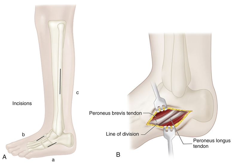
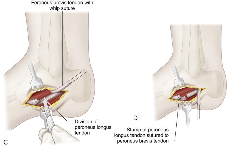
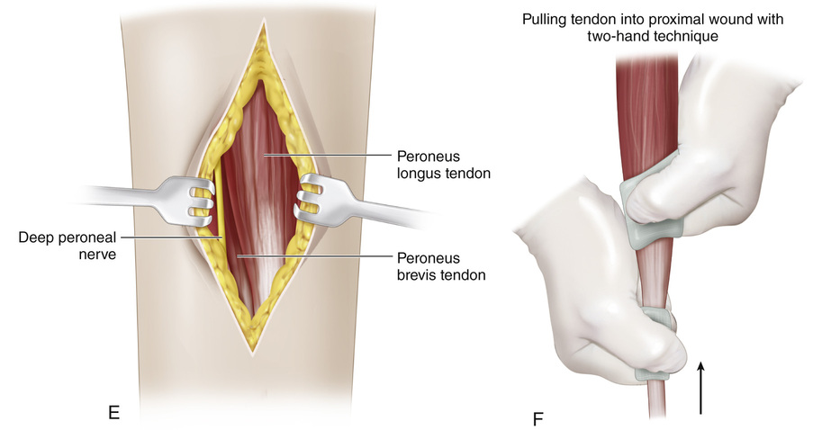
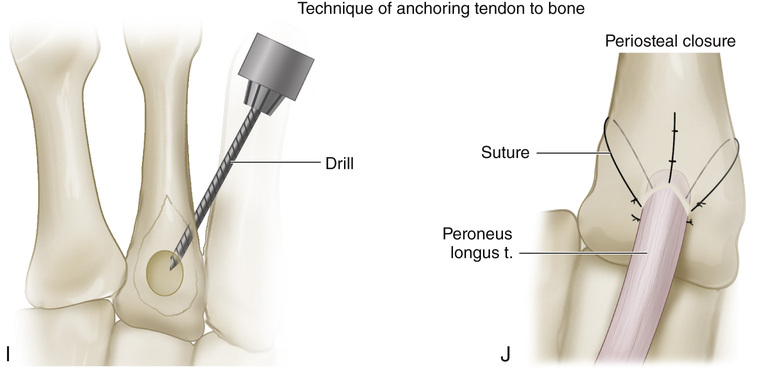
The patient is placed in a semilateral position with a sandbag under the hip on the affected side. A, A 3- to 4-cm-long incision (a) is made over the lateral aspect of the foot from the base of the fifth metatarsal to a point 1 cm distal to the tip of the lateral malleolus. Subcutaneous tissue is divided, and the tendons of the peroneus longus and brevis are exposed. A second incision (c) is then made over the fibular aspect of the leg; it begins 3 cm above the lateral malleolus and extends proximally for a distance of 7 cm. Subcutaneous tissue and deep fascia are incised, and the peroneal tendons are exposed by dividing their sheath. The peroneus longus tendon lies superficial to that of the peroneus brevis. The muscle is inspected to ensure that it is of normal gross appearance. B, Next the peroneus brevis muscle is detached from the base of the fifth metatarsal and a whip suture is inserted into its distal end. C and D, The peroneus longus tendon is divided as far distally as possible. The peroneus brevis tendon is sutured to the distal stump of the peroneus longus tendon to preserve the longitudinal arch and depression of the first metatarsal. E and F, The peroneus longus tendon is mobilized and, with a two-hand technique, gently pulled into the proximal wound in the leg. The origin of the peroneus brevis tendon from the fibula should not be disrupted. An adequate opening is made in the intermuscular septum with care taken not to injure any neurovascular structures. G and H, A 2- to 3-cm-long longitudinal incision is made over the dorsum of the foot (incision b in part A), centered over the base of the second metatarsal. The deep fascia is divided, and the extensor tendons are retracted to expose the proximal fourth of the second metatarsal. The periosteum is divided longitudinally and the cortex of the recipient bone is exposed. With an Ober tendon passer, the peroneus longus tendon along with its sheath is passed into the anterior tibial compartment, deep to the cruciate crural and tarsal ligaments, and delivered into the incision on the dorsum of the foot. We do not recommend a subcutaneous route. A direct line of pull of the peroneus longus tendon from its origin to its insertion should be ensured. I and J, A drill hole is made in the base of the second metatarsal. A star-head hand drill is used to enlarge the hole to receive the tendon adequately. The peroneus longus tendon is passed through the recipient hole and sutured on itself under correct tension. If the peroneus longus tendon is not of adequate length, two small holes are made 1.5 cm distal to the large hole at each side of the metatarsal shaft. The silk sutures at the end of the tendon are passed from the large central hole to the lateral distal small holes and the tendon is securely sutured to the bone. The ankle joint should be in neutral position or 5 degrees of dorsiflexion. The pneumatic tourniquet is released and hemostasis is obtained. The wounds are closed in routine manner. A long-leg cast is applied with the ankle in 5 degrees of dorsiflexion and the knee in 45 degrees of flexion. Postoperative care follows the guidelines outlined in the section on the principles of tendon transfer.
Anterior Transfer of the Peroneus Longus Tendon to the Base of the Second Metatarsal
Operative Technique

Related posts:
 26 Shelf Acetabular Augmentation
51 Correction of Hammer Toe by Resection and Arthrodesis of the Proximal Interphalangeal Joint
41 Sacro-Iliac Screws
58 Arthrodesis of the Ankle Joint via the Anterior Approach Without Disturbing the Distal Tibial Growth Plate
12 Ganz Periacetabular Osteotomy
66 Posterior Release of Elbow Extension Contracture
26 Shelf Acetabular Augmentation
51 Correction of Hammer Toe by Resection and Arthrodesis of the Proximal Interphalangeal Joint
41 Sacro-Iliac Screws
58 Arthrodesis of the Ankle Joint via the Anterior Approach Without Disturbing the Distal Tibial Growth Plate
12 Ganz Periacetabular Osteotomy
66 Posterior Release of Elbow Extension Contracture
Stay updated, free articles. Join our Telegram channel

Full access? Get Clinical Tree


