Submuscular Plating for Pediatric Femur Fractures
Indications
 This procedure is indicated for patients ≥ 5 years until skeletal maturity.
 Submuscular plating is ideally suited for comminuted or long-oblique unstable femur fractures.
 Submuscular plating is also a good option for proximal-third or distal-third femur fractures. In the proximal-third and distal-third fractures, there needs to be enough room for two to three screws in the proximal or distal diaphysis.
Examination/Imaging
 Anteroposterior (AP) and lateral radiographs of the femur are necessary. It is also critical to have clear radiographs of the ipsilateral hip and knee to evaluate the extent of the fracture and rule out a femoral neck or knee fracture.
 Figure 1 shows an AP radiograph of an unstable comminuted femur frature. Submuscular bridge plating is a clear treatment option in this fracture.
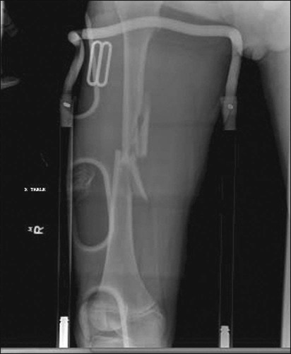
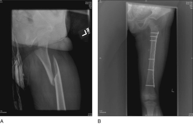
 Figure 2A presents an example of a long-oblique unstable femur fracture for which submuscular bridge plating is a good treatment option. The postoperative AP radiograph in Figure 2B shows the result after submuscular plating.
Surgical Anatomy
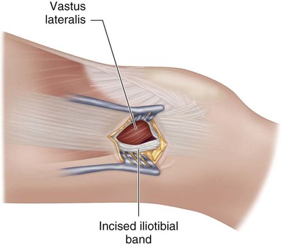
 The distal margin of the vastus lateralis muscle is deep to the iliotibial fascia in line with the proximal pole of the patella.
 The fibers of the distal vastus lateralis are oblique. The muscle is not attached to the bone, and there is a plane between the muscle and the lateral periosteum of the femur that is easily explored for plate insertion (Fig. 3).
Positioning
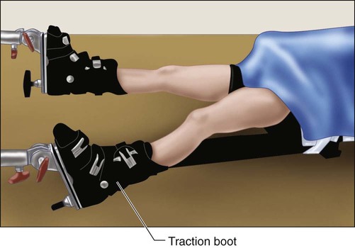
 Patients are positioned supine on a fracture table. The well leg is extended and slightly abducted to allow a true lateral fluoroscopic image of the fractured femur. Alternatively, a “well-leg” holder may also be used.
 Provisional reduction restoring femoral length and rotation is obtained with boot traction and verified fluoroscopically. Figure 4 shows patient positioning on the traction table with boot traction. In this case, the legs are scissored in the anterior-posterior direction to give the C-arm access to obtain clear AP and lateral images.
 In comminuted fractures, special attention should be given to rotation. Final alignment is performed with plate fixation as described later.
Portals/Exposures
 A small (4- to 7-mm) incision is made at the distal lateral thigh. The incision is midway anterior-posterior and as distal as the proximal pole of the patella. The incision is similar to the lateral incision for retrograde insertion of elastic nails.
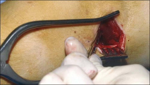
 As the dissection is advanced through the tensor fascia lata, the distal oblique fibers of the vastus lateralis muscle will be exposed (Fig. 5). Blunt dissection is performed deep to the distal muscle fibers to enter the plane between the vastus lateralis and lateral femur periosteum. This plane is easily entered and allows proximal plate advancement with minimal force.
Musculoskeletal Key
Fastest Musculoskeletal Insight Engine


