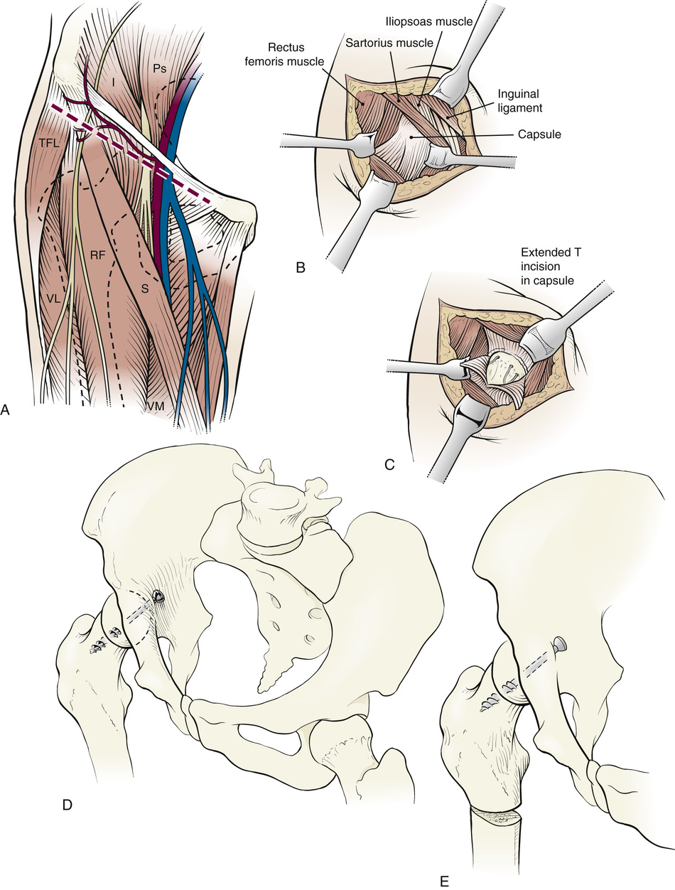
Intraarticular Hip Fusion for Avascular Necrosis
A, The patient is positioned on a radiolucent tabletop with the entire affected lower extremity draped free and with C-arm fluoroscopy available. An anterior approach to the hip is best with this technique, exposing the anterior femoral capsule and subsequently the inner aspect of the ilium for insertion of the screws across the ilium into the femoral head and neck. I, Iliac muscle (m.); Ps, psoas major m.; RF, rectus femoris m.; S, sartorius m.; TFL, tensor fasciae latae m.; VL, vastus lateralis m.; VM, vastus medialis m.
B, The anterior capsule of the hip is exposed broadly through this approach. The iliac muscle is also stripped from the inner wall of the pelvis.
C, The femoral head is exposed and dislocated from the acetabulum. Necrotic bone is removed from the femoral head with rongeurs and cup arthroplasty reamers. The acetabulum is similarly prepared with curets and cup arthroplasty reamers, removing all articular cartilage and sclerotic bone.
D, The denuded femoral head is reduced into the acetabulum into a “best fit” position. One or two cannulated screws are then inserted from the inner wall of the pelvis across the joint into the femoral head and neck. Fluoroscopic visualization may be required for optimum insertion and control of the depth of insertion into the femoral neck.
E, An intertrochanteric or subtrochanteric osteotomy is made to allow repositioning of the leg in a position of slight abduction and external rotation. Normally the leg should be placed in a position of extension in the supine position. The upper femur may be exposed through a separate lateral incision, or by extending the anterior exposure and reflecting the vastus lateralis from the anterior aspect of the femur. If the osteotomy is unstable, an intramedullary rod (e.g., a Rush rod) may be inserted into the medullary canal for partial control of the femoral fragments.
Postoperative Management
The patient is placed in a one-and-one-half-hip spica cast until union of the fusion and femoral osteotomy. Alternatively, external fixation of the pelvis to the lower femoral fragment may be used.

Stay updated, free articles. Join our Telegram channel

Full access? Get Clinical Tree


