Diagnostic imaging has been an essential component of chiropractic practice since the early days of the profession. Imaging studies allow the clinician deeper insight into the structural aspects of the body, permitting a degree of specificity in neuromusculoskeletal diagnosis that would otherwise be impossible. Plain-film radiography and other imaging methods allow chiropractors to rule out the presence of dangerous pathologic conditions before proceeding with manual chiropractic procedures and to refer appropriate cases for medical care or co-management.
All chiropractic colleges provide extensive training in radiology as part of the required curriculum, sufficient to prepare the graduate to own and operate an x-ray machine and to interpret basic radiographic findings. All North American state and provincial licensing laws include radiology within the chiropractor’s scope of practice, as do those in many other nations.
Chiropractic practice, with its neuromusculoskeletal focus, requires significant knowledge in skeletal radiology. Recent research by Taylor and colleagues1 has evaluated students, clinicians, radiology residents, and radiologists from the chiropractic and medical professions in their interpretation of abnormal lumbar spine radiographs. The researchers found no significant difference between the performance of chiropractic radiologists and medical skeletal radiologists or between chiropractic and medical clinicians. Moreover, the chiropractic radiologists, chiropractic radiology residents, and chiropractic students scored significantly higher than the corresponding medical categories (general medical radiologists, medical radiology residents, and medical students, respectively).
HISTORY OF DIAGNOSTIC IMAGING
1895—On November 8, 1895 in Würzburg, Germany, Wilhelm Conrad Roentgen discovers x-rays while investigating the properties of cathode ray (Crookes) tubes. Shortly thereafter, Roentgen takes an x-ray of his wife Bertha’s hand, an exposure that requires more than 15 minutes.
1898—Thomas A. Edison creates the fluoroscope whereby x-rays are projected on an intensifying screen coated with calcium tungstate as the fluorescent material. Thus human anatomy can be seen in real time. Fluoroscopy was the first widespread application for x-ray technology, allowing visualization of the beating heart, diaphragmatic movement, and intestinal peristalsis. Permanent photographic images were more challenging to obtain because the image was supported on a glass plate; the final image was heavy and subject to breakage. Unfortunately, practitioners were unaware of the whole-body radiation dose delivered to the patient and the physician during such procedures.
Because the deleterious effects of ionizing radiation were unknown at the dawn of the twentieth century, no protection was provided for the patient or physician. Radiation physicians tested the hardness (quality) of the x-ray beam by putting their own hands into the x-ray beam and viewing the resulting image. Within a few short years, some of the adverse biologic effects (skin cancer, thyroid cancer, and cataracts) of radiation exposure became manifest in these physicians who were excessively exposed to this type of radiation on a daily basis.
Fluoroscopy is currently used for contrast studies of the gastrointestinal and genitourinary tracts. The use and interpretation of fluoroscopy in assessing cervical spinal motion is being evaluated.
1901—Roentgen receives the Nobel Prize in physics for the discovery of x-rays.
1910—Palmer School of Chiropractic in Davenport, Iowa, purchases the first x-ray unit in the chiropractic profession.
1913—The Coolidge hot-filament x-ray tube is developed. Belgian glass was in short supply during World War I, thus the Eastman Kodak Company produced a cellulose-based film whereby the emulsion that contains the actual image was coated on one side only.
1917—The Potter Bucky moving grid is introduced, improving contrast on images of large body parts and making larger film sizes (14 × 17 inches) practical. This introduction signals the end of glass plates as film base material.
1922—Arthur Holly Compton describes the scattering of x-rays (Compton effect) for which he later receives the Nobel Prize in 1927.
1929—The rotating anode tube is introduced. Rotating the anode during x-ray production spreads out the heat that is produced and allows for greater radiographic technique (milliampere seconds [mAs]) to be used. By this time, virtually all chiropractic colleges have courses in radiology.
1935—The first standing, full-spine film is taken by New York City chiropractor Warren Sausser using a specially made film, 14 × 36 inches, from the Eastman Kodak Company.
1936—Tomography is introduced whereby reciprocal movement of the x-ray tube and film blur the anatomy above and below the level of interest within the patient’s body, allowing the anatomy at the plane of interest to be visualized in greater detail.
1936—The National Council of Chiropractic Roentgenologists (NCCR) is founded. Members were chiropractors in general practice with particular interest in radiography and spinography.
1946—Purcell and colleagues and Bloch and colleagues publish the principles of nuclear magnetic resonance. The use of magnetic fields for chemical spectroscopy is applied in the analysis of compounds in solution and for determining the structure of biologic macromolecules. 2,3
1946—National Council of Chiropractic Roentgenologists (NCCR) holds its first meeting at the Palmer House Hotel in Chicago. Speakers include Drs. Leo Wunsch, John Teranel, Fred Baier, Ted Vladeff, and Warren Sausser. 4
1951—The first nuclear medicine scans of the thyroid gland are produced by Cassen and Curtis. 5
1956—Eastman Kodak Company introduces the first automatic roller transport film processor.
1958—The American Chiropractic Board of Roentgenology (ACBR) is formed for specialty certification of chiropractic radiologists. The original qualifications to sit for the certifying examination included 200 hours of lecture and maintaining an active chiropractic practice with at least 5-years’ use of x-rays. The first three chiropractors to pass the certifying examination were Drs. Joseph Janse, Lester P. Rehberger, and Earl A. Rich. The professional designation for candidates who pass the examination is Diplomate of the American Chiropractic Board of Roentgenology (DACBR).
1964—The first annual gathering of chiropractic radiologists takes place at Lincoln Chiropractic College, Indianapolis, Indiana.
1966—Diagnostic ultrasound, which grew out of sonar technology in World War II, is commonly available.
1967—The first resident training program in chiropractic radiology is instituted at the National College of Chiropractic in Lombard, Illinois. Three academic years, encompassing 4400 hours of training, is required before eligibility for certification is possible. Drs. James F. Winterstein and Donald B. Tomkins are the first resident-trained candidates to become certified as DACBRs after training with chiropractic radiologist Leonard Ritchie.
1968—The American Chiropractic College of Roentgenology (now the American Chiropractic College of Radiology) is formed.
1973—Hounsfield creates the first computed tomography (CT) scanner, demonstrating human anatomy in a series of axial “slices.” CT begins to replace most plain-film tomography applications. 6
1973—Raymond Damadian and Paul Lauterbur produce the first nuclear magnetic resonance images (now known as magnetic resonance imaging [MRI]), demonstrating the distribution of charged polar protons (primarily hydrogen protons). 7,8
1974—Rare earth intensifying screens are introduced. Rare earth screen phosphors are doubly efficient at converting x-ray photon energy into light energy as compared with non-rare earth compounds such as calcium tungstate. A significant reduction in patient exposure dose is realized from this innovation in x-ray image production. 9
1980—The first commercial superconducting MRI magnet is introduced by Technicare.
1983—The Eastman Kodak Company develops the first tabular grain film emulsion. Tabular grain films allow for less silver to be used per film, as well as thinner emulsions that contain the actual image. Thinner emulsions result in thinner films, thus reducing the parallax effect and producing more detailed images.
1985—Medium-frequency x-ray generators (6 kHz) are made commercially available, and in 1987, 100-kHz true high-frequency generating systems are developed. These systems are supplied by single-phase electrical power sources but have radiographic power equal to 3-phase generating systems found in hospitals, making this technology affordable for private practices. An added benefit is a significant radiation dose reduction for patients.
1990—Toshiba introduces spiral CT, which allows for a single but continuous exposure of a large body part, such as the chest or abdomen, in a single breath-hold. The total time in the scanner for the patient is reduced, and greater patient throughput is possible.
1998—Multiple rows of detectors (from 4 to 16 rows) and slice acquisition times under 1 second make multislice CT practical for imaging large body cavities such as the chest or abdomen.
CHIROPRACTIC APPLICATIONS FOR DIAGNOSTIC IMAGING
Diagnostic imaging in chiropractic practice includes imaging studies (primarily plain-film x-ray) performed or supervised by the chiropractor and other studies (primarily MRI, CT, and diagnostic ultrasound) performed by chiropractic or medical radiology specialists on referral from the treating chiropractor. At present, most chiropractic general practices have an on-site x-ray machine, but very few have an on-site CT or MRI facility. Therefore patients needing such specialized procedures are referred to larger radiology facilities.
X-Ray
Radiography is the production of photographic images by passing x-rays (a form of electromagnetic energy with a short wavelength) through the body. X-rays should be taken only when a clinical indication exists that they are needed. Radiographs may be indicated to rule out contraindications to adjustment/manipulation, such as fracture, dislocation, tumor, or infection, and to evaluate structure and function. This use is sometimes considered a “rule-out” situation in which a single diagnosis is not obvious from the patient’s history and physical examination. In a typical chiropractic setting, an older patient might present with localized pain in the neck that radiates into one of the upper extremities. A common cause for this situation might be disk degeneration with or without bone spur (osteophyte) formation at the Luschka joint, resulting in intervertebral foraminal stenosis (Fig. 11-1). In such a case, a series of neck x-rays should be obtained that would include oblique views showing the intervertebral foramina. If the oblique films demonstrate the foramina to be narrowed by osteophytes, this would confirm a diagnosis of degenerative foraminal stenosis and would be the likely source of the patient’s complaint of pain radiating from the neck.
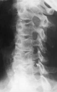 |
| Fig. 11-1 This oblique x-ray of the cervical spine demonstrates degenerative narrowing of the C5-C6 foramen resulting from degenerative hypertrophy of the Luschka joint (arrow). (From Yochum TR, Rowe LJ: Essentials of skeletal radiology, ed 2, Baltimore, 1996, Williams & Wilkins. Reprinted with permission of Lippincott-Williams & Wilkins.) Lippincott-Williams & Wilkins |
Another patient may present with a productive cough and an elevated temperature. This presentation suggests the potential for pneumonia. Obtaining posteroanterior and lateral chest x-rays to see if a pulmonary consolidation (fluid accumulation in the airspace of the lung) is present would be most prudent, thus confirming a diagnosis of pneumonia. If consolidation is not evident on the radiographs, then another cause for the symptoms must be sought.
Other clear indications for a chiropractor to order radiographs include concern for the presence of spondylolisthesis, congenital or functional short leg, scoliosis, intervertebral fixations, congenital anomalies and variants, and clinical manifestations of vertebral subluxation complex.
Flexion, extension, and lateral bending views of the cervical or lumbar spine may follow static (neutral posture) radiographs, enabling the clinician to evaluate global range of motion, intersegmental biomechanics, and intersegmental stability. Cervical flexion and extension views are commonly obtained. Lateral bending of the cervical spine, lumbar flexion and extension, and lumbar lateral bending views are also obtained.
Fluoroscopy
Fluoroscopy is an x-ray procedure that allows real-time visualization of physiologic motion within the human body. During fluoroscopy, the clinician can watch the progression of a bolus of swallowed barium moving through the esophagus and stomach or peristaltic waves moving iodinated dye down the ureter. Rarely, fluoroscopy is used to watch the cervical spine move in flexion, extension, and lateral bending.
Although the radiation dose associated with traditional fluoroscopy is substantial, it is currently the only practical means to see the dynamics of intestinal and genitourinary function.
Gastrointestinal Fluoroscopy
Upper Gastrointestinal Study
An adult male patient may present with neck pain and difficulty swallowing. Diagnostic considerations for such a presentation include compression of the upper esophagus by large osteophytes on the anterior aspect of the lower cervical vertebrae or possibly a mass arising from the upper esophagus or the surrounding soft tissues. Lateral x-rays of the cervical spine may demonstrate thickening of the soft tissues in front of the C6 and C7 vertebrae, lateral displacement of the tracheal air shadow on the anteroposterior (AP) film, or both. These radiographic findings suggest a tumor of the upper esophagus or adjacent soft tissues.
To further investigate this finding, the chiropractor would be obligated to refer the patient for a barium study that evaluates the upper gastrointestinal (GI) system. For this study, the patient drinks a barium mixture that, when x-rayed, shows the cavity of the esophagus or stomach as white. Displacement of the barium within the esophagus would then confirm the presence of the tumor, and referral to a gastroenterologist would be indicated for treatment of the tumor.
Another example would be a case in which, responding to a local television appeal, a 60-year-old man obtains a screening test from the local pharmacy and discovers that occult blood is present in the stool. This man is a chiropractic patient and reports this finding. The chiropractor would then order a confirmatory laboratory test, which also comes back positive. Occult blood in the stool suggests that the bleeding has originated high in the GI system (esophagus or stomach). The most common reason for this bleeding includes gastric ulcer or neoplasm. Referring the patient for an upper GI study to localize the origin of the bleeding would be reasonable and prudent.
If the study shows evidence of a gastric ulcer, the chiropractor may counsel the patient on diet and lifestyle changes to reduce stress while adjusting the spine for any subluxations. However, if the study showed evidence of a tumor of the stomach, the chiropractor would initiate a medical referral for appropriate treatment.
Lower Gastrointestinal Study
Diseases of the lower intestine may express sequelae in the spine. Seronegative enteropathic diseases (certain bowel diseases that do not express a positive rheumatoid factor in the blood) such as Crohn’s disease and ulcerative colitis may also cause spinal arthritic disease. These changes include inflammatory bone spurs on the vertebral bodies called syndesmophytes and, in some cases, inflammatory changes affecting the sacroiliac, hip, and shoulder joints.
A chiropractor may first find these distinctive spinal and sacroiliac changes on spinal x-rays and then have to refer the patient for a lower GI study to document any associated intestinal disease.
Genitourinary Fluoroscopy
Kidney disease can be a cause of back pain. To rule out the genitourinary system as the primary cause of back pain, a chiropractor may choose to order fluoroscopy as a specific imaging test to rule out kidney disease or obstruction of the outflow of urine.
Intravenous Pyelogram
Occasionally, on a lumbar spine x-ray, a calcification will be seen lateral to the L2 or L3 vertebra on the AP film, and the calcification superimposes on these same vertebral bodies on the lateral view. The radiographic appearance and anatomic location suggests a kidney stone. To differentiate a kidney stone from a calcified lymph node, referral for an intravenous pyelogram (IVP) would be necessary.
To obtain an IVP study, an iodinated dye is injected into the bloodstream, and the dye is then filtered by normally functioning kidneys. If the calcification is shown to displace the dye as it collects in the renal pelvis, the renal stone is confirmed. Once the kidney stone is confirmed, then referral to a nephrologist or internist is appropriate.
Related posts:

Stay updated, free articles. Join our Telegram channel

Full access? Get Clinical Tree


