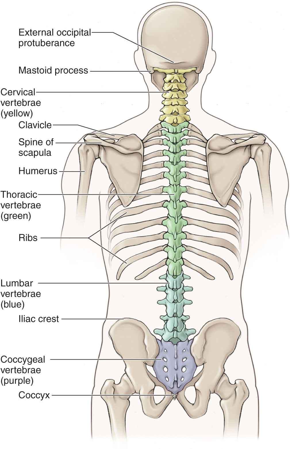
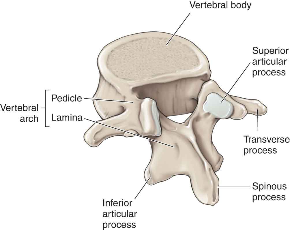
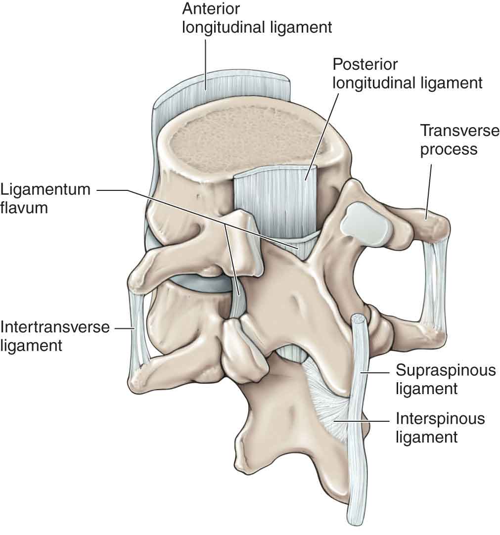
The design specification for the human vertebral column is the provision of structural stability affording full mobility as well as protection of the spinal cord and axial neural tissues.1 While achieving these seemingly disparate objectives, the spine also contributes to the functional requirements of gait and to the maintenance of static weight-bearing postures (see Chapter 6).1 At the component level, the basic building block of the spine is the vertebra. The vertebra serves as the weight-bearing unit of the vertebral column, and it is well designed for this purpose. Although a solid structure would provide the vertebral body with sufficient strength, especially for static loads, it would prove too heavy and would not have the necessary flexibility for dynamic load bearing.2 Instead, the vertebral body is constructed with a strong outer layer of cortical bone and a hollow cavity, the latter of which is reinforced by vertical and horizontal struts called trabeculae. The term vertebral column describes the entire set of vertebrae excluding the ribs, sternum, and pelvis (Fig. 22-1). The normal vertebral column is made up of 29 vertebrae (7 cervical, 12 thoracics, 5 lumbar, and 5 sacral) and three or four coccygeal segments. The adage that “function follows form” is very much applicable when studying the vertebral column. Although all vertebrae have similar characteristics, each has specific details that reflect its unique function. The overall contour of the normal vertebral column in the coronal plane is straight. In contrast, the contour of the sagittal plane changes with development. At birth, a series of primary curves give a kyphotic posture to the whole spine. With the development of the erect posture, secondary curves develop in the cervical and lumbar spines, producing a lordosis in these regions. The curves in the spinal column provide it with increased flexibility and shock-absorbing capabilities.2 FIGURE 22-1 The vertebral column. (Reproduced, with permission, from Chapter 1. Back. In: Morton DA, Foreman K, Albertine KH. eds. The Big Picture: Gross Anatomy. New York, NY: McGraw-Hill; 2011.) Functionally, the vertebral column consists of anterior and posterior columns. The anterior column is the hydraulic and weight-bearing portion that provides the vertebral column with its shock-absorbing capability and consists of the vertebral bodies and intervertebral disks (IVD).3 The posterior column consists of the structures that provide the gliding mechanism for movement, the articular processes, and the zygapophyseal (facet) joints.3 A motion segment in the vertebral column is defined as two adjacent vertebrae and consists of three joints. One joint is formed between the two vertebral bodies and the IVD of the anterior column. The other two joints are formed by the articulation of the superior articular processes of the inferior vertebra and the inferior articular processes of the superior vertebra (Fig. 22-2) of the posterior column. FIGURE 22-2 The vertebral body. (Reproduced, with permission, from Chapter 1. Back. In: Morton DA, Foreman K, Albertine KH. eds. The Big Picture: Gross Anatomy. New York, NY: McGraw-Hill; 2011.) The vertebral column contains 24 pairs of zygapophyseal joints, which project from the neural arch of the vertebrae. The regional characteristics of the zygapophyseal joints are described in the relevant chapters. Mechanically, zygapophyseal joints are classified as plane joints as the articular surfaces are essentially flat.4 The articular surfaces are covered with hyaline cartilage and, like most synovial joints, have small fatty or fibrous synovial meniscoid-like fringes that project between the joint surfaces from the margins.5 These intra-articular synovial folds act as space fillers during joint displacement and actively assist in the dispersal of synovial fluid within the joint cavity.1 The articular processes act as a mechanical barricade, particularly against excessive torsion and shear, permitting certain movements while blocking others4: Most zygapophyseal joint surfaces are oriented somewhere between the horizontal and vertical planes. In the cervical spine, the zygapophyseal joints are relatively horizontal while progressively increasing toward 45 degrees to the horizontal from the upper to the lower segments.6–9 In the thoracic region, the joints assume an almost vertical direction while remaining essentially in a coronal orientation, which facilitates axial rotation and resists anterior displacement.10 In the lumbar spine, the zygapophyseal joints are vertical with a curved, J-shaped surface predominantly in the sagittal plane, which restricts rotation and also resists anterior shear.1 Understanding the variable structure and function of the human zygapophyseal joints and their relationship with the other components of the vertebral column is an important requirement for the examination and intervention of individuals with mechanical spinal pain disorders.1 The IVDs of the vertebral column lie between the adjacent superior and inferior surfaces of the vertebral bodies from C2 to S1 and are similar in shape to the bodies (Fig. 22-3). The IVD forms a symphysis or amphiarthrosis between two adjacent vertebrae and represents the largest avascular structure in the body. Each disk is composed of an inner nucleus pulposus (NP), an outer annulus fibrosus (AF), and limiting cartilage end plates. The annulus and end plates anchor the IVD to the vertebral body. In cervical and lumbar regions, the IVDs are thicker anteriorly, and this contributes to the normal lordosis. In the thoracic region, each of the IVDs is of uniform thickness. Phylogenetically, the IVD is a relatively new structure. In the human spinal column, the combined heights of the IVDs account for approximately 20–33% of the total length of the spinal column.11 The presence of an IVD not only permits motion of the segment in any direction up to the point that the disk itself is stretched, but also allows for a significant increase in the weight-bearing capabilities of the spine.12 A normally functioning disk is extremely important to permit the normal biomechanics of the spine to occur and to reduce the possibility of mechanical interference among any of the neural structures. Although the disk is incapable of independent motion, movement of the disk does occur during the clinically defined motions of flexion–extension, side bending, and axial rotation.13 The major stresses that must be withstood by the IVD are axial compression, shearing, bending, and twisting, either singly or in combination with each other. FIGURE 22-3 The intervertebral disk and ligamentous structures. (Reproduced, with permission, from Chapter 1. Back. In: Morton DA, Foreman K, Albertine KH. eds. The Big Picture: Gross Anatomy. New York, NY: McGraw-Hill; 2011.) The role of the IVD is unique because it operates as an osmotic system, holding neighboring vertebral bodies together while simultaneously pushing them apart. As such, the IVD is a dynamic structure that responds to stresses applied from vertebral movement or from static loading. There are regional differences within the spine, each with its own specific demands and function. As discussed, all vertebral disks have traditionally been described as being composed of three parts: the AF, the vertebral end plate, and a central gelatinous mass, called the NP (Fig. 22-1). However, this description is based on the anatomy of a lumbar disk, the region where most research has occurred and for which many authors have extrapolated the anatomy to all IVDs.14 An appreciation of the differing anatomy of the IVDs, throughout the spine, which are described in the separate chapters, is important when developing clinical examination and intervention models.14 The spine contains four junctions, each of which is different in posterior element orientation and spinal curvature. Transitional vertebrae can occur at any of the junctions. The transition can be “complete” but is more commonly partial. These junctions, described by Schmorl and Junghanns15 as ontogenically restless, are often rich in anomalies16: Although highly variable, the line of gravity acting on a standing person with ideal posture passes through the mastoid process of the temporal bone anterior to the second sacral vertebra, posterior to the hip, and anterior to the knee and ankle (see Chapter 6).4 In the vertebral column, the line of gravity is on the concave side of the apex of each region’s curvature. As a consequence, ideal posture allows gravity to produce a torque that helps maintain the optimal shape of each spinal curvature.4 The movements of the vertebral column occur in diagonal patterns as a combination of flexion or extension with a coupled motion of side bending and rotation. Since the spine can move from top-down or bottom-up, the motion at a functional unit is defined by what is occurring at the anterior portion of the vertebral body of the superior vertebra. For example, at the L3–4 segments, flexion involves an anterior motion of the L3 vertebra relative to the L4 vertebra, and extension involves a posterior motion of the L3 vertebra relative to the L4 vertebra. Movements of the spine, like those elsewhere, are produced by the coordinated action of nerves and muscles. Agonistic and synergistic muscles initiate and perform the movements, whereas the antagonistic muscles control and modify the movements. The amount of motion available in each region of the spine is a factor of a number of variables. These include the following: The type of motion available is governed by: Although the range of motion (ROM) at each vertebral segment varies, the relative amounts of motion that occur at each region is well documented11,17 Although various motion patterns have been proposed for the sacroiliac joint,18–21 the precise model for sacroiliac motion has remained fairly elusive (see Chapter 29).22–24 Postmortem analysis has shown that until an advanced age, small sacroiliac movements are measurable under different load conditions.25,26 Including translations and rotations around three different axes, the spine is considered to possess 6 degrees of freedom. These include flexion/extension, side bending, rotation, compression/distraction, anterior/posterior shear, and lateral shear.
CHAPTER 22
Vertebral Column


 Horizontal articular surfaces favor axial rotation.
 Vertical articular surfaces (in either sagittal or frontal planes) act to block axial rotation.

SPINAL JUNCTIONS
 The craniovertebral junction is located between the cervical spine and the atlas, axis, and head. This region is covered in Chapter 23.
 The cervicothoracic junction represents the region where the mobile cervical spine and the relatively stiffer superior segments of thoracic spine meet, and where the powerful muscles of the upper extremities and shoulder girdle insert. The cervicothoracic junction is described in Chapters 25 and 27.
 The thoracolumbar junction is located between the thoracic spine, with its large capacity for rotation, and the lumbar spine, with its limited rotation. This region is described in Chapter 27.
 The lumbosacral junction is located between the lumbar spine, with its ability to flex and extend, and the relative stiffness of the sacroiliac joints. This region is described in Chapters 28 and 29.
SPINAL MOTION
 Disk–vertebral height ratio
 Compliance of the fibrocartilage
 Dimensions and shape of the adjacent vertebral end plates
 Age
Age
 Disease
 Gender
 The shape and orientation of the articulations
 The ligaments and muscles of the segment, and the size and location of its articulating processes
Flexion/Extension
At the segmental level, flexion produces a combination of an anterior roll and an anterior glide of the vertebral body in the sagittal plane. The anterior portion of the vertebral bodies approximate, and the spinous processes separate. The reverse occurs with extension.
Side Bending
Side bending, which occurs in the frontal plane, is a complex and highly variable movement involving side bending and rotatory movements of the interbody joints and a variety of movements at the zygapophyseal joints.27 During side bending, the lateral edges of the vertebral bodies approximate on the side toward which the spine is bending, compressing the IVD, and separate on the opposite side, distracting the IVD.
Rotation
Irrespective of whether the movement occurs from the pelvis upward, or from the head down, rotation to the left results in a relative movement of the body of the superior vertebra to the left, and its spinous process to the right. The opposite occurs with rotation to the right. During rotation, torsion of the IVD occurs.
Compression/Distraction
Compression (approximation) or distraction (separation) occurs with a longitudinal force.
Anterior/Posterior Shear
An anterior shear occurs when the body of the superior vertebra translates anteriorly on the vertebra below. A posterior shear occurs when the body of the superior vertebra translates posteriorly on the vertebra below.
Lateral Shear
A lateral shear occurs when the body of the superior vertebra translates sideways on the vertebra below. This commonly occurs during side bending.
In general, the human zygapophyseal joints of the spine are capable of only two major motions: gliding upward and gliding downward. If these movements occur in the same direction, flexion or extension occurs. If the movements occur in opposite directions, side bending occurs. Although rotation does occur as a motion component within intervertebral segments, it is always coupled and never an isolated motion.28 Indeed during functional rotation of the spine, the actual motion occurring at any given zygapophyseal joint is a linear glide. Because the orientation of the articular facets of the zygapophyseal joints does not correspond exactly to pure planes of motion, pure motions of the spine occur very infrequently.29 In fact, most motions of the spine occur three-dimensionally because of the phenomenon of coupling. Coupling involves two or more individual motions occurring simultaneously at the segment and has been found to occur throughout the lumbar, thoracic, and cervical regions. Descriptions of the types of coupling that occurs in these regions are provided in the respective chapters. All normal motions in the cervical, thoracic, and lumbar regions involve both sides of the segment moving simultaneously around the same axis. That is to say, a motion on the right side of a segment produces an equal motion on the left side of that same segment. If both sides of a vertebral segment are equally impaired (equally hypomobile or hypermobile), there is no change in the axis of motion except in the case where it ceases to exist, as in a bony ankylosis. Where a symmetric impairment of motion exists, there is no noticeable deviation from the path of flexion or extension (impaired side bending and rotation), but rather, the path is shortened with a hypomobility (producing decreased motion) or lengthened with a hypermobility (producing increased motion).
An alteration of the structures of the motion segment can result in a loss of motion, a loss of segment integrity (instability), or a loss of function. These changes result mainly from injury, developmental changes, fusion, fracture healing, healed infection, or surgical arthrodesis.17 The loss of motion segment integrity, which can be measured with flexion–extension roentgenograms, is defined as an anteroposterior motion of one vertebra over another that is greater than 3.5 mm in the cervical spine, greater than 2.5 mm in the thoracic spine, and greater than 4.5 mm in the lumbar spine.11
Fryette’s Laws of Physiologic Spinal Motion
Although listed as laws, Fryette’s30 descriptions of spinal motion are better viewed as concepts because they have undergone review and modifications over time. These concepts serve as useful guidelines for the evaluation and intervention of spinal dysfunction and are cited throughout many texts describing spinal biomechanics.
Fryette’s First Law
“When any part of the lumbar or thoracic spine is in neutral position, side bending of a vertebra will be opposite to the side of the rotation of that vertebra.”
The term neutral, according to Fryette, is interpreted as any position in which the zygapophyseal joints are not engaged in any surface contact, and the position where the ligaments and capsules of the segment are not under tension. This law describes the coupling for the thoracic and lumbar spines. The cervical spine is not included in this law because the zygapophyseal joints of this region are always engaged. When a lumbar or thoracic vertebra is side bent from its neutral position, the vertebral body will turn toward the convexity that is being formed, with the maximum rotation occurring near the apex of the curve formed.
Dysfunctions that occur in the neutral range are termed by osteopaths as type I dysfunctions.
Fryette’s Second Law
“When any part of the spine is in a position of hyperextension or hyperflexion, the side bending of the vertebra will be to the same side as the rotation of that vertebra.”
Put simply, when the segment is under load, the coupling of side bending and rotation occurs to the same side. The term non-neutral, according to Fryette, is interpreted as any position in which the zygapophyseal joints are engaged in surface contact, the position where the ligaments and capsules of the segment are under tension, or in positions of flexion or extension. This law describes the coupling that occurs in the C2–T3 areas of the spine.
Dysfunctions occurring in the flexion or extension ranges are termed by osteopaths as type II dysfunctions.
Fryette’s Third Law
Fryette’s third law tells us that if motion in one plane is introduced to the spine, any motion occurring in another direction is thereby restricted.
Combined Motions
It would appear that, irrespective of the coupling that occurs, there is a great deal of similarity between a motion involving flexion followed by left side bending, and a motion involving left side bending followed by flexion. However, although both motions have the same end result, they use different methods to arrive there. The same could be said of the following combined motions:
 Flexion and right side bending followed by right side bending and flexion
 Extension and right side bending followed by right side bending and extension
 Extension and left side bending followed by left side bending and extension
By using combined motions, the clinician can often reproduce a patient’s symptom that was not reproduced using the planar motions of flexion, extension, side bending, and rotation.32–34 However, care should be taken when utilizing combined motions, especially with the acute and subacute patient, when a reduction of symptoms through modalities and gentle exercise might be preferable to exacerbating the patient’s condition through a comprehensive movement examination.
Using a biomechanical model, a restriction of extension, side bending, and rotation to the same side of the pain is termed a closing restriction, whereas a restriction of the opposite motions (flexion, side bending, and rotation to the opposite side of the pain) is termed an opening restriction. Motions that involve flexion and side bending away from the symptoms tend to invoke a stretch to the structures on the side of the symptoms, whereas motions that involve extension and side bending toward the side of the symptoms produce a compression of the structures on the side of the symptoms.32–34 An example of a stretching pattern would be a pain on the right side of the spine that is increased with either flexion followed by a left side-bending movement or a left side-bending motion followed by a flexion movement. A compression pattern would involve pain on the right side of the spine that is increased with a movement involving either extension followed by right side bending or right side bending followed by the extension.
The symptom reproduction that occurs with combined motions usually follows a logical and predictable pattern. However, there are situations in which illogical patterns are found. Because the vertebral column consists of many articulating segments, movements are complex and usually involve several segments resulting in restrictions that may be complex and apparently illogical. An example of such a pattern would be a pain on the right side of the spine that is increased with a flexion and right side bending combination but decreased with an extension and right side bending combination. The movements just described involve a combination of stretching and compression movements. These illogical patterns typically indicate that more than one structure is involved.32–34 Of course, they could also indicate to the clinician that the patient does not have a musculoskeletal impairment.
SPINAL STABILITY
Spinal stability is characterized by the behavior of the spinal column and the coordination of muscles that surround the spine. There are two types of stability:
 Static. This type of stability refers to a state of equilibrium where the velocity of the object or body is zero. When a person is standing perfectly still with no swing back and forth, the person said to be in static equilibrium.
 Dynamic. This type of stability implies a change over time, but the velocity is constant. Theoretically, if a perturbation is applied to the spine, and the spine behaves as it did in its undisturbed state, it is said to be stable, whereas if the spine’s kinematic behavior changes as a result of the disturbance, it is unstable.35 States requiring dynamic stability are far more common than those requiring static stability. Thus, from the mechanical point of view, the spinal system is inherently unstable. Panjabi36,37 indicates that there are three subsystems that contribute to spinal stability:
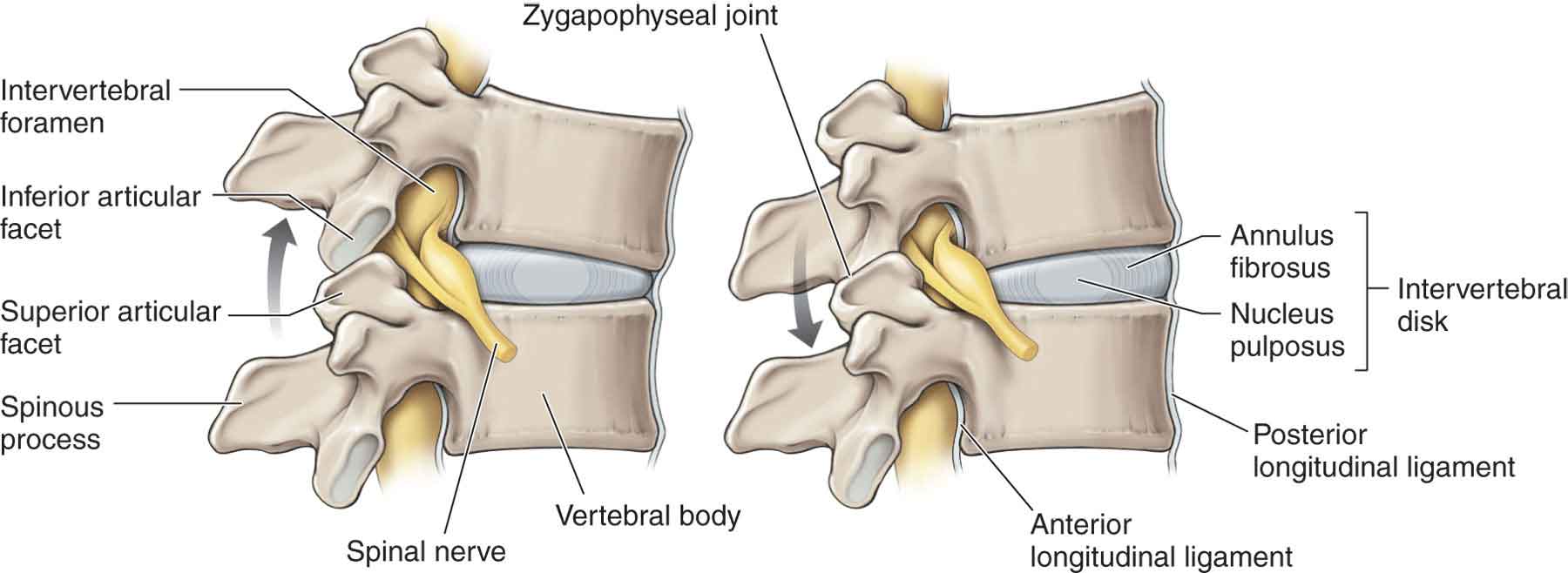
 Passive system. The spinal column, which includes the vertebrae, IVDs, zygapophyseal joints, joint capsules, and ligaments (Fig. 22-4) are the load-bearing units and the source of passive stiffness for stabilizing the spine. Passive stiffness of the ligaments and joint capsules is mainly a factor at the extremes of the ROM. The effectiveness of the passive support system is a factor of the ability of its structures to resist the forces of translation, compression, and torsion.

FIGURE 22-4 The passive system of the spine. (Reproduced, with permission, from Chapter 1. Back. In: Morton DA, Foreman K, Albertine KH. eds. The Big Picture: Gross Anatomy. New York, NY: McGraw-Hill; 2011.)
 Active system. Muscles, which serve to reduce or prevent movement and are the source of active stiffness (i.e., muscles act like stiff springs) by using stored elastic energy and the level of activation or force.36,38 Because the passive stiffness is mainly engaged at the extremes of range, the primary source of stiffness for stability during movements in healthy individuals is from the active stiffness of the trunk muscles and not the passive stiffness of ligaments. A large number of muscles have a mechanical effect on the spine and pelvis, and all muscles are required to maintain optimal control.38 This muscle activity must be coordinated within a hierarchy of interdependent levels: control of intervertebral translation and rotation, control of spinal posture/orientation, and control of the body with respect to the environment, in addition to maintaining a number of homeostatic functions such as respiration and continence.38–40 The concept of different trunk muscles playing differing roles in the provision of dynamic stability to the spine was proposed by Bergmark41and later refined by others.42–47 The specific muscles that provide stability and their interactions are described in the relevant chapters.
 Central nervous system (CNS), which utilizes feedforward (anticipatory) control to generate active muscle stiffness and uses the feedback (reflex) control to augment the stiffness.48 The CNS must continually interpret the status of stability, plan mechanisms to overcome predictable challenges, and rapidly initiate activity in response to unexpected challenges.38
The neutral zone is a term used by Panjabi36 to define a region of laxity around the neutral resting position of a spinal segment. The neutral zone is the position of the segment in which minimal loading is occurring in the passive structures (IVD, zygapophyseal joints, and ligaments) and the active structures (the muscles and tendons that control spinal motion), and within which spinal motion is produced with minimal internal resistance.2 Panjabi et al.49 have studied the effect of intersegmental muscle forces on the neutral zone and ROM of a lumbar functional spinal unit subjected to pure moments in flexion–extension, side bending, and rotation. Simulated muscle forces were applied to the spinous process of the mobile vertebra of a single motion segment using two equal and symmetric force vectors directed laterally, anteriorly, and inferiorly. The simulated muscle force maintained or decreased the motions of the lumbar segment for intact and injured specimens, with the exception of the flexion ROM which increased.49
The interaction between the passive and active systems of the spine changes when there has been prolonged tension or cyclic loading of the passive system resulting in a change to the muscle responses in the form of a delayed response. Furthermore, it is hypothesized that these changes in muscle activation may lead to increased spinal compression forces, which have been recognized as a risk factor for vertebral end-plate fracture, especially if applied repetitively.53,54 An alternate consequence is that muscle insufficiency resulting from fatigue may shift the loading to the passive tissues,55,56 which may put the spine at increased risk of injury.
Under normal circumstances, when large perturbations are expected, preparatory coactivation of the spinal muscles enhances active stiffness, and reflexes augment the stiffness at the appropriate time.59 These anticipatory postural adjustments and corrective responses are part of a motor control strategy executed by the CNS that was learned from previous experience when performing similar movements or activities.60–63
The emphasis on spinal stability exercises should be to:
 Strengthen the trunk muscles so that they are able to produce sufficiently large forces and active stiffness. The timing and sequencing of muscle activity, coupled with the appropriate magnitude of muscle activation, produces smooth, accurate, and efficient movement behavior that is adjusted to the immediate demands and consequences within the environment.48
 Increase the endurance of the trunk muscles so that the force output of the muscles does not deteriorate.
 Incorporate sound motor learning principles to address impaired motor control strategies.
The various strategies to incorporate into the intervention for spinal instability are addressed in the appropriate chapters.
EXAMINATION OF THE VERTEBRAL COLUMN AND PELVIS
The examination of the spine is complicated by the number of conditions that can cause pain in this region of the body. In addition, there is little scientific evidence for the establishment of many of the diagnostic labels attributed to spinal pain, such as instability, degenerative disk disease, and subluxation.64
The purpose of the examination is to identify correctly those patients who will benefit from a physical therapy intervention. The correct identification of a diagnosis requires the use of evidence-based measuring tools that are valid, specific, and sensitive. There are numerous methods for examining and evaluating the spine, and each of these results in a conclusion that determines the course of the intervention.65,66 Unfortunately, many of the procedures that are used today to examine the spine demonstrate methodologic shortcomings.64–66 The interventions for spinal conditions fair no better. Despite the ever-increasing numbers of randomized controlled trials evaluating interventions for low back pain (LBP),67 treatment outcomes remain less than optimal.68 One reason for this may be the fact that many trials test interventions in a heterogeneous population of patients with LBP and such a diverse group of patients may not correspond to the one-treatment approach.69 This hypothesis has led to the design of classification systems to identify subgroups of patients who may respond preferentially to certain treatments.70
Use of Traditional Classification Systems
In recent years, attempts have been made to use a variety of methods to classify spinal pain, particularly LBP, into syndromes. The term syndrome implies that the specific diagnosis is unknown. A syndrome is a collection of signs and symptoms that, collectively, characterizes a particular condition. In the spine, where determining a specific diagnosis has historically been proven to be extremely difficult, syndromes have become popular. By classifying syndromes, it is proposed that a patient is more likely to respond to a type of intervention unique to that syndrome. The criteria that have thus been used to categorize a syndrome include the following:
 Pathoanatomy.71,72 This strategy involves using correlations to produce categories. The disadvantage of using pathoanatomy is the difficulty in identifying a relevant pathoanatomic cause for most patients.73
 Presence or absence of radiculopathy.74,75
 Location and type of pain.76
 Duration of the symptoms (acute, subacute, or chronic).77
 Activity and work status.75,78
 Impairments identified during the physical examination.
 Direction of motion that reproduces, peripheralizes, or centralizes the symptoms.79
The more common classifications are outlined next. The use of such classification systems may have some prognostic value and can direct clinicians to specific interventions.80
Osteopathic System
Osteopaths rely on the results of the active motion tests and position tests to determine their intervention approach. Note: Osteopaths use the term “side flexion” instead of “side bending.”
Position Testing
Position testing involves palpation of the soft tissues over paired transverse processes of the spine, to detect palpable positional irregularity and altered tissue tension at a segmental level when the spine is positioned in flexion or extension compared with neutral. To locate the transverse process in the lumbar spine, the clinician first locates the spinous process to determine the level and then moves slightly laterally and superiorly by placing the thumbs on either side of the spinous process. Thus, the clinician must be very familiar with so-called layer palpation to be sure that the palpating fingers are monitoring the positions of the transverse processes at a particular segmental level. Vertebral dysfunctions occur as a combination of movements in the three planes. The key movement is the rotation. Theoretically, the rotational dysfunction, which is a result of the altered axis of rotation produced by the stiffer of the two sides of the segment, will be palpated as a much firmer end-feel to the palpation on that side. The direction of the rotation is named after the more posterior of the two transverse processes, and the positional name is an osteokinematic one having no established relationship with any joint.
Position Testing in Extension
If a marked segmental rotation is evident at the limit of extension, this would indicate that one of the facets is unable to complete its inferior motion (i.e., it is being held in a relatively flexed or superior position). The direction of the resulting rotation (denoted in terms of the anterior part of the vertebral body) informs the clinician as to which of the facets is not moving. For example, if the segment is rotated to the left when palpated in extension, the right zygapophyseal joint is not moving normally. This impairment can be described in one of the following three ways:
- The right zygapophyseal joint cannot extend or “close.”
- The right zygapophyseal joint is flexed (F), rotated (R), and side-flexed (S) left (L) around the axis of the right zygapophyseal joint.
- The right zygapophyseal joint is unable to perform any motions that require an inferior glide, such as extension, right side bending, and right rotation.
FRS or extension impairments are more evident in the positional tests than ERS impairments (described next) because there is less overall motion available into extension.
Position Testing in Flexion
If a marked segmental rotation is evident in full flexion, this indicates that one of the facets cannot complete its superior motion. For example, if the segment is found to be left-rotated when palpated during flexion, the left zygapophyseal joint is not moving normally.
This impairment can be described in one of the following three ways:
- The left zygapophyseal joint cannot flex or “open.”
- The left zygapophyseal joint is extended (E), rotated (R), and side-flexed (S) left (L).
- The left zygapophyseal joint is unable to perform any motion that requires a superior glide such as flexion, right rotation, and right side bending.
Position Testing in Neutral
Positional testing is performed in the neutral spine position for three reasons as follows:
- If a rotational impairment of a segment exists only in neutral and is not evident in either full flexion or full extension, the cause of the impairment is probably not mechanical in origin but rather neuromuscular. These neuromuscular impairments are usually found at the spinal junctions, particularly the thoracolumbar and cervicothoracic junctions.
- If a marked rotation is evident at a segment and this rotation is consistent throughout flexion, extension, and neutral, then the cause is probably an anatomic anomaly (e.g., scoliosis) rather than an articular problem.
- If the cause of the rotational impairment is articular (zygapophyseal joint), the positional testing in neutral gives the clinician an idea as to the starting position of the corrective technique.
The terminology used to describe the rotational disruption of the pure spinal motions (ERS or FRS) describes the positional and kinetic impairments only. It does not indicate what the pathology might be. Reasons for these impairments, other than movement dysfunctions, may include bony anomalies such as a deformed transverse process, compensatory adaptation, structural scoliosis, or a hemivertebra.
The analysis of the change in the rotational impairment between full flexion and full extension theoretically gives clues as to the pathology. Thus, in conjunction with other tests, such as active motion testing, this analysis can assist in ascertaining a biomechanical diagnosis.
Position testing has been found to be very reliable when used by experienced clinicians to identify segmental levels based on the relative position of spinous processes.81 The results for interrater reliability have been mixed when clinicians are asked to determine lumbar segmental abnormality using position testing,82,83 and poor when attempting to determine the segmental level of a marked spinous process.84
