Wrist Orthopaedic Tests
Wrist Palpation
Anterior Aspect
Descriptive Anatomy
Six wrist and digit flexor tendons cross the wrist (Fig. 7-1):
Flexor carpi ulnaris
Palmaris longus
Flexor digitorum profundus
Flexor digitorum superficialis
Flexor pollicis longus
Flexor carpi radialis
Procedure
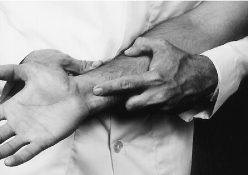
Palpate each tendon just proximal to the flexor retinaculum, noting any tenderness or calcific deposits (Fig. 7-2). Tenderness may indicate tenosynovitis of the suspected flexor tendon.
Descriptive Anatomy
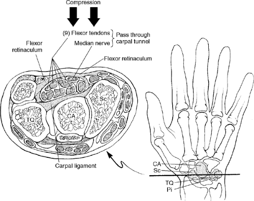
The carpal tunnel is deep to the palmaris longus at the anterior surface of the wrist. It is bound by the pisiform and the hook of the hamate medially, the tubercle of the scaphoid and tubercle of the trapezium laterally, the flexor retinaculum anteriorly, and the carpal bones posteriorly (Fig. 7-3). Inside the tunnel lie the median nerve and the finger flexor tendons from the forearm to the hand. This tunnel is a common site of compression neuropathy.
Procedure
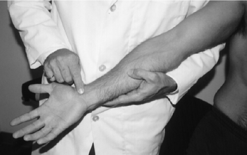
The actual tunnel and structures within the tunnel are not palpable. The borders of the tunnel should be palpated for deformity and/or tenderness (Fig. 7-4). The area over the tunnel should be palpated for increase in symptoms, such as numbness, tingling, pain, and weakness in the hand. These symptoms may indicate carpal tunnel syndrome.
Descriptive Anatomy
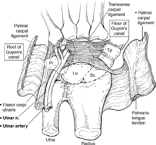
The tunnel, or canal, of Guyon is between the pisiform and the hook of the hamate. It contains the ulnar nerve and artery (Fig. 7-5). It is also a common site of compression neuropathy.
Procedure
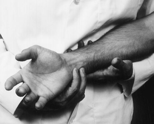
The ulnar artery and nerve are not palpable in the tunnel. Palpating over the tunnel may increase tenderness to the area and the symptoms to the ulnar distribution of the hand (Fig. 7-6).
Descriptive Anatomy
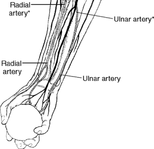
The radial and ulnar arteries are the two branches of the brachial artery that supply the hand with blood flow. The radial artery lies lateral at the anterior lateral aspect of the wrist, and the ulnar artery is at the anterior medial aspect of the wrist (Fig. 7-7).
Procedure
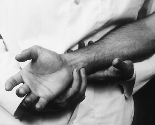
Palpate each artery individually and determine the amplitude of both pulses bilaterally (Figs. 7-8 and 7-9). A decrease in amplitude may indicate a compression of the respective artery between the elbow and the wrist if the brachial artery is palpated and not compromised. A common site of compression of the ulnar artery is the tunnel of Guyon.
Posterior Aspect
Descriptive Anatomy
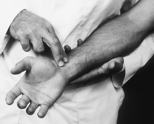
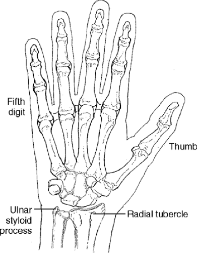
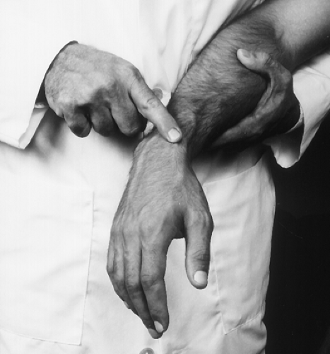
The ulnar styloid process is at the posterior aspect of the wrist proximal to the fifth digit. The radial tubercle is at the posterior aspect of the wrist proximal to the thumb (Fig. 7-10).
Procedure
Palpate the ulnar styloid process and radial tubercle for tenderness, pain, swelling, or deformity (Figs. 7-11 and 7-12). Pain at the radial tubercle following trauma may indicate a fracture, such as Colles’, a fracture of the distal radius with dorsal angulation. Pain at the ulnar styloid may be associated with a distal ulnar fracture. Tenderness, swelling, or deformity at either site may indicate rheumatoid arthritis.
Descriptive Anatomy
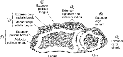
There are six fibro-osseous tunnels at the posterior aspect of the wrist. The extensor tendons to the hand pass through these tunnels, which are bound by the extensor retinaculum superficially and are lined with a synovial sheath. From the thumb laterally, these are the tunnels and their respective tendons (Fig. 7-13):
Tunnel 1 Adductor pollicis longus, extensor pollicis brevis
Tunnel 2 Extensor carpi radialis longus and brevis
Tunnel 3 Extensor pollicis longus
Tunnel 4 Extensor digitorum and extensor indexes
Tunnel 5 Extensor digiti minimi
Tunnel 6 Extensor carpi ulnaris
Procedure
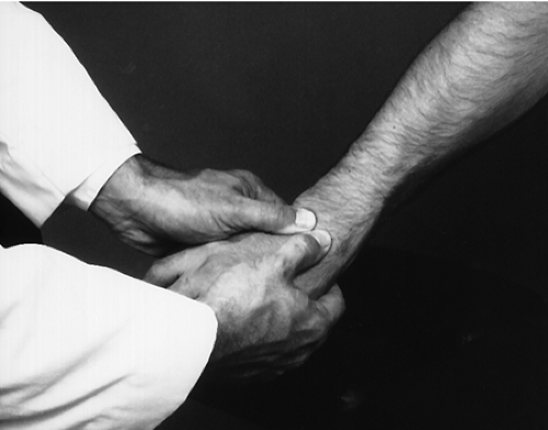
Support the patient’s hand with your fingers while palpating the wrist with both your thumbs (Fig. 7-14). Note any crepitus or restriction of movement. Crepitus may indicate tenosynovitis of one of the extensor tendons.
Wrist Range of Motion
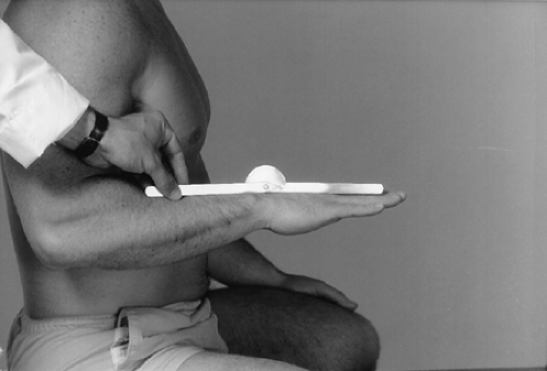
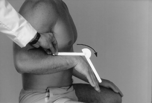
With the patient’s wrist in the neutral position, place the goniometer in the sagittal plane with the center at the ulnar styloid process (Fig. 7-15). Instruct the patient to flex the wrist downward, and follow the hand with one arm of the goniometer (Fig. 7-16).
Normal Range
Normal range is 75 ± 7.6° or greater from the 0 or neutral position (2).
| Muscles | Nerve Supply |
|---|---|
| Flexor carpi radialis | Median |
| Flexor carpi ulnaris | Ulnar |
With the patient’s wrist in the neutral position, place the goniometer in the sagittal plane with the center at the ulnar styloid process (Fig. 7-17). Instruct the patient to extend the wrist backward while you follow the hand with one arm of the goniometer (Fig. 7-18).
Stay updated, free articles. Join our Telegram channel

Full access? Get Clinical Tree


