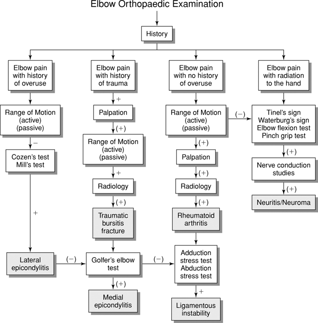
Elbow Orthopaedic Tests
Elbow Palpation
Medial Aspect
Descriptive Anatomy
The ulnar nerve is a branch of the medial cord of the brachial plexus. It passes in the groove between the medial epicondyle and the olecranon fossa (Fig. 6-1).
Procedure
With your index finger, palpate the groove between the medial epicondyle and the olecranon process. Notice whether the nerve is tender to palpation or thickened (Fig. 6-2). This can indicate nerve compression or scar tissue on the nerve leading to paresthesia into the forearm and/or loss of interosseous muscle strength.
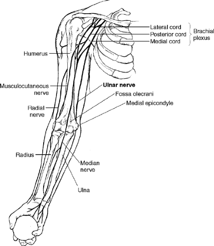 Figure 6-1 |
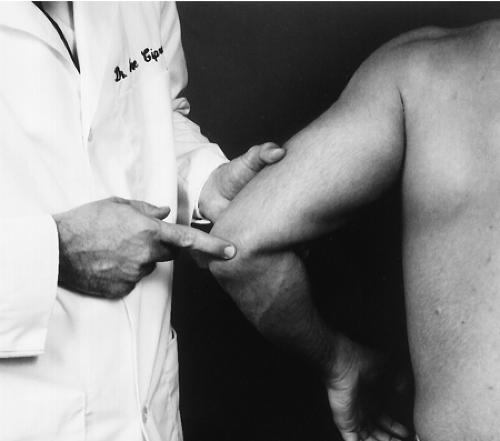 Figure 6-2 |
Descriptive Anatomy
The medial epicondyle is a relatively large protuberance at the medial distal end of the humerus. Attached to the condyle are the wrist flexor and pronator group muscles. This group consists of the pronator teres, flexor carpi radialis, palmaris longus, and flexor carpi ulnaris. All of these muscles originate from the medial epicondyle as a common tendon (Fig. 6-3).
Procedure
With the elbow flexed to 90°, palpate the epicondyle and its tendons with your index finger; look for tenderness, inflammation, and temperature elevation (Fig. 6-4). This may indicate a strain of one or more of the previously mentioned tendons or an inflammation of the medial epicondyle caused by various activities, such as golf or tennis.
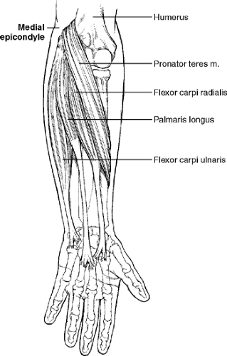 Figure 6-3 |
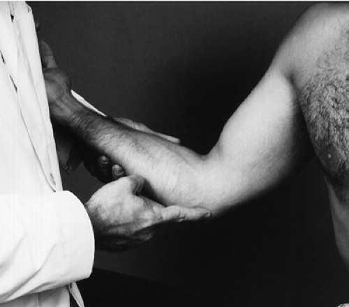 Figure 6-4 |
Descriptive Anatomy
The ulnar collateral ligament attaches the medial epicondyle to the medial aspect of the ulna at the trochanteric notch (Fig. 6-5) and stabilizes the humeroulnar articulation medially.
Procedure
With your index finger, palpate the area of the ulnar collateral ligament (Fig. 6-6). Ordinarily it is not palpable. You should check for tenderness, which may indicate a sprain caused by forced valgus stress.
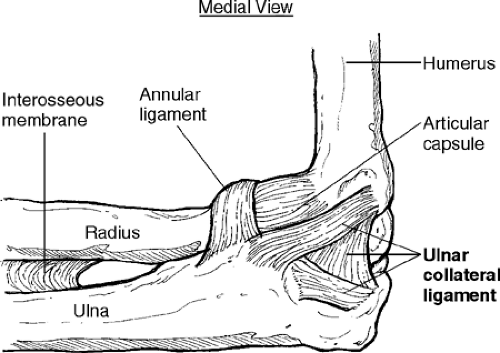 Figure 6-5 |
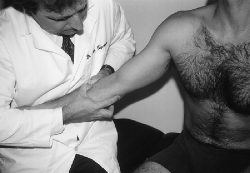 Figure 6-6 |
Lateral Aspect
Descriptive Anatomy
The lateral epicondyle is a relatively large protuberance at the lateral distal end of the humerus. Attached to the condyle is the common extensor tendon. From this tendon arise the carpi radialis brevis, extensor digitorum, extensor digiti minimi, and extensor carpi ulnaris. The brachioradialis and extensor carpi radialis longus and brevis are attached superior to the lateral epicondyle at the supracondylar ridge (Fig. 6-7).
Procedure
With the patient’s elbow flexed to 90°, palpate the lateral epicondyle and supracondylar ridge with your index and middle fingers (Fig. 6-8). Note any tenderness, inflammation, and temperature elevation at either location. These signs may indicate an inflammation of the lateral epicondyle (epicondylitis) or a strain of the extensor tendons of the wrist.
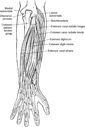 Figure 6-7 |
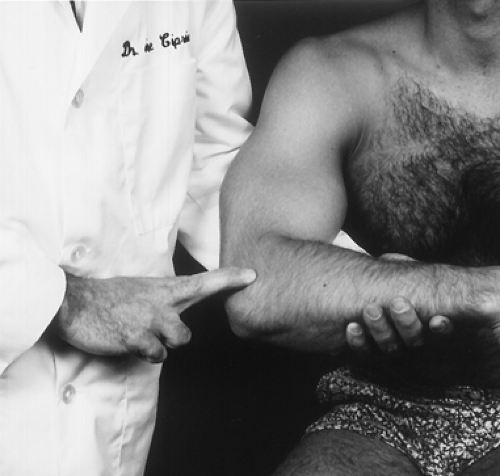 Figure 6-8 |
Descriptive Anatomy
Radial collateral ligament and annular ligaments are thick structures that extend from the lateral epicondyle of the humerus to the annular ligament and lateral aspect of the ulnar. The annular ligament encircles the radial head (Fig. 6-9). The radial collateral ligament stabilizes the humeroulnar articulation laterally.
Procedure
With your index and middle fingers, palpate the area of the radial collateral ligament from the lateral epicondyle to the annular ligament (Fig. 6-10). Check for tenderness, which may indicate a sprain caused by forced varus stress.
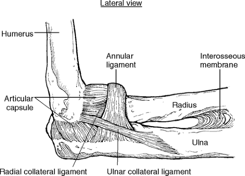 Figure 6-9 |
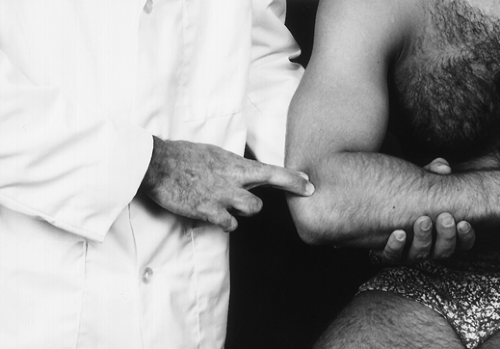 Figure 6-10 |
Posterior Aspect
Descriptive Anatomy
The olecranon process is posterior to the elbow at the proximal end of the ulnar. It is covered by the olecranon bursa, which is not normally palpable (Fig. 6-11).
Procedure
With the patient’s elbow flexed to 90°, palpate the olecranon process and bursa for tenderness, inflammation, and increased temperature (Fig. 6-12). A thick, boggy feeling may indicate olecranon bursitis. Check the posterior aspect of the olecranon border for rheumatoid nodules, which indicate rheumatoid arthritis.
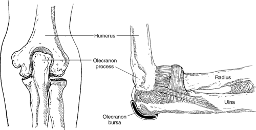 Figure 6-11 |
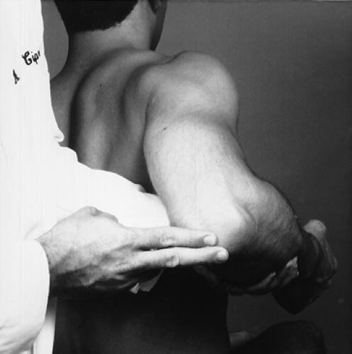 Figure 6-12 |
Triceps Muscle
Descriptive Anatomy
The triceps muscle has three heads; the long head crosses both the glenohumeral joint and the elbow joint, and it inserts into the olecranon process (Fig. 6-13).
Procedure
With the patient’s elbow slightly flexed, have the patient lean on a table. This will facilitate the palpation of the muscle. With your thumb and index finger, palpate the length of the muscle down to the olecranon process, looking for any tenderness or defects secondary to trauma (Fig. 6-14). This may indicate a strain or active trigger points of the triceps muscle. A hard mass may indicate myositis ossificans secondary to repeated trauma.
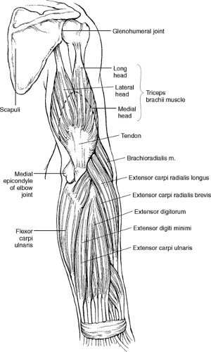 Figure 6-13 |
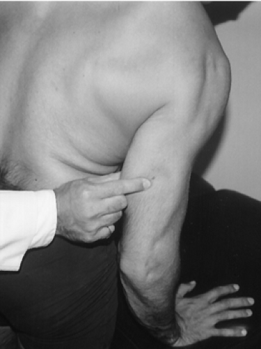 Figure 6-14 |
Anterior Aspect
Location
The cubital fossa is the triangular space bordered by the brachioradialis laterally and the pronator teres medially. The base is an imaginary line between the two epicondyles. The structures that pass between the fossa are the biceps tendon, brachial artery, median nerve, and musculocutaneous nerve (Fig. 6-15).
Procedure
With the patient’s elbow slightly flexed and the patient resisting flexion, palpate the cubital fossa with your index finger, looking for the biceps tendon, which lies medial to the brachioradialis muscle (Fig. 6-16). Tenderness may indicate a strain in the musculotendinous junction. A ruptured tendon is not palpable in the fossa, and a bulbous gathering of muscle is evident in the upper arm.
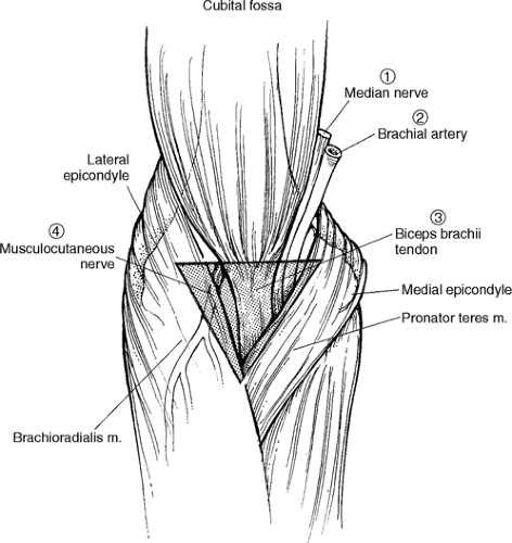 Figure 6-15 |
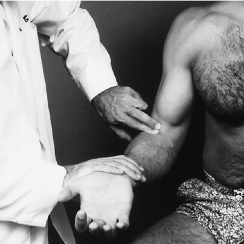 Figure 6-16 |
Elbow Range of Motion
With the patient seated, elbow extended, place the goniometer in the sagittal plane with the center at the elbow joint (Fig. 6-17). This is the neutral position for the elbow joint for flexion and extension. Instruct the patient to flex the arm as far as possible while following the forearm with one arm of the goniometer (Fig. 6-18).
Normal Range
Normal range is 141° ± 4.9° or greater from the 0 or neutral position (2).
| Muscles | Nerve Supply |
|---|---|
| 1. Brachialis | Musculocutaneous |
| 2. Biceps brachii | Musculocutaneous |
| 3. Brachioradialis | Radial |
| 4. Pronator teres | Median |
| 5. Flexor carpi ulnaris | Ulnar |
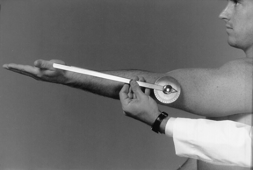 Figure 6-17 |
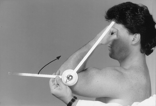 Figure 6-18 |
Related posts:

Stay updated, free articles. Join our Telegram channel

Full access? Get Clinical Tree


