Wound Care


Wound Care


Christine M. Wietlisbach


Introduction


Therapists specializing in upper extremity rehabilitation see and treat a wide variety of wounds, including abrasions, lacerations, skin tears, blisters, punctures or penetrations, human-animal-insect bites, de-gloving, surgical incisions, open surgical wounds, burns, and skin grafts. The significance of this aspect of hand therapy cannot be understated. Understanding how to properly care for a hand wound gives you the ability to reduce pain and swelling, speed wound closure, and minimize superficial scar tissue formation. When these factors are controlled, your client is much more likely to participate in all other aspects of the hand therapy program that lead to successful outcomes.


Both the American Occupational Therapy Association (AOTA) and the American Physical Therapy Association (APTA) definitively state that wound care is within the scope of practice of occupational therapists and physical therapists, respectively.12 Many state practice acts also define wound care as within our scope of practice, but states vary with regard to what specific wound care procedures can be performed by occupational and physical therapists. As such, you should always consult your state licensing board on the legal parameters of providing this service in your state.


This chapter introduces the basics of proper wound care. You will learn how wounds heal, the many factors that can influence wound healing, how to cleanse and debride wounds, and how to choose dressings that maintain the optimal environment for wound healing. Additionally, you will learn how to measure, describe, and document wounds along the healing continuum. This chapter does not cover the specifics of treating burns or skin grafts. These are special-circumstance wounds and while many principles of basic wound care will also apply to burns and skin grafts, the appropriate handling of these wounds is more complicated and beyond the scope of this chapter.


Diagnosis


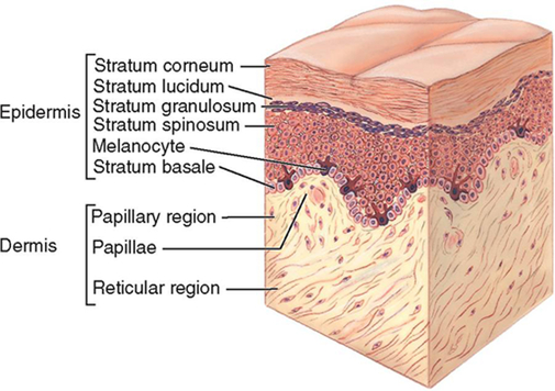
For the purposes of this text, we will consider a wound as any sort of open traumatic or surgical damage to the skin. The skin consists of two layers: the epidermis and the dermis (Figure 21-1).


The epidermis is the outermost surface of the skin. It is avascular and made up primarily of keratinocytes—cells that produce the protein keratin. Although it is only about 0.5 mm thick, the epidermis is a rather dense and tough covering that helps shield the body from infection, trauma, and rapid dehydration.


The dermis lies directly below the epidermis, is about 3 mm thick, highly vascular, and made up mostly of fibroblasts. Fibroblasts are cells that produce collagen and elastin, which give skin strength and flexibility. Other cells found in the dermis include macrophages that destroy debris and bacteria within the skin and mast cells that secrete substances that initiate inflammation to help fight infection. The dermis helps regulate body temperature, protects the body from infection when foreign substances break through the epidermis, and provides sensory information via receptors located there.


Timelines and Healing


Normal Wound Healing


When the skin is wounded, it normally undergoes a predictable sequence of events consisting of three overlapping phases: inflammation, proliferation, and maturation (also called remodeling). Collectively, these three phases are known as the wound healing process. This process is a very complicated vascular and cellular response to tissue injury. What follows here is a very basic description of these events. Understanding the wound healing process can assist you in evaluating and planning a course of treatment for upper extremity wounds.


Inflammation is the initial response to tissue injury. During the inflammatory phase of wound healing, the body is working to control blood loss and to clean the wounded area. Blood loss is controlled, in part, when the blood vessels in the injured area immediately constrict. This is known as vasoconstriction. At the same time, a protein-based fluid leaks out of the vessels and swelling begins at the injury site. Additionally, the body begins sending specialized clean-up cells to the area that work to break down and destroy damaged tissue, foreign matter, and bacteria. Within about 30 minutes, mast cells release histamine, which causes blood vessels to open, or vasodilate. This vasodilation pushes more fluid into the tissue spaces and causes increased pain, swelling, and discoloration (red, blue, or purple) in and around the wound.


The inflammatory phase begins the moment of injury and continues until the injured area is free of debris and bacteria. It is a normal and necessary phase of tissue healing. The acute inflammatory phase usually lasts 2 days to 2 weeks. During this time, physicians and therapists should strive to control only excessive inflammation and edema, because these issues result in severe pain and potentially decreased circulation, which can cause further tissue damage. However, it is important to remember that some amount of inflammation and swelling is normal and necessary to prepare the wound for the proliferative phase of healing.


The proliferative phase of wound healing begins once the injured area is clean and free from damaged tissue, foreign matter, and bacteria. At this point, the body starts repairing the open space created by the wound. Proliferation consists of granulation, angiogenesis, wound contraction, and epithelialization. Granulation occurs when the body forms a matrix of connective tissue, including collagen, in the wound bed. This tissue is known as granulation tissue and builds on itself to fill the “hole” of the wound. Angiogenesis is the growth of new blood vessels. Very small capillary networks are formed in the granulation tissue of the wound and give healthy wounds their distinctive pink-red color. Wound contraction occurs when specialized cells in the wound bed act to pull the edges of the wound together. Finally, epithelial cells migrate across the top of the granulation tissue and completely cover the wound. This is known as epithelialization. The proliferative phase may take up to a few weeks to complete. It is completed more rapidly in a moist and protected environment, which becomes important for you to remember when choosing appropriate wound dressings.


Newly epithelialized wounds are still quite fragile and must go through the maturation phase of wound healing (also called the remodeling phase). During this final phase, water and amino acids are squeezed out of the granulation tissue matrix as collagen fibers continue to be produced. This new tissue, dense with collagen, is called scar tissue. Scar tissue is not as elastic or as cosmetically appealing as skin, but it serves as an adequate wound cover.


Factors Affecting Wound Healing


Although most wounds progress through the three phases of healing without incident, there are certain factors that can influence both the rate and quality of healing. These factors include:


Neither the therapist nor the client has control over all factors affecting wound healing. However, to the extent that it is possible, all factors that can be modified to improve wound healing should be addressed. Familiarizing yourself with these variables will help you and your clients create an optimal environment for wound healing. Understanding these variables will also help explain why some wounds take longer to heal.


Adequate circulation is essential to wound healing. Wounds will not heal unless an adequate supply of oxygenated blood reaches the wound bed. Many things can reduce blood flow to the hands, such as peripheral vascular disease, diabetes, smoking,3 excessive edema, and mechanical stress from orthotics or dressings. It is the therapist’s responsibility to assure that all wound dressings and orthotics fit correctly. Dressings that are wrapped too tightly or orthotics that cause pressure areas can affect circulation in the hand.


In addition to having adequate circulation, a wound must be clean before it can heal. A wound is considered “clean” when it is free from infection and debris. Wound debris is anything embedded in the wound bed that should not be there: sutures, gauze fibers, dog hair, dead tissue, and so on. When a wound is infected or contains debris, the body’s natural reaction is to clean it up by initiating an inflammatory response, sending specialized clean-up cells to the area that work to destroy foreign matter and bacteria. The wound will remain stuck in the inflammatory phase of healing until it is clean. As a hand therapist, you play an important role in monitoring wounds for infection and in removing debris from the wound bed to facilitate more rapid healing.


Chemical stress occurs when a toxic substance makes contact with granulation tissue forming in the wound bed. Cells that make up granulation tissue are very fragile and must be treated with care. When chemical stress occurs, the new cells die, and this slows the rate of wound healing. Many products that have traditionally been used to clean wounds, such as hydrogen peroxide and povidone-iodine, are cytotoxic and can kill tissue cells.4 You must use care in choosing wound cleansing products, and educate your clients to do the same.


The temperature of the wound bed also influences healing. Wounds heal best when the wound surface temperature is kept relatively constant and close to the normal core body temperature range of 36° C to 38° C (96.8° F to 100.4° F).5 The temperature of the wound bed drops, on average, 2 degrees Celsius (3.6° F) when the dressings are removed and the wound is cleansed with room temperature saline. It can take up to 3 hours for the temperature of a wound to return to pre-dressing change temperature once a new dressing is applied.6 Strategies that reduce the frequency of dressing changes help keep the wound warm and the temperature more constant, and this assists with healing.


Another factor that assists with wound healing is moisture balance. A wound that is either too wet or too dry will not heal as quickly as a properly-balanced moist wound. A moist wound provides the optimal environment for cell growth and migration of epithelial cells over the wound bed. Wounds with a high level of exudate (drainage) can become too wet, and this often leads to breakdown of the wound bed and surrounding skin. Wounds with little or no exudate can become too dry, and this slows the action of regenerative cells in the wound.7


Finally, there are a few factors specific to the uniqueness of each client that will interact and affect wound healing. Wounds in younger clients tend to heal more quickly than wounds in older clients. This is generally due to the presence of medical conditions, medications, and inadequate nutritional intake more likely to be found in older clients. However, when these factors are present in younger clients, the effect on wound healing is the same. Chronic diseases, such as peripheral vascular disease and diabetes, will slow wound healing. Clients with cancer or AIDS or those with autoimmune disorders who require immunosuppressive drug therapy will also demonstrate slower wound healing.8 Additionally, clients with chronically poor nutritional intake will demonstrate less efficient wound healing because malnutrition affects cell production, collagen synthesis, and wound contraction.9


Non-Operative Treatment: Basic Wound Management


With so many variables affecting wound healing, what can you do to help heal your clients’ wounds? We already know that some factors affecting healing are out of both the therapist’s and the client’s control. However, there are a few factors that we can manipulate to facilitate better wound healing. It is helpful to think of basic wound management in terms of the three hallmarks of therapy-assisted wound care: 1) wound debridement, 2) proper wound cleansing, and 3) maintenance of proper moisture balance in and around the wound.


Wound Debridement


Debridement is the removal of necrotic tissue from a wound so that healthy tissue is exposed in the wound bed. Remember, a wound will not heal as long as it contains debris. It will be stuck in the inflammatory phase of wound healing until it is clean. Dead tissue is a type of wound debris, and it can take two forms: slough or eschar. Slough is a moist composite of fibrin, bacteria, dead cells, and exudate. It is whitish or yellowish in color and usually somewhat adhered to the wound in the form of stringy tissue. Eschar is dead tissue that is usually hard and dry but will occasionally be moist in appearance. It is black in color and firmly attached to the wound.


Both slough and eschar are breeding grounds for bacteria and increase the risk of wound infection. Removal of this devitalized tissue will both speed healing and reduce the risk for wound infection, so it is generally one of the first actions that should be considered as part of the wound care plan. However, not all wounds should be debrided. Any wound in an area where blood flow is impaired should be debrided cautiously, if at all. You should always discuss with the physician any plans to debride a wound in an area with compromised circulation.


There are four traditional methods of wound debridement: autolytic, enzymatic, sharp, and mechanical. Autolytic debridement is the method of choice in the hand therapy clinic. Occasionally, you may need to resort to enzymatic or sharp debridement. Mechanical debridement is no longer used by most therapists and physicians, and it should be avoided.


Autolytic debridement occurs when the body breaks down necrotic tissue on its own. We can encourage this by choosing wound dressings that keep the wound moist and trap the body’s natural enzymes that break down dead tissue. Film and hydrogel dressings are excellent for promoting autolytic debridement. A discussion of these and other dressings will be covered later in this chapter. Autolytic debridement is comfortable and usually effective, but it can take longer to accomplish than the other methods of debridement. Therapists should take care to maintain a proper moisture balance when using this method. A wound that is too wet can result in macerated wound edges, which is a weakening of healthy tissue around the wound caused by too much fluid absorption. Macerated skin can easily break down and cause the wound to enlarge.


Enzymatic debridement uses topical enzymes to break down slough and eschar. Therapists should check their state practice act for legal guidance on the application of topical medications. Some states do not allow therapists to do this. The most widely-used enzymatic treatment is collagenase ointment, which is sold under the brand name Santyl and is available with a physician’s prescription. Collagenase ointment is applied to necrotic tissue once or twice daily and covered with a dressing. Very dry eschar should be cross-hatched first to help the enzymes penetrate the tissue. Enzymatic debridement is selective, in that it breaks down necrotic tissue without harming healthy granulation tissue in the wound bed. This method is very effective, but may cause some discomfort and irritation in clients who are hypersensitive to the enzyme.


Sharp debridement is the use of any sharp instrument, such as scissors or a scalpel to selectively remove necrotic tissue. Again, therapists should refer to their state practice act for legal guidance on this. Some states allow occupational and/or physical therapists to perform sharp debridement; other states do not. Sharp debridement is the fastest and most effective method of debridement, but it should only be done by a skilled clinician. Once you cut something out, it is permanent. It can sometimes be difficult for an inexperienced therapist to distinguish between adipose tissue, slough, and tendon. If in doubt, never cut! This method of debridement can also be uncomfortable for the client and may require topical or local anesthetic. Because of these factors, many therapists defer sharp debridement to the physician.


Mechanical debridement is the removal of dead tissue using methods, such as whirlpool agitation, high pressure fluid irrigation, or wet-to-dry dressings. Wet-to-dry dressings are made by wetting gauze and inserting it into the wound. The dressing is allowed to dry inside the wound, and the gauze is then ripped out quickly so that tissue within the wound is pulled out along with the gauze. This method will, of course, remove some necrotic tissue every time it is performed. The problem is that any new granulation tissue forming in the wound bed is also pulled out with the gauze. Removing healthy tissue along with dead tissue is called non-selective debridement, and it is disruptive to wound healing. Mechanical debridement methods, especially wet-to-dry dressings, slow wound healing and are unnecessarily painful for clients. Therefore, they are not recommended. There are much more comfortable and effective methods to debride wounds in the hand clinic.


Sometimes you will utilize a combination of debridement methods. For example, the surgeon may surgically remove most of the necrotic tissue, and you will remove the rest with autolytic or enzymatic debridement. No matter what form of debridement you employ, the goal is always the same. You want to remove all devitalized tissue so that only a healthy, well-vascularized, pink-red wound bed remains.


There is one more thing to mention about preparing the wound for healing through the removal of unwanted tissue. A healthy wound bed is pink-red. However, there is a certain type of tissue that forms in the wound bed that is red-colored but undesirable. This tissue develops when there is an overgrowth of granulation tissue, possibly due to infection or excessive moisture in the wound.10 It is called hypergranulation tissue.


Hypergranulation tissue normally looks like shiny, deep-red balls of tissue that grow taller than the wound margins. Some therapists think it looks like little red raspberries (Figure 21-5). The tissue is soft and will often bleed easily when touched. This tissue must be treated in order for the wound to heal normally. An effective way to treat this tissue is to apply silver nitrate to the hypergranular areas.10,11 Use silver nitrate sticks and roll the treatment end of the stick over the abnormal tissue. As you treat the tissue, it will turn gray in color. After treating with silver nitrate, you can bandage the wound as you normally would. Repeat the procedure at each dressing change until the hypergranulation tissue is controlled.


Wound Cleansing


Wounds should be cleansed every time the dressing is changed. The purpose of cleansing the wound is to remove loose debris and surface contaminants from the wound bed. Gauze fibers, loose sutures, liquefied necrotic tissue, and bacteria are all commonly found in wounds and must be washed out. Ideally, this should be done without causing trauma to any new tissue forming in the wound bed. The therapist must decide two things: 1) what solution to use to cleanse the wound, and 2) how to apply the solution.


The best solutions for cleansing wounds in the hand clinic include normal saline, sterile water, and drinkable tap water.12 Avoid using solutions such as hydrogen peroxide, Dakin’s solution, povidone iodine/Betadine, soap, or bleach on clean wounds. These solutions contain chemicals that are toxic to granulation tissue, and use of these solutions will slow wound healing. Using a product like hydrogen peroxide is really only appropriate in the home setting for cleaning cuts and scrapes immediately after an injury. Hydrogen peroxide is fine for cleaning a dirty superficial wound at home once or twice. However, once that wound is free of injury-related debris like dirt, asphalt, and grass, it should be cleansed with saline, sterile water, or drinkable tap water so as not to impede the healing process.


The method you use to apply the cleansing solution is also important. You want to use enough pressure to remove surface debris and contaminants without causing trauma to any new tissue forming in the wound bed. To date, research has failed to identify the ideal method of wound cleansing.13 However, most practitioners have discarded the practice of whirlpool wound cleansing and replaced that method with syringe irrigation. Syringe irrigation is more convenient and carries less risk of cross-contamination between clients than whirlpool cleansing. Much of the available literature indicates that wounds seen by the hand therapist should be irrigated with pressures at or below 8 psi.13,14 A 35 mL medical syringe with a 25-gauge needle will produce 4 psi, and a 35 mL/19 gauge combination will produce 8 psi.15 This equipment is readily available in most health care settings. You can cleanse all wounds in the clinic with either of these syringe/needle combinations filled with saline, sterile water, or drinkable tap water.


Maintenance of Moisture Balance


Moisture in the wound bed is critical to efficient wound healing. A moist wound will heal much faster than a wound that is either too wet or too dry. Maintaining an appropriate moisture balance depends on one thing: your choice of wound dressing. Every wound is different and will require different dressings or dressing combinations to keep it moist. If a wound has a lot of exudate, you will want to choose a dressing that can absorb the drainage. On the other hand, if a wound is too dry, you will want to choose a dressing that will add moisture to the wound bed.


Choosing the right dressing is somewhat of an art form that requires practice. The more wounds you see, the better you will get at selecting the right dressing. However, understanding the different categories of available dressings, and the characteristics of each, will help guide you.


Dressings are sometimes described as non-occlusive, semi-occlusive, or totally occlusive. This nomenclature has to do with the relative ability of a dressing to block water, water vapor, and bacteria from passing through the dressing. A completely non-occlusive dressing will allow the free passage of water, vapor, and bacteria. A completely occlusive dressing will not allow any passage of water, vapor, and bacteria. A semi-occlusive dressing falls in the middle of this continuum—generally allowing the passage of water vapor, but not water or bacteria. In truth, there is no completely occlusive or completely non-occlusive dressing, but dressings are described with the term that most accurately describes its ability to keep water, vapor, and bacteria from passing through.


When occlusive dressings first arrived on the scene, many physicians feared that their use would cause infection. This myth is still prevalent in some settings. However, research has shown that occlusive dressings do not increase the risk for infection.17 The goal is to find a dressing that will keep bacteria out, retain some moisture, but still absorb any excess fluid if needed.


Standard dressing choices generally fall into the following categories: transparent films, impregnated low-adherence dressings, hydrogels, hydrocolloids, gauze, foams, and alginates. There are other types of specialty dressings available, but the typical hand therapist will make his or her selection from these standard dressing choices. Every medical supply company has these types of dressings available, but they will be identified by different brand names. When looking for a specific type of dressing, ask your supplier for the names of the available dressings in that category. The dressing categories described below are listed in order from least absorptive to most absorptive.1821


Transparent Films


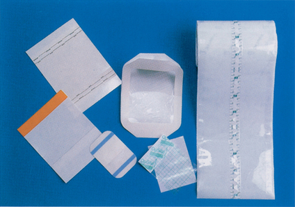
These versatile dressings are exactly what they sound like: thin, see-through films (Figure 21-2). Films come in a variety of shapes and sizes and easily conform to the contours of the hand. They adhere right to the skin and can be used either as a primary dressing (making contact with the wound) or as a secondary dressing (holding a primary dressing in place). Transparent films are semi-occlusive in that they are permeable to water vapor, but impervious to liquids and bacteria.19 Therefore, they are waterproof in the shower and good at keeping bacteria out of the wound. Another advantage of transparent film dressings is our ability to see the wound without removing the film. Films are non-absorptive, and because these dressings hold most moisture in, they are excellent for promoting autolytic debridement of slough or moist eschar. Transparent films must be changed if too much fluid builds up under the film and starts to leak out the edges. However, dry or very low draining wounds can tolerate a film dressing in place for up to 7 days.


Brand names of transparent films include OpSite (Smith & Nephew), Tegaderm Transparent Film (3M), and Suresite (Medline).

Stay updated, free articles. Join our Telegram channel

Sep 9, 2016 | Posted by in MANUAL THERAPIST | Comments Off on Wound Care

Full access? Get Clinical Tree

Get Clinical Tree app for offline access