Watson-Jones Approach
Darwin Chen
Richard Berger
The original Watson-Jones anterolateral approach to the hip, as popularized by Sir Reginald Watson-Jones in the 1930s, was described through the interval between the tensor fascia lata and the gluteus medius (1). Further modified by Charnley (2), Harris (3), and Muller (4), this intermuscular interval allows for excellent exposure of the acetabulum and proximal femur for fracture surgery and arthroplasty, without the need for trochanteric osteotomy. Most versions of the Watson-Jones anterolateral approach involve varying degrees of detachment of the abductor mechanism; however, abductor-sparing modifications have also been developed (5). The main advantage of this approach is a lower dislocation rate secondary to preservation of the posterior structures (6,7,8,9). The main disadvantage of this approach is violation of the abductor musculature, potentially leading to a Trendelenburg limp and abductor weakness (8). The Watson-Jones approach, in its various forms and iterations, is versatile and can range from being highly extensile to minimally invasive.
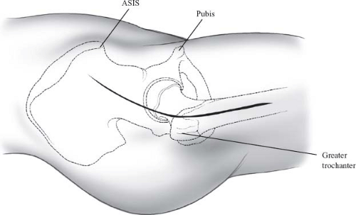
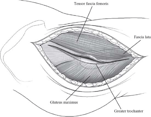
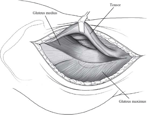
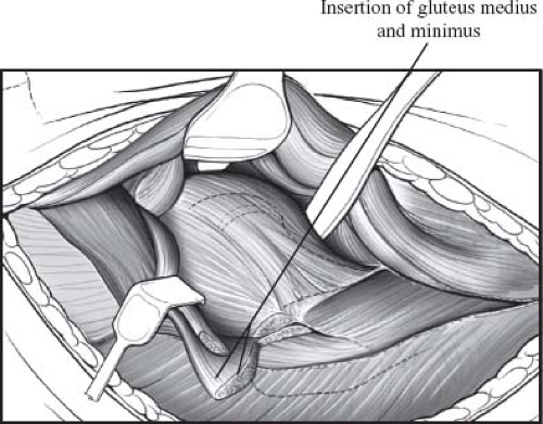
As originally described (1), the initial incision begins at the midpoint between the anterosuperior iliac spine and the tip of the greater trochanter, proceeds toward the greater trochanter in a slightly curvilinear fashion, and continues distally parallel to the shaft of the femur (Fig. 4.1). Following subcutaneous dissection, a fascial incision is made in the iliotibial band, in line with the incision (Fig. 4.2). The abductor complex, greater trochanter, and vastus lateralis are now visible (Fig. 4.3). Retraction of the gluteus medius and minimus posteriorly reveals the anterior hip capsule, covered by its surrounding pericapsular fat (Fig. 4.4).
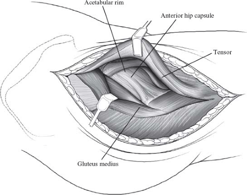 Figure 4.4. Retraction of the gluteus medius and minimus reveals the pericapsular fat surrounding the anterior hip capsule. |
The anterior one-third of the gluteus medius insertion is released off of the tip of the greater trochanter by means of a short transverse incision. It is important to note that the muscle fibers of the anterior one-third of the gluteus medius run antero-obliquely, serving a hip flexor function in addition to hip abduction. Retracting the remainder and bulk of the gluteus medius posteriorly, the gluteus minimus tendon
is identified and released, leaving a 1-cm cuff of tendon on bone for later repair (Fig. 4.5).
is identified and released, leaving a 1-cm cuff of tendon on bone for later repair (Fig. 4.5).
The anterior hip capsule is now exposed by placing curved, Hohmann-type retractors extracapsularly around the superior and the inferior femoral neck. Another retractor is placed medially over the anterior acetabular rim, underneath the iliopsoas and the direct head of the rectus femoris. Care must be taken to place this retractor directly over the bone onto the anterior column and underneath the muscle, so as not to injure the femoral neurovascular bundle. A capsulotomy (T- or H-shaped) or capsulectomy (square shaped along superior and inferior borders of the femoral
neck, intertrochanteric ridge, and anterior acetabular rim) can be performed at this point, exposing the femoral head and neck. The hip is dislocated anteriorly with the aid of a hip skid by externally rotating and extending the leg. After properly identifying the level of the femoral neck osteotomy as per the preoperative plan, the neck is cut with an oscillating saw and the head–neck fragment is removed.
neck, intertrochanteric ridge, and anterior acetabular rim) can be performed at this point, exposing the femoral head and neck. The hip is dislocated anteriorly with the aid of a hip skid by externally rotating and extending the leg. After properly identifying the level of the femoral neck osteotomy as per the preoperative plan, the neck is cut with an oscillating saw and the head–neck fragment is removed.
Extensile exposure of the proximal femur can easily be achieved by extending the skin incision and fascia lata split distally along the femoral shaft, essentially incorporating a lateral approach to the femur. The vastus ridge and vastus lateralis muscle are identified. The vastus lateralis can either be split in line with its fibers, or can be elevated off the intermuscular septum and retracted anteriorly. While releasing the vastus lateralis off the intermuscular septum, the surgeon must be cautious to maintain hemostasis, as multiple perforating vessels exist in this interval. Proceeding distally, the entire lateral aspect of the femoral shaft can be exposed down to the knee.
Extensile exposure of the acetabulum through the anterolateral approach is more difficult, but feasible. This can be achieved by releasing additional gluteus medius fibers off of the greater trochanter, mobilizing the femur and allowing it to be retracted further posteriorly during acetabular preparation and reaming. Care must be taken not to disrupt the superior gluteal neurovascular bundle supplying the abductors. The posterior fascia lata flap can also be extended further proximally by creating an oblique incision in the interval between the gluteus maximus and the fascia lata. Alternatively, a trochanteric osteotomy can be performed, completely mobilizing the abductor complex while preserving its attachments to the greater trochanter. A Chevron-type osteotomy of the trochanter allows for more stable fixation than a straight or oblique osteotomy and increases surface area for healing of the osteotomy site. In addition, release of the inferior hip capsule will also mobilize the femur further to allow for more extensile acetabular exposure.
Minimally Invasive, Modified Watson-Jones Operative Technique
Stay updated, free articles. Join our Telegram channel

Full access? Get Clinical Tree


