CHAPTER 44 Vertebroplasty
OVERVIEW
Fracture of the intervertebral body represents structural failure of its osseous components, causing compression deformity.1 Although compression fractures can be quite painful and incapacitating, many are asymptomatic and heal undetected.2 Uncovering a vertebral body compression fracture must alert the treating clinician to properly diagnose the responsible etiology. Vertebral fracture is the most common complication of osteoporosis.3 The lifetime risk of a clinically relevant vertebral fracture due to osteoporosis is approximately 16% in white women, and this proportion varies depending on the radiographic definition utilized in the study.4 Age, history of fracture and osteoporosis, and decreased height and physical activity have all been associated with vertebral fractures in both genders.5 Less common etiologies of vertebral compression fractures include multiple myeloma, lymphoma, metastatic disease, and benign vascular lesions. Malignant compression fractures account for approximately 25% of nontraumatic vertebral compression fractures.6 Hemangiomas are even less common, occurring with an incidence of 10–12%, many of which are asymptomatic and discovered incidentally.7
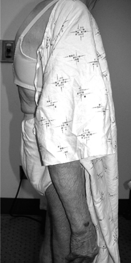
Vertebral body compression fractures typically manifest as episodes of acute and frequently explosive axial pain precipitated by routine activities of daily living. Movement is limited by pain, and flexion is restricted more than extension. This spinal pain is exacerbated by axial loading during sitting or standing, and alleviated by recumbent positioning. The process of changing position is particularly uncomfortable, leaving the afflicted patient with the desire to remain in a position of comfort; particularly resting supine in bed. Thoracic fractures can result in thoracic kyphosis (dowager’s hump) (Fig. 44.1) leading to a discrepancy between standing height and arm span, and pulmonary dysfunction. Involvement of the lumbar spine may lead to progressive loss of normal lumbar lordosis, leading to early satiety and abdominal bloating. Neurologic involvement is rare in osteoporotic compression fractures because the posterior cortical wall is rarely breached.8 When involvement of the spinal cord or cauda equina occur, it should suggest other conditions that can lead to a compression fracture such as infection, metastatic or primary bone tumors, myeloma, Paget’s disease, or lymphoma.9
Pain and disability consequent to osteoporotic vertebral fractures tax the health care, industrial, and societal systems. Approximately 750 000 new osteoporotic vertebral fractures occur in the United States each year,3 and over one-third of these become chronically painful.10 Vertebral fractures in patients aged 65 years or older account for 150 000 hospital admissions per year11 with an estimated total cost of 1.5 billion dollars.3 Patients aged 45 years or older account for 161 000 physician office visits and more than 5 million restricted-activity days per year.12 Hence, osteoporotic vertebral compression fractures are a leading cause of disability and morbidity in the elderly.13 Such outcomes are due to chronic pain, progressive vertebral collapse, and spinal kyphosis.14 Osteoporotic compression fractures have been shown to adversely affect quality of life, physical functioning, mental health, and survival.15
TRADITIONAL TREATMENT APPROACH
Traditional treatment pursuits strive to achieve pain relief, reduction of the sequelae of deconditioning, and maximal function. These objectives are largely achieved by conservative measures including relative rest, narcotic analgesics, antiosteoporotic medications, spinal orthotics, and gradual extension-biased exercises.3,16 Rarely, these fractures are associated with neurologic compromise or spinal instability necessitating surgical repair. Bed rest is prescribed in the acute phase, but must be minimized to 2 days to reduce the occurrence of contractures,17 worsening osteoporosis,18 muscle weakness and atrophy,19 and thromboembolic complications.20 Each of these entities will increase the morbidity and mortality resulting from a compression fracture. In the majority of cases, the pain will abate within 2–4 weeks to as many as 8–12 weeks. However, a significant number will remain painful,15 possibly due to incomplete healing with progressive bony collapse, altered spinal kinematics resulting from deformity, or development of a pseudoarthrosis at the involved segment.14 Analgesic medications are poorly tolerated by elderly patients due to confusion, increased fall risk, and constipation. Bracing may not be well tolerated due to decreased diaphragmatic excursion, and restriction of abdominal movement. Overall, these treatment interventions aim to ameliorate the symptoms associated with osteoporotic compression fractures, while not addressing underlying tissue injury.
Historical overview of vertebroplasty
Over the past two decades, vertebroplasty, the percutaneous injection of polymethyl methacrylate (PMMA) directly into the fractured vertebral body, has been increasingly utilized to stabilize vertebral body compression fractures. This technique was invented in 1984 by two French physicians, Galibert and Deramond, to treat symptomatic vertebral angiomas, and their findings were subsequently published in 1987.21 Nine years later, Cotton et al.22 and Weill et al.,23 in two independent studies, established the efficacy of vertebroplasty in patients suffering from metastatic spine disease or multiple myeloma. Jensen et al. published the first American trial in 1997 of applying vertebroplasty to treat osteoporotic vertebral compression fractures.24 Hence, European physicians have served the pioneering role in establishing the utility of vertebroplasty, acquiring greater experience with this new technology in pursuit of primarily treating painful spinal tumors. In contrast, North American physicians have witnessed a growth in deploying this procedure to treat painful osteoporotic patients who themselves served as the impetus for exploring this treatment option.25 Consequently, vertebroplasty has been promoted as a viable option to treat painful vertebral compression fractures in the absence of sound methodologic prospective trials supporting this contention. Although conclusive evidence is lacking, diligent studies have been and are being performed to explain vertebroplasty’s therapeutic benefit, substantiate its safety, demonstrate the durability of its efficacy, and refine its technique.
PATHOPHYSIOLOGY OF VERTEBRAL BODY COMPRESSION FRACTURE
Mechanical failure of the vertebral body represents disrupted structural integrity. The vertebral body’s ability to function properly to bear and transmit loads is a function of the health of its trabecular bone; its structural design as dictated by shape, size, and organization; and the loading conditions involving the energy it absorbs.1
Intrinsic factors
The strength of the trabecular bone is a key attribute in determining a vertebral body’s risk of fracture. Trabecular bone functions to carry the axial load transmitted to the vertebral body from the adjacent intervertebral discs. Trabecular bone will fail if its strength is less than the working stresses experienced within the vertebral body during physiologic or traumatic loads. Whole bone fractures are the consequence of such failures. Trabecular bone strength is determined by apparent density and its architecture. Reductions in apparent density occur with aging, osteoporosis, and tumor infiltration. The compressive mechanical strength of trabecular bone is proportional to the apparent density squared. Thus, a decrease in the later will cause a disproportionate reduction in trabecular bone strength.1
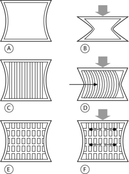
Vertebral trabeculae are arranged in vertical and horizontal columns, and the vertical columns outnumber the horizontal ones at any given density.1 Compressive loads are initially borne by the vertical trabecular struts, and when these struts begin to bow their horizontal counterparts act as cross-beams to restrain this displacement (Fig. 44.2).26 Consequently, an axial load is sustained by a combination of vertical pressure and transverse tension within the trabeculae. The ability to transfer load from vertical pressure to transverse tension confers resilience to the vertebral body.26 As the number of trabeculae decrease with aging, osteoporosis, or space-occupying lesion, the altered architecture reduces the vertebral body’s resiliency, thus lowering its threshold for fracture under load.1
A strong, positive correlation between lumbar spine bone mineral density (BMD) and failure force required to fracture a lumbar vertebra has been unequivocally established.27 Hence, BMD can be used as a convenient and specific predictor of the risk of vertebral body failure under compressive loads. This correlation appears to be a continuum without a single fracture threshold value for BMD at which a vertebral body will fail.1 A given vertebra will fatigue with repetitive loading, allowing it to fail at a lower load force than would be required during a single load application.28 The phenomenon of vertebral fatigue may be due to intraosseous crack initiation, crack growth, and final failure.29 Indeed, such bony deficits have been observed in human vertebral bone, irrespective of age, lending credence to the presence of such microdamage reducing vertebral fracture resistance.30
Extrinsic factors
Extrinsic factors such as loading conditions are as integral to vertebral body fracture as the already discussed intrinsic factors. Fractures occur at a point when external loads result in internal stresses that exceed the strength of the bone. Clinical recognition of the features of osteoporotic vertebral compression fractures can be difficult. Cooper et al. reviewed charts from 341 fracture patients over a 5-year period and found that 16% of patients were diagnosed incidentally, 33% after a fall, and 9% after routine lifting.2 Within this study cohort, approximately 50% of the vertebral fractures were reported as spontaneous or incidental,2 perhaps a result of vertebral fatigue.1 In a smaller study, Myers et al. recorded a history of a fall in 50% of osteoporotic acute fracture patients over 60 years old, a history of acute fracture in 20% of patients with routine activities of daily living, with the remainder of patients not able to recall a specific event at the time of injury.31 The apparent disparity among the causes of osteoporotic vertebral compression fractures might be explained by a combination of intrinsic and extrinsic factors. A single traumatic event leading to a load sufficient to fracture a weak vertebra might explain a fracture due to osteoporosis or a tumor. A fracture associated with routine activity or no apparent cause may represent structural fatigue due to age, osteoporosis, or tumor-related vertebral changes. A ‘factor of risk’ has been established incorporating an individual patient’s BMD to forecast the risk of vertebral fracture consequential to some routine activities of daily living.32 Factors of risk are the ratio of the estimated forces applied to the spine to the vertebral failure threshold based on lumbar BMD. A ratio equal to or greater than 1 represents a strong likelihood of fracture.1,32 Individuals with low BMD routinely operate near a ratio of 1, and women with below average BMD who lift a toddler experience a factor of risk near 1.1 For women with very low BMD, innocuous activities such as tying one’s shoes increases the risk of fracture.1
Implicit in the dynamic relationship between routine activities and fracture risk is a role for posture and neuromuscular conditioning, and degenerative disc disease. Evidence has recently been published substantiating this contention.33,34 An involutional loss of functional muscle motor units occurs with aging and predisposes an individual to poor postural control.35 A 2-year structured back extension exercise program has been shown to reduce the incidence of vertebral compression fractures and increase bone mineral density in healthy, postmenopausal women, up to 8 years after cessation of the program.33 Thus, extension-based exercise helps maintain bone mineral density and prevent vertebral compression fractures, perhaps due to improved posture and spinal stabilization.
Pollitine et al., in an elegant study, defined the relative loads experienced by the three columns of the spine in patients with degenerative discs.34 Twice as much load is assumed by the posterior column than by the anterior vertebral body in the erect standing position in patients with degenerative intervertebral discs. In contrast, the anterior vertebral body’s axial load increases almost three times during forward bending in the presence of degenerative discs. A relative ‘off-loading’ of the anterior vertebral body during upright posture will result in bone loss due to a lack of routine dynamic strain.36,37 Ultimately, the weakened anterior vertebral body is abnormally loaded above its counterpart in spines containing healthy, nondegenerate discs. The findings of Pollitine et al. suggest that in a person with severely degenerated intervertebral discs, the load placed on the anterior vertebral body increases by greater than 300% in flexion as compared to standing erect.34 Sinaki et al.’s reduction in compression fractures may be due to their patients’ increased spinal extensor conditioning as well as an independent increase in BMD. Their treatment arm utilized extension exercises without specific land-based weight-bearing exercises.33 The increased extensor conditioning may have decreased the load incurred by the anterior vertebral body during flexion activities, and may have inadvertently increased the anterior vertebral body’s resilience to fracture. However, Sinaki et al. did not remark about the presence or absence of disc degeneration.
An accurate comprehension of the pain generating mechanisms of vertebral compression fractures is indispensable if successful treatment is to be effectuated. Traditional treatment interventions assume adequate tissue healing with time, which seems to be logical given the natural history of gradual resolution of pain within 2–12 weeks. However, chronic pain may develop due to microfracture nonunion,14,38 pseudoarthrosis,3 sensitization of the basivertebral nerve,39 or spinal deformity14 leading to painful paraspinal muscle spasm. The first three of these probable pathophysiologic mechanisms can be addressed with the percutaneous injection of PMMA. Although kyphotic deformity is not maximally corrected by vertebroplasty, reduction in pain allowing extension-based spinal stabilization may reduce painful myofascial dysfunction, thus reducing kyphosis.
MECHANISM OF ACTION OF VERTEBROPLASTY
The pain relief afforded to patients having undergone vertebroplasty has not yet been successfully attributed to a primary mechanism. Thermal,40–43 chemical,44,45 and mechanical46 effects have all been reported as possible mechanisms. However, the mechanical effects of vertebroplasty have been regarded as the critical factor leading to symptom reduction.46–48 Tohmeh et al., in 1999, demonstrated increased strength and stiffness in osteoporotic cadaveric compression fractures utilizing either a unipedicular or bipedicular approach.46 However, the authors did not investigate the distribution or volume of PMMA injected relative to these mechanical parameters. Liebschner et al. subsequently established that symmetric deposition of PMMA is ideal to prevent single-sided load transfer, and that overall a small amount is needed to restore stiffness and strength.49 Belkoff et al. provided preliminary guidance for the amount of injected PMMA necessary to restore stiffness and strength;47 however, Molloy et al. later established the approximate percentage of vertebral body volume required to be filled to restore adequate strength and stiffness.48 A weak correlation has been noted between percentage of fill and restored strength and stiffness,48 which echoes previous clinical observations of minimal correlation between pain relief and the amount of PMMA injected.22 If an intravertebral cleft is present, there is a trend toward greater pain relief if these clefts are opacified during vertebroplasty.50 These findings suggest that either the singular factor of achieving a minimum threshold of stabilization is needed to abate micromotion or malunion induced pain, or that an alternate pain-reducing mechanism is yet to be demonstrated.
The polymerization process of PMMA is exothermic and its effects have been studied utilizing cadaveric vertebral bodies.42,51 Deramond et al. observed sufficiently elevated and sustained intraosseous temperatures in the central and anterior vertebral bodies during PMMA injection, and lack thereof at the anterior wall of the spinal canal, to cause thermal damage to intraosseous neural structures.42 Belkoff and Molloy subsequently observed higher temperature elevation in vertebral specimens during intraosseous injection of PMMA, perhaps due to a different temperature recording technique, but these higher temperatures did not occur at the spinal canal.51 The authors were led to believe that the risk of thermal necrosis may have been underappreciated. Yet histologic examination of four vertebral bodies, previously treated with percutaneous injection of PMMA, retrieved from human spines during spinal surgery, identified rare foci of necrosis.52 Unfortunately, histologic assessment was not performed on the intraosseous neural structures of these four samples. Further, a vast network of substance-P containing nerves has been documented within the vertebral body.39 Consequently, thermal and/or chemical denervation may contribute to the pain-relieving effect of vertebroplasty.
INDICATIONS AND CONTRAINDICATIONS
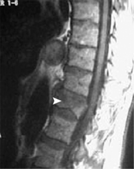
Vertebroplasty was first developed to treat symptomatic vertebral body hemangiomas.21 Since 1984, the conditions for which percutaneous injection of PMMA has been undertaken have been expanded to include primary and metastatic spinal fractures (Fig. 44.3),22,23 osteoporotic compression fractures (Fig. 44.4),24 vertebral fractures due to osteogenesis imperfecta,53 recurrent fracture or pain at a previously treated segment,54 and pseudoarthroses associated with noninfected avascular necrosis of the vertebral body.55
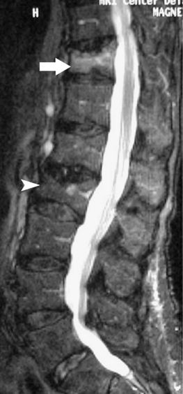
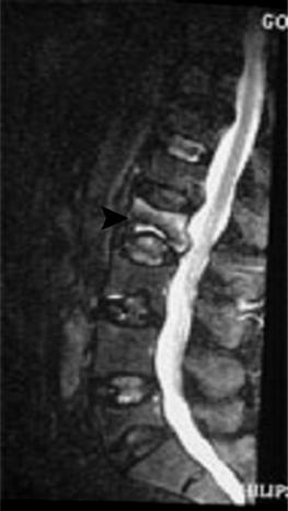
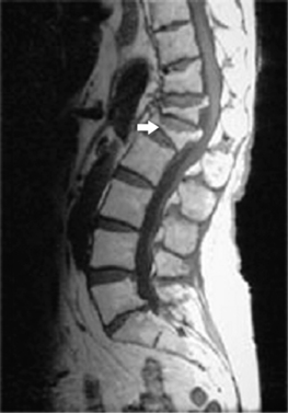
The universally accepted indications for performing percutaneous vertebroplasty (PVP) are severe, focal, intractable spinal pain, in the absence of radicular pain, recalcitrant to traditional treatment measures due to acute compression fractures.3,14,22,25,24,56–72 An acute fracture is substantiated by a typical history and evidence of new or progressive fracture by magnetic resonance imaging (MRI) (Fig. 44.5). Alternatively, persistent pain, despite appropriate conservative care, greater than 3 months due to an imaging confirmed compression fracture is a relative indication.73 In a retrospective observational investigation, Kaufmann et al.73 found that fracture age at the time of PVP was not independently associated with postprocedural pain or activity. In subsequent retrospective study, Brown et al.74 found that 80% of 41 osteoporotic fracture patients with symptom duration of greater than 12–24 months had post-PVP pain improvement; in comparison, 92% of 49 control patients, those with symptoms less than 1 year, experienced pain relief. Although the number of patients achieving partial or complete pain relief was not statistically different between groups, complete pain relief was more frequent in the control group. The ideal time frame during which to intervene with PVP to treat painful vertebral compression fractures has yet to be determined in well-controlled prospective studies. However, some contend that the likelihood of improvement probably decreases over time and appears to be low for remote, 6 months or older, fractures.3
Contraindications to PVP include a fracture that breaches the posterior vertebral wall, retropulsed bone fragments, gross spinal instability, clinical evidence of myelopathy or radiculopathy, osteoblastic metastatic lesions, uncorrectable coagulopathy, systemic or spinal infection, or medical conditions that preclude safe emergent surgical decompression.3 A loss of vertebral body height of greater than 75%, has been cited as a relative contraindication to PVP (Fig. 44.6).23,56 However, Peh et al. evaluated 37 patients who underwent PVP to treat vertebra plana, defined as a collapsed vertebra to less than one-third of its original height.57 Ninety-four percent of these patients experienced pain reduction, with 51% of the 37 patients reporting complete pain relief.57 No major complications were reported, and the authors concluded that PVP of severe osteoporotic vertebral body compression fractures should not be withheld from this category of patient.57 At this point, it seems most prudent to regard severe vertebral compression fractures as relative indications in the absence of myelopathy, instability, or disruption of the posterior vertebral body wall.
EFFICACY OF PERCUTANEOUS VERTEBROPLASTY
Successful clinical outcomes have been reported since the inception of PVP in 1984.21 A plethora of studies have been published documenting the therapeutic benefit of PVP in treating painful vertebral body compression fractures.3,14,21–25,53–58–65,75 The excitement generated by a treatment intervention that achieves complete or near complete pain relief has fueled many investigations attesting to its efficacy. Many of these reports have evaluated pain reduction, and less commonly quality of life or patient satisfaction. Only one study included a control group, and no studies to date have employed a prospective, randomized trial incorporating a control group. Hence, the natural history of a certain vertebral compression fracture has not been concurrently compared to the clinical course of this condition after treatment with PVP. Rather, several investigations have seemingly established PVP’s safety and efficacy at reducing disruptive pain symptoms due to vertebral body compression fractures.
Osteoporotic compression fractures
Vertebral compression fracture is a common clinical manifestation of osteoporosis, but rarely leads to neurologic compromise.9 However, osteoporotic vertebral compression fractures are invariably associated with significant impairment in functional, physical, and psychosocial indices.76 Quality of life suffers after the first osteoporotic compression fracture and decreases further with subsequent fractures.77 Afflicted individuals experience pain, disability, loss of activity, fear of falling, and embarrassment over their appearance.78 Increased mortality has been observed after an initial osteoporotic compression fracture, and progressively increases with advancing time after the initial injury.79 As our society continues to age, refining the appropriate role of PVP in treating osteoporotic vertebral compression fractures has emerged as the multifaceted impetus for a variety of investigations.
Jensen et al. first addressed the efficacy of PVP to treat osteoporotic vertebral compression fractures in the late 1990s.24,75 Ninety percent of 29 patients treated with PVP reported significant reduction in pain within 24 hours after the procedure.24 At a mean follow-up of 281 days, 23 of these 26 patients who initially experienced significant improvement reported sustained pain reduction and increased mobility.75 In a larger study of 245 osteoporotic patients, Evans et al.58 reported significant reduction in pain, and improved ambulatory status and activities of daily living at a median follow-up interval of 7 months. Yet the 245 cases were reviewed retrospectively and represented the only cases out of 488 available for follow-up. The authors did not use a validated disability assessment tool, and it was administered at varying times after treatment intervention. Long-term observations were conducted by Grados et al.59 who documented a statistically significant reduction in visual analog scale (VAS) pain scores in 96% of osteoporotic compression fracture patients treated with PVP. The mean duration of follow-up was 48 months with a range of 12–84 months. The authors concluded that the results obtained by PVP are durable over time, as their findings did not deteriorate from 1 month to a mean of 48 months after treatment intervention. However, 15 of the original cohort of 40 were not available at follow-up, the methodology was retrospective, and clinical significance, such as quality of life or disability, of the reduced VAS ratings was not assessed. These findings have been corroborated by those of other retrospective studies incorporating smaller subgroups of osteoporotic patients with significant pain relief occurring in 75–95% of treated patients.56,60–63
Peters et al.64 prospectively observed a statistically significant decrease in VAS ratings in 84% of 42 osteoporotic compression fracture patients treated with PVP at 56 levels, and 7 patients reported no change in their pain level. All 42 patients were available at follow-up at 6 weeks, and had failed to improve despite a 6-week course of undefined medical management. The authors employed a bipedicular approach to inject PMMA into each fractured vertebrae except for six that were treated with a unipedicular technique. No relationship was disclosed between unipedicular technique and failure of pain reduction, and follow-up occurred at 6 weeks. Focusing on the efficacy of PVP in treating severe (>70% vertebral body collapse) osteoporotic compression fractures, Peh et al.57 prospectively found complete or partial pain relief in 97% of 38 patients undergoing 48 PVPs. Just 30 of the initial 38 patients were available for follow-up at a mean of 11 months (range 3–24 months). Pre-intervention symptoms ranged from 2 weeks to over 1 year. Bipedicular injection was competed in 20 levels, and 28 underwent unipedicular injection. McGraw et al.65 prospectively followed 100 patients undergoing 156 PVPs for osteoporotic (n=92), neoplastic (n=5), concomitant spinal stenosis (n=2), and osteogenesis imperfecta (n=1) related fractures. Ninety-nine patients were available for follow-up at a mean interval of 21.5 months (range 6–44 months), and 93% of patients reported improvement in their back pain with a statistically significant reduction in VAS ratings. However, the authors did not report the pre-PVP symptom interval, stratify results according to diagnostic category or how many levels were treated using a bipedicular technique. All three investigations used similar inclusion criteria including painful osteoporotic compression fractures refractory to medical management, proven by imaging and history and physical examination. The severity of collapse varied among the studies, and all three inquiries used pain reduction and mobility as the outcomes measures without incorporating a valid disability tool.
Cortet et al.66 prospectively evaluated the efficacy of PVP in treating 20 osteoporotic vertebral compression fractures in 16 patients suffering from intractable pain despite 3 months of medical management. A statistically significant improvement was noted in VAS ratings, McGill-Melzack scoring system, and five out of six dimensions of the Nottingham Health Profile at 1, 3, and 6 months after PVP. Zoarski et al.67 prospectively followed 30 patients utilizing verbal pain scores and the Musculoskeletal Outcomes Data Evaluation and Management Scale (MODEMS) spinal intervention questionnaire. All patients demonstrated a lack of improvement after a minimum of 4 weeks of conservative care prior to PVP. Twenty-nine patients reported pain relief within hours of the procedure, and at 2 weeks 80% reported feeling better and 90% reported they would undergo the procedure again. All four modules of the MODEMS data (treatment score, pain and disability, physical function, and mental function) improved with statistical significance at 2 weeks. Ninety-six percent of 23 available patients at follow-up at 15–18 months reported continued pain relief and satisfaction with their outcome. All 30 patients underwent bipedicular injection of PMMA. Findings in a long-term, prospective study by Perez-Higueras et al.68 seem to support the durability of the results afforded to patients by PVP. Thirteen out of 17 original patients treated for 39 osteoporotic vertebral compression fractures were available at a mean follow-up of 65 months (range 60–71). All patients had failed to improve after a minimum of 3 months of conservative care. Outcomes were measured by VAS ratings and the short McGill questionnaire and all 13 demonstrated significant improvement at 3 months which increased slightly at the 5-year follow-up.
These prospective studies have helped to provide greater insight regarding the efficacy of PVP in treating osteoporotic vertebral compression fractures. However, none incorporated a control group for comparison to the treatment arm. Implicit in this methodology is that a regression toward the mean has already occurred during the period of conservative care. Hence, any treatment intervention after this time frame would be attributed to the intervention itself. However, none of these studies recorded such statistical analysis. To date, one prospective study has incorporated a control group of patients treated with conventional therapy.69
In a nonrandomized fashion, Diamond et al.69 compared the clinical outcomes of 55 osteoporotic patients who underwent PVP with 24 patients who refused PVP and were conservatively managed. Inclusion criteria were acute severe vertebral compression pain unrelieved by 1–6 weeks of oral analgesics, densitometric evidence of osteoporosis, and acute fracture activity as indicated by MRI or technetium-99 m bone scan. No differences existed between the two groups regarding smoking history, alcohol intake, corticosteroid use, thyroxine therapy, vitamin D and parathyroid hormone levels, bone density, or degree of vertebral collapse. More importantly, both groups had statistically similar baseline levels of pain and disability. Fifty-three patients in the PVP group and 21 of the conservative group were available at a mean follow-up of 215 days (range 57–399), and outcomes were measured by VAS ratings and the Barthel index level of function. Statistically significant improvement in pain and physical functioning was noted at 24 hours in the PVP compared to the conservatively treated group. No differences were observed at 6 weeks or 6–12 months. A 40% reduction in the mean length of hospitalization was observed in the PVP group. These findings suggest, but do not prove, that PVP provides effective pain relief in the short term. The absence of a clinical separation between the two groups at longer follow-up may be due to the natural history of most compression fracture symptoms abating over 6–12 weeks. Hence, these cases may have been destined to clinically improve within 6 weeks after initiation of the study due to the natural history of the condition itself.
Stay updated, free articles. Join our Telegram channel

Full access? Get Clinical Tree


