Clinical: Superficial (cutaneous/subcutaneous) hemangiomas are reddish-wine-colored painless lesions, generally present at birth. Intramuscular hemangioma arises within the belly of a single muscle; only in the hand and foot it may expand between the fascia, muscles, and tendons. It is possible to observe pain and swelling associated with venous stasis. Pain is sharp and becomes more intense with tension of the muscle. Shortening of muscles causes first joint dysfunction and then joint deformity. In the hand and foot, an increase of skin temperature, of the superficial venous reticulum, telangiectasia, cyanosis, and hyperhidrosis are observed.
The majority of epithelioid hemangioma presents as subcutaneous masses of a year or less in duration. The process is usually uninodular, but multinodularity, generally in contiguous areas, can be present. Dermal examples are less frequent, and deep-seated cases are rare. Epithelioid hemangioendothelioma develops often as a painful nodule in either superficial or deep soft tissue. Deeply situated tumors may be associated with focal ossification that can be detected on plain films.
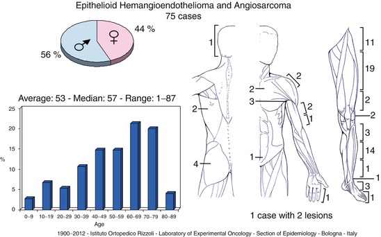
Soft tissue angiosarcoma develops as enlarging mass, in 1/3 of patients associated with other symptoms such as coagulopathy, anemia, persistent hematoma, or bruisability.
Imaging: X-rays are usually negative although small round granular calcifications with a smooth surface and concentric stratifications (phleboliths) can be seen. Vascular tumors present an inhomogeneous pattern on MRI. The lesion often appears as a “bunch of grapes,” occasionally with a serpentine or tubular pattern. On T1, angiomatous tissue has an intermediate intensity between that of muscle and fat, but areas of stagnant blood and hemorrhage can cause high signal intensity. On T2, vascular spaces seen are hyperintense, but fibrous septa and calcified foci are hypointense. Fluid-fluid levels can be appreciated. Malignant vascular lesions often show nonspecific characteristics. The presence of intratumoral necrosis may be demonstrated by the use of contrast agents.
Histopathology: Benign hemangiomas can be cavernous, capillary, or pseudovenous. Cavernous hemangiomas are the most frequent. Grossly they are made of a bunch of scattered nodules, from dull red to bluish, with internal cloistering. Histologically, they are composed of extremely dilated vessels with a very thin wall of flattened endothelium and collagen membrane, filled with blood. Capillary hemangiomas are more compact and pinkish. They are composed of many small vessels regularly coated with endothelium spreading into collagen stroma. Pseudovenous hemangiomas appear spongelike with fibrous cloisters and intercommunicating lacunae only partially filled with blood, thrombi, and phleboliths. They are composed of craggy, ramified, labyrinthic cavities with very irregular, thick walls with a pseudovenous fibromuscular structure. Intramuscular hemangiomas have been traditionally classified according to vessel size in small (capillary), large (cavernous), and mixed.
Epithelioid hemangioma is usually 0.5–2.0 cm in size, generally with a rather nonspecific nodular appearance. Subcutaneous examples of epithelioid hemangioma are histologically characterized by a prominent proliferation of small, capillary-sized vessels lined by plump, epithelioid endothelial cells, with a typical immature appearance, sometimes lacking a well-defined lumen. These vessels are rimed by a single cell endothelium layer with an intact myopericytic/smooth muscle layer. The process is usually well demarcated from the surrounding soft tissue, and commonly, it is associated with or centered around a larger vessel, usually a muscular artery. An inflammatory milieu rich in eosinophils and lymphocytes is generally present. Dermal examples of epithelioid hemangioma generally show a more mature appearance with a well-canalized lumen, and endothelial cells are somewhat less plump, frequently more cobblestone, or hobnail-like in appearance. In addition, dermal examples are less circumscribed and are not usually associated with a larger central vein or muscular artery. Epithelioid hemangioendothelioma generally arises as a fusiform intravascular mass that may resemble an organizing thrombus. Histologically, they are composed of short strands, cords, or solid nests of epithelioid eosinophilic endothelial cells, sometimes with intracytoplasmic lumina (vacuoles) containing erythrocytes. Cells appear quite bland with little or no mitotic activity and are embedded in a distinctive matrix that varies from light blue (chondroid-like) to deep pink (hyaline) in color.
Related posts:

Stay updated, free articles. Join our Telegram channel

Full access? Get Clinical Tree


