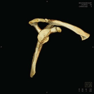
Chapter 16 A certain degree of anterior inferior glenoid bone loss has been reported in up to 90% of patients with recurrent anterior instability.1 Bone loss patterns may be traumatic, with a bony fragment present from an acute dislocation, or may be attritional, with erosion of a previous fragment or the anterior glenoid surface.2,3 In either case, the glenoid morphology changes over time, usually progressing to more significant bone loss with repeated instability events.4 Glenoid bone loss represents the progressive loss of a key static stabilizer in glenohumeral stability because there is a smaller surface area to resist axial loads, less articular length, and decreased depth of articular conformity.3,4 All of this contributes to increased instability events. It is well documented that when glenoid bone loss reaches a key threshold level of 20% to 30%, significant instability ensues.2–6 To adequately reconstitute the glenoid bony arc and restore stability, bony augmentation is required for adequate glenohumeral stability. Options for restoration of glenoid bone traditionally have included coracoid transfer consisting of one of the variants of the Bristow-Latarjet procedure, iliac crest autograft, or allograft glenoid.1,7–12 Recently, a novel technique for glenoid bone and cartilage restoration using distal tibia allograft has been proposed.13 The following technique describes the procedure in detail, as well as preoperative considerations and intraoperative pearls and pitfalls. This technique offers several unique advantages over previous glenoid bone augmentation procedures: no donor site morbidity, abundant availability, and anatomic reconstitution of the glenoid arc with a robust intra-articular chondral surface with a foundation of abundant, dense corticocancellous bone that can be conformed to each individual’s degree of bone loss.13 Patients typically report recurrent anterior instability. A detailed history should be taken to determine their activity level as well as the details of their initial dislocation event. Patients with significant glenoid bone loss (15% to 30%) typically have a history of either a high-energy axial load event or low-energy traumatic dislocation with progressive ease of subluxation.3,4 Instability is usually in the midabduction (20 to 60 degrees) ranges of motion.4,12 They describe instability with daily activities and inability to participate in athletic activities. Many patients have already undergone a previous soft tissue anterior stabilization and have recurrent instability or dislocation events. Examination should begin with a careful inspection of both shoulders for deltoid or rotator cuff atrophy, scapular dyskinesis, pectoralis minor tightness, and deformity. A careful neurologic examination should also be conducted. Active and passive range of motion should be tested along with rotator cuff strength, with any side-to-side asymmetry noted. Special attention should be directed toward subscapularis testing because this may be either split or taken down and repaired during the operative procedure.4 Provocative testing should evaluate for long head of biceps pathology, labral tears, and subacromial pathology. Instability testing should focus on anterior subluxation or apprehension at the midranges of abduction (20 to 60 degrees) in addition to full abduction and external rotation (the 90-90 position).4,12 Patients with glenoid bone loss generally do not have multidirectional instability, and this should be carefully evaluated before any surgical procedure is considered. Imaging should begin with plain radiographs of the shoulder to include standard anteroposterior (AP), glenoid profile AP, scapular-Y, and axillary views. Additional views focused on the glenoid face include the West Point, Didiee, and apical oblique views.4 Plain radiographs are only mildly effective at demonstrating glenoid bone loss; however, they will demonstrate loss of the sharp cortical density at the inferior glenoid, which is replaced by a bony shadow.14 Magnetic resonance imaging (MRI) and magnetic resonance arthrography (MRA) are also useful for demonstrating soft tissue pathology including rotator cuff tears and labral degeneration and tears and can demonstrate bone loss with a high-quality 3-tesla magnet on a sagittal oblique cut of the glenoid.4 Computed tomography (CT) with three-dimensional reconstruction is the most accurate method for determining glenoid bone loss and should be included in the standard workup for recurrent anterior instability1 (Fig. 16-1). A best-fit circle is drawn over the inferior two thirds of the glenoid on the glenoid image with the humerus digitally subtracted.15 The amount of bone loss is then measured as a percent of total surface area of the previously drawn circle.4 There are multiple techniques described for measuring glenoid bone loss; however, the quickest method is simply measuring the bony deficit anteriorly and dividing by the total circle diameter. Typically a 6- to 7-mm deficit represents approximately 25% glenoid bone loss.2,5,6 The usual pattern of bone loss is parallel to the long axis of the glenoid and generally occurs between the 2:30 and 4:20 positions with reference to a clock face.16
Treatment of Recurrent Anterior Inferior Instability Associated with Glenoid Bone Loss
Distal Tibial Allograft Reconstruction
Preoperative Considerations
Physical Examination
Imaging
![]()
Stay updated, free articles. Join our Telegram channel

Full access? Get Clinical Tree


Treatment of Recurrent Anterior Inferior Instability Associated with Glenoid Bone Loss: Distal Tibial Allograft Reconstruction


