Treatment: Manual therapy modes

Chapter 8 Treatment


Manual therapy modes


Introduction


This chapter provides an introduction to the body tissues typically affected by the ‘malalignment syndrome’ and a selection of manual treatment approaches available to correct any abnormal biomechanical forces and help these tissues recover. The best treatment approach should be based on the structure primarily contributing to the problem, the individual’s expectations, philosophy and interests, as well as the availability of manual therapists in your area. It is the authors’ opinion that the connection of the patient with the therapist can be as important as the type of treatment chosen, thus we encourage trying different practitioners or approaches to find the right match.


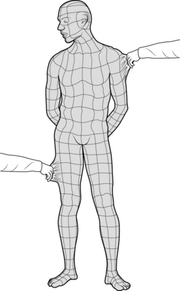
Manual therapy can be practised by numerous specialists such as physiatrists, osteopaths, physiotherapists, chiropractors, massage therapists, naturopathic doctors and athletic trainers. The techniques described in this chapter can be applied by some or all of these practitioners depending on their education and training (see Ch. 7 regarding the Canadian programme for post-graduate training of physiotherapists in the theory and practice of manual therapy, leading to designation as a ‘Fellow of the Canadian Association of Manual Therapists’ or ‘FCAMT’). Patients should not hesitate to ask their local practitioners what post-graduate training they have had in manual therapy!


The previous chapters describe muscle energy techniques for treatment in particular of pelvic malalignment. When the pelvis is out of alignment, tissues around the pelvis must compensate, altering the optimal biomechanics and tissue length-tension relationships. There are four main systems recognized to influence pelvic dysfunction: articular, neural, visceral and myofascial (Lee 2007c, 2011). These tissues are discussed as to their anatomy, biomechanics, and the manual therapy techniques that can directly affect them.


Manual therapy is an all-encompassing term for ‘hands-on’ therapeutic interventions performed by various health practitioners to alleviate ailments and treat specific etiologies. The definition of manual therapy varies among health professionals and seems to be influenced by their scope of practice and how their techniques have evolved. This chapter will not attempt to review all treatment techniques available but to highlight some of the most common approaches used today. Each therapy will be introduced with a brief summary of its history and its originator(s), the physiology or biomechanics of how it functions, and a generalized description of the patient experience. The information provided is only a short description of each technique; references for further reading and resources are provided for those who want to pursue learning more about the individual techniques.


Tissue types


Connective tissue


Connective tissue usually refers to fascia, ligaments, and tendons, which are primarily composed of collagen and/or elastin.


Collagen makes up about 25-35% of our entire body’s protein content. This long fibrous structure contributes to the tensile strength of fascia, cartilage, ligaments, tendons, bone and skin and is a major component of the extracellular matrix supporting most of our tissues.


Elastin, unlike collagen, can stretch easily and has almost perfect recoil. With age, it loses some of its elasticity and may even calcify. The fibres are composed of protein elastin fibrils which branch and rejoin loosely. Elastin is not as organized as collagen and is usually thinner and less abundant.


Fascia


Fascia is made up of collagen, elastin and a polysaccharide gel complex forming a three-dimensional cobweb covering the body’s tissues (Fig. 8.1). It has three continuous layers: (1) a superficial layer under the skin, (2) a deeper, ‘ensheathing’ layer which covers muscles, bone, nerves, blood vessels, and organs right down to the cellular level, and (3) the deepest layer, which connects with the dura of the central nervous system. The muscle is intimately connected with fascia, hence the term ‘myofascia’. Fascia functions to support our body, absorb shock, enhance cellular respiration and metabolism, and assist with fluid and lymphatic flow.


Malfunction of the fascial system can occur from poor or prolonged postures causing asymmetrical loading, trauma, and repetitive strain. Changes in fascial mobility occur when the fascia is bound down or restricted in areas. Due to its continuous nature it causes a global effect by pulling on the body at points distal to the primary site of dysfunction (see Chs 2, 3). This stimulates nociceptors, causing pain, and reduces the body’s ability to absorb and distribute forces that may be incurred with sudden trauma or repetitive loading. It can also alter an individual’s biomechanics and influence his or her posture and dynamic balance. Injuries to the fascial system do not result in typical nerve root or trigger point referral patterns and standard imaging tests (i.e. X-rays, CT scans, MRI, etc.) will not pick up fascial dysfunction, so typically these malfunctions usually go undiagnosed.


Therapeutic interventions can alter our fascia by: stretching its elastic component, shearing the cross-links that may have developed, changing the viscosity of the ground substance by increasing the production of hyaluronic acid, and increasing its mobility and fluid flow. Techniques which have a direct affect on fascia include myofascial release, visceral manipulation, cranial osteopathy, craniosacral therapy, Bowen therapy, acupuncture and IMS.


Contractile tissue


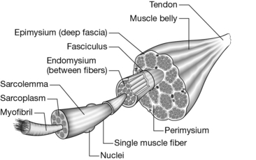
Muscles are comprised of bundles of fasciculi that, in turn, are made up of bundles of muscle fibres (Fig. 8.2; the latter) themselves are formed from bundles of myofilaments (myofibrils) composed of two main proteins, actin and myosin: the contractile elements of the muscle. Each layer of a bundle is separated by fascial tissue that helps transmit forces from muscle to tendon and bone.


Muscles are always in some degree of contraction known as ‘muscle tone’. The tone of a muscle is determined by impulses coming from the spinal cord which depend on the amount of alpha activity in nerves. This activity is influenced by one’s psychological state, neurochemistry, and feedback from receptors within the muscles. There are two types of muscle receptors:


These muscle receptor mechanisms work in opposite ways to help control muscle tone. Muscle spindles use a positive feedback cycle to continuously monitor muscle length and rate of length change by stimulating the anterior horn cells in the spinal cord which, in turn, provides the stimuli for increasing muscle tone. Conversely, Golgi tendon organs use a negative feedback system to protect the muscle from sudden forces and tearing. They excite an inhibitory interneuron in the spinal cord to suppress the alpha motor neuron and allow the muscle to relax. This is believed to be the mechanism behind trigger point and acupressure release techniques.


When joint receptors are damaged by injury, the damage will alter the body’s position sense – or proprioception – and also affects muscle tone. When the antagonist muscles are unable to relax or are pathologically shortened, the normal range of motion is restricted which will again influence muscle tone. Certain muscles are over-active and tend to shorten, while others are underactive and tend to lengthening or atrophy. These are factors that contribute to the ‘malalignment syndrome’, where the pattern of muscular ‘facilitation’/’inhibition’ and strength is noted to be asymmetrical with an ‘upslip’ and ‘rotational malalignment’ (see Boxes 8.1, 8.2 for an example of muscles which become shortened or lengthened).


With muscle imbalances or the ‘malalignment syndrome’ there is a higher tendency for injury as there is less flexibility, strength and endurance in certain structures. Treatment techniques that affect muscle tone in particular include joint mobilization, muscle energy techniques, myofascial release, and counter-strain technique (discussed below).


Neural tissue


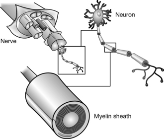
The nervous system is a continuous network of specialized cells that communicates information from the body to the brain and vice versa (Fig. 8.3). These messages or electrical signals, including perception and movement, are responsible for interaction with the environment. The nervous system is composed of neurons and glial cells which together generate electrical impulses that travel between our central nervous system (CNS) and peripheral nervous system (PNS). Generally, our CNS processes information in our spinal cord and brain and then sends signals back to our PNS where sensory, motor, and autonomic nerves leave the spinal cord to innervate our muscles, articular structures, organs, and glands. The neurons communicate with each other via electrochemical signals or neurotransmitters to transmit impulses from one neuron to the next.


The nervous system is continuous from the CNS to the peripheral nerves. Stress placed on one part of the system can be transmitted to other parts during movement (Breig 1978). In normal conditions, pain does not occur with movement or normal compression. However, the neural tissue can produce adverse symptoms with even minor stimuli if irritation due to a mechanical (compression or tension) and/or chemical (inflammation or ischemia) cause is present. Dahlin & Mclean (1986) showed that:


Common sites of injury include soft tissue, osseous tunnels (i.e. a spinal nerve exiting through an intervertebral foramen), nerve branches, and tension points (C6, T6, L4, posterior knee, and anterior elbow) where nervous tissues are relatively fixed.


Neural tissue has some elastic properties and can tolerate up to 20% elongation. If compression and traction occur together, only 15% elongation is required to stimulate the nociceptors and produce pain (Sunderland 1978). In addition, neural tension can cause venous stasis and stop intraneural blood flow at approximately 15% elongation (Ogata & Naito 1986; Rydevik et al. 1981). If the nerve’s distal axon shows evidence of degeneration or is injured, it will be more susceptible to mechanical and chemical irritants. Both compression and elongation contribute to the body’s physiological response by causing inflammation (Dahlin & Mclean, 1986) and/or ischemia (Rydevik et al. 1981).


Treatment of the nervous system is geared to restoring proper neural mobility, reducing chemical irritation, and minimizing any mechanical pressure or tension. David Butler (2000) advocated that mobilization of the nervous system can have a mechanical effect on the vascular system by dispersing intraneural oedema. Altering the mechanical restraints on the axoplasm and improving circulation will increase the energy available for axonal transport. Mobilization may also help the axons to regenerate by allowing better contact guidance (Lundborg 1988), and causing micro-trauma that may stimulate neurite promoting factors (Heumann et al. 1987). When there is rapid improvement of the nerve, it may be due to increased blood supply or changes in availability of CSF as nerves and nerve roots get at least half their metabolic requirements from these fluids.


Articular structures


The main components of the articular structures (the body’s joints) are bones, cartilage, synovial fluid, and joint receptors. While these components are predominantly responsible for allowing movement, they have a direct influence on posture and muscle length-tension relationships which can lead to malalignment and development of the ‘malalignment syndrome’.


Joint receptors


Joint receptors are mechanoreceptors which send signals to the body about injury and positional sense. According to Wyke’s classification of articular receptors, there are four types, all of which differ in structural compositions and how they influence the body.


Type 1 receptors are found in the superficial layers of a capsule with a greater density in the proximal joints. They are stimulated by proprioception, vibration, and discrimination of touch, and can sense static positions, pressure changes, velocity and the direction of a person’s movement. Because they have a low threshold, are slow to adapt, and fire almost continuously, they can be clinically influenced by changes of posture and positioning.


Type 2 receptors are found in the deep layers of the capsules and fat pads with a greater density in the distal joints. They are also stimulated by proprioception, vibration, and discrimination of touch. However, they only sense dynamic changes in position, such as occur with acceleration/deceleration, and are inactive at rest. Because Type 2 receptors have a low threshold and are quick to adapt, they are clinically influenced by movement such as exercise and joint mobilizations and manipulations.


Type 3 receptors are found in ligaments near bony attachments but not in the longitudinal ligaments of the spine (Fig. 3.68). These receptors are stimulated by extremes of dynamic movement when the ligaments are stressed. They are dynamic receptors that are inactive at rest, have a high threshold and are slow to adapt. Clinically, these receptors can be stimulated by providing a strong, prolonged distraction on a joint to help reduce tone in the surrounding muscles.


Type 4 receptors are a plexus of free nerve endings found in fibrous capsules, articular fat pads, ligaments, the periosteum, and the walls of blood vessels. They are pain receptors and are stimulated by mechanical and chemical factors. While they have a high threshold, they are a protective mechanism and are, thus, non-adapting and inactive at rest. Stimulation of Type 4 receptors will produce a gamma withdrawal reflex in an attempt to remove the joint from possible or further injury.


Visceral tissue


Visceral tissue is specialized tissue unique to each organ/viscera in the body, all of which are encapsulated in a fascial covering. With the support of fascia and specialized ligaments, viscera and organs are suspended in the body, decreasing gravitational strain on these important structures (Fig. 4.43). Posture, alignment of the bones and muscle balance all play a role in ensuring the proper force through the fascia required to maintain the position of the organs and viscera. When the ‘malalignment syndrome’ is present, fascial pull through the body can affect organ position and cause stress to affect its function. A detailed explanation of viscera and organ tissue is beyond the scope of this book. It is suggested the reader refer to more specialized anatomy and physiology texts.


Manual therapy treatment techniques


Joint mobilization


Joint mobilization is a therapeutic technique which applies a manual force to a specific joint and causes the surfaces of joints to glide parallel to each other or gap perpendicularly. This can be done passively or with the help of the patient to reduce muscular restriction and produce movement into the joint’s capsular barrier. The mechanisms of mobilization are still being investigated; however, there are several proposed mechanical and neurophysiological effects which are felt to act by influencing:


Connective tissue is influenced by cellular modulation. Mobilization causes the release of enzymes to break down cross-links in collagen, increasing inter-fibre distance. It stimulates fibroblast synthesis of collagen proteoglycans which bind with water and increase inter-fibre lubrication. In addition, it creates a piezoelectric current which aids in the alignment of new fibres and realignment of old ones.


Neural tissues are influenced in a way which decreases pain perception. Joint mobilization stimulates Type 1 and 2 mechanoreceptors, inhibiting nociceptive impulses and altering the afferent input to trigger efferent output or gamma bias, thereby relaxing the muscle (Sterling et al. 2000). Mobilization can also stimulate the release of endorphins and enkephalins, natural pain relievers produced by the body.


Articular structures can be mobilized to increase joint mobility by changing capsular elasticity. A ‘fixated’ or ‘locked’ joint can be corrected by moving a joint inclusion, such as a meniscoid or loose body, that is restricting the mobility. In addition, movement of the joint improves the articular cartilage nutrition and circulation which, in turn, can increase the supply of materials required for healing and aid in the removal of chemical irritants. It also alters joint lubrication; the resulting change is more significant with higher grades of mobilization.

Stay updated, free articles. Join our Telegram channel

Sep 11, 2016 | Posted by in SPORT MEDICINE | Comments Off on Treatment: Manual therapy modes

Full access? Get Clinical Tree

Get Clinical Tree app for offline access