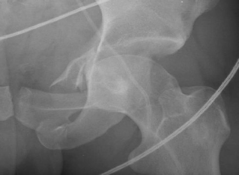
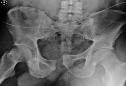
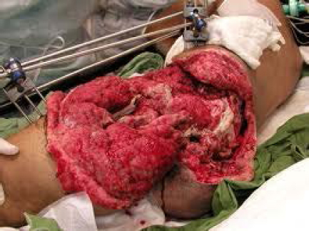
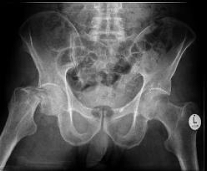
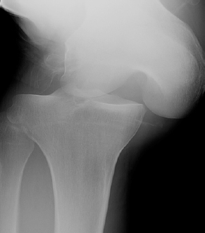
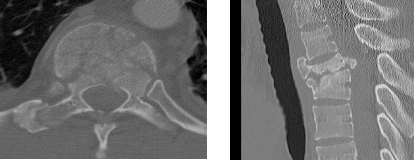
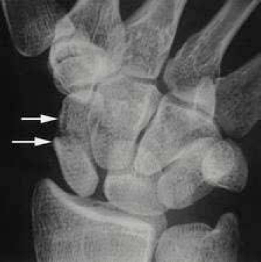
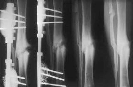
Trauma 1. ACETABULAR FRACTURE. Your registrar has asked you to come to A&E and help him manage this patient who was involved in a RTA. This is an isolated closed injury. Take me through what you are going to do. What will you do? This is an AP pelvic radiograph demonstrating an acetabular fracture, with both column involvement and a fracture of the acetabular floor, as demonstrated by the incongruity of both the ilioischial and ilioinguinal line. This is a high energy injury and I would: 1. Assess the patient according to ATLS protocol, and would identify any concurrent life threatening injuries. 2. With regards this specific fracture, I would undertake a thorough assessment of the neurovascular status. 3. I would investigate this fracture with further imaging, such as CT scan, and discuss the management of this patient with my local pelvic unit. Which acetabular fractures require emergent surgery? 1. Open 2. NV injury-40% chance of sciatic injury with posterior column fracture. 3. Irreducible posterior dislocation What are the indications for non-operative management? 1. Undisplaced fracture 2. Secondary congruence 3. Anterior column fracture 4. Low transverse fracture 5. Osteoporosis What are the indications for ORIF? The indications for ORIF depend on patient’s factors and fracture characteristics. Patient characteristics: Age, Medical comorbidities, pre-injury functional status. Fracture chararacteristics: A- Incongruity 1. Displacement-more than 4mm gap/more than 2mm step 2. Fragment in joint 3. Displaced fracture femoral head 4. Soft tissue interposition B- Instability 1. Fracture dislocation-(Posterior wall and posterior coloumn) PW and PC/(Anterior wall and anterior coloumn)AW and AC (40% of Posterior wall fracture needs surgery as unstable). 2. Roof arc angle greater than 45 degree. What is the roof arc angle?- 2 lines- drop vertical from centre and another 45 degrees to it (like Centre edge angle). A fracture in the roof suggests an unstable fracture pattern. What is the optimal timing of operation? Ideally, not longer than two weeks. What are the principles of fixation in acetabular fractures? 1. Anatomical reduction 2. Stable internal fixation 3. Early mobilisation What approaches do you know for acetabular fracture fixation? I would base my approach based on the fracture configuration: 2. If dislocation with femoral head fracture – anterior approach to fix femoral head fracture. How will you manage femoral neck fracture with dislocation? Lateral approach- to provisionally stabilise neck with K – wires. Reduce dislocation-and subsequently undertake definitive fixation of neck. Do you know of any classification for acetabular fractures? Letournal and Judet Simple-Anterior wall/Anterior coloumn/posterior wall/posterior colomn Associated-T shape/Posterior wall plus posterior coulomn/transverse plus posterior wall/Anterior colomn plus posterior hemitransverse/Both colomn What factors have been shown to influence the outcome in acetabular fractures? 1. Age 2. Osteoporosis 3. Type-Anterior wall fracture/femoral head fracture/articular damage/Posterior wall multifragmentarity. 4. Early surgery in less than two weeks 5. Minimal surgical approach 6. Quality of reduction 25 year old front seat driver, presents to A&E following RTA. How will you manage this patient? This is an AP pelvic radiograph demonstrating an open book pelvic injury. According to the Young and Burgess classification, this is an (anteroposterior compression)APC 3 fracture, with complete disruption of the sacroiliac joint and anterior pelvic diastasis. I would undertake my initial assessment according to ATLS protocol as this is a high energy trauma. The principles of my initial management – would be to follow along agreed BOAST guidelines. 1. Assess haemodynamic status and fracture stability. 2. If haemodynamically unstable (major bleed) I would consider: -Early application of pelvic binder/ sheet or external fixator -Resuscitate –Blood/FFP/platelets. – Determine the need to initiate a massive transfusion protocol. -Aim to get a primary clot. -Assess chest/abdomen by surgeons- plain radiograph chest/FAST scan, to rule out other sources of bleeding. 3. If still unstable– consider angio suite for emergency embolisation OR open pelvic packing after ex-fixator. 4. CT scan pelvis- only after haemodynamic stabilisation. 6. Assess for other associated injuries –Genito urinary/GIT You notice blood on PR, and blood at penile tip. What else would you consider doing at this stage? I would be worried about associated genitourinary and gastrointestinal tract injury. I would therefore involve an urologist and a general surgeon early. – Urologist to consider urgent contrast studies (cysto/urethrogram) to assess for: – extra peritoneal injury –such as urethral laceration/rupture. – intra peritoneal injury- open bladder laceration. – open pelvic injury with wound to the groin/buttockvagina/rectum- need to consider draining bladder with cystostomy. -posterior urethral injury- supra pubic catheter and repair. -open pelvis with bowel injury- needs bowel diversion with end colostomy upper quadrant. When would you consider applying a temporary external fixator on before laparotomy? This is controversial, but one of the indications would be to stabilise the pelvis during transfers. Management of Open Pelvic Fractures: 1. Assess –Age/Injury severity score/wound/Heamodynamic instability/fracture instability/ other injuries- rectum, vagina, urethra. 2. Treat like open fracture- antibiotics 3. Multidisciplinary team needed -Surgeon- to assess bleeding from abdomen/chest/rectum. -Urology – urethral injuries -Plastics – soft tissue cover -Orthopaedic- fracture stabilisation 4. Temporary pelvic stability-Pelvic binder/sheet/external fixator/C clamp 5. If still haemodynamically unstable- blood products, then angiographic embolisation. 6. If still unstable- external fixator in theatre, then – laparotomy and exploration -ligate and pack -wound debride+ external fixator if contaminated/open reduction and internal fixation if clean -Urologist – urethral and bladder injuries -Surgeon- bowel injury/diverting colostomy -ICU care 7. Sepsis control –IV Antibiotics 8. Think about compartment syndrome Do you know of any classification for pelvis fractures? Tiles –stable/rotationally unstable/both vertically and rotationally unstable Young and Burgess –Lateral compression(LC)/Anteroposterior compression(APC)/Vertical shear(VS) What factors have been shown to affect the prognostic factors for pelvic fractures? 1. Heamodynamically unstable – 40% mortality (5% if stable) 2. Posterior disruption- 37% mortality 3. Open fracture 4. Old age 5. High Injury severity score(ISS) 6. Vertical shear -25% mortality What are the indications for surgical fixation in pelvic fractures? 1. Symphysis pubis diastasis more than 2.5 cm 2. Sacrum diastasis more than 1cm 3. Limb length discrepancy more than 1.5 cm 4. Instability- rotational and vertical 3. Neck of femur fracture in young pt: This is a 65 year old who fell down some steps and sustained this injury. Tell me about this injury and what other key features of the history do you need, to direct your management? This is a displaced intracapsular fracture of the right neck of femur. This is a Garden IV fracture, as there is complete displacement. I would like to know about any other associated injuries, timing of this injury and the patient’s medical status and co-morbidities. I would like to know the patient’s pre-morbid functional status. What is your initial management? I would manage this patient along the recent BOAST guidelines. I would ensure adequate analgesia, IV access and hydration, routine blood tests and ECG, and arrange for admission to the ward. How would you treat this patient? I would discuss the treatment options with the patient, taking into consideration the above points, and propose a total hip replacement (THR). This is based on evidence that patients do functionally well with THR, and reoperation rates are lower. This is also being advocated by NICE guidelines. Ref-Keating JF. JBJSAm 2006 Feb; 88(2):249-60. RCT of displaced intracapsular fractures in healthy older patients. How would you treat this fracture if the patient were 40 years old? I would aim to conserve the femoral head by reducing the fracture by open reduction and internal fixation with 3 screws. If this patient presented at 11pm, would you operate that night? I would operate the next morning, as evidence suggest no difference in avascular necrosis or non-union rates between less than 12 hours or more than 12 hours. These patients however cannot be delayed more than 48 hours. Ref-Parker et al Injury 2005- Complication after intracapsular fractures in young adults- meta-analysis. Upadhyay A et al JBJS Br 2004 Sep. Prospective RCT. Reports that a delay of more than 48 hours before surgery did not influence the rate of union or avascular necrosis when compared with surgery within 48 hours of injury. No difference in outcomes in closed vs open reduction techniques. The single most important factor to ensure a good prognosis is achieving good anatomic reduction. Reference- Jameson et al. Patient and implant survival following 4323 total hip replacements for acute femoral neck fracture. 2012.94B.1557-1566. Avery et al. Total Hip replacment in mobile, independent patients with displaced intracapsular fracture neck of femur. Aseven to ten year follow-up report of a prospective randomised trial. JBJS)Br)93B-8:1045-48 This are the plain radiographs of a 25 year old patient brought in by ambulance following a rugby tackle. How will you assess, this patient? 1. Initial assessment according to ATLS protocol 2. Immediate reduction needed – in A & E, – splint in 20 degree flexion. – Postero lateral dislocations may need open reduction, as the medial femoral condyle may have button holed through the capsule. 3. Assess vascular injury -30-40% -Assess at regular intervals if pulse normal -Angiography if (ankle brachial pressure index)ABPI less than 0.9 -Maximum ischaemic time -6-8 hours Popliteal artery tethered – proximally in Hunters canal under the fibrous arch of the adductor muscles – distally under fibrous arch of soleus – in middle by 5 geniculate arteries 4. Assess neurology -10-30% 5. Get radiographs to assess associated fractures 6. Get MRI/MR arthrogram to assess ligament damage Do you know of any Classification? Shenck Anatomic Classification 1992 Five types -1/2/3/4ligaments/ Fracture dislocation with or without neurovascular injury. When would you consider emergency surgery? Absolute indications for emergency surgery are: 1. Arterial injury – external fixator and vascular surgery 2. Irreducibility – open reduction – apply external fixator if unstable in brace 3. Open dislocation / washout – do not perform early ligament reconstruction – apply external fixator if unstable in brace 4. Compartment syndrome When could you consider options for dealing with this injury? Non operative Brace 30 – 40 for six weeks – for elderly patients with multiple co-morbidities. – external fixator for obese patient – Disadvantage: stiffness Levy et al Arthroscopy 2009 Systematic Review -showed improved outcomes with operative management. Early versus Late Surgery? Levy et al Arthroscopy 2009 Systematic Review – early treatment consistently results in higher outcome scores. – easier identification of tissues – some tissue amenable to repair Timing for definitive surgery – Allow swelling to settle Beyond three weeks, collateral and posterolateral structures too scarred for early repair and will need late reconstruction. Repair versus Reconstruction? 1. Postero-lateral corner (PLC) – usually treated early with repair of avulsions. – reconstruction for midsubstance injuries Stannard et al Am J Sports Med 2005 – repair v reconstruction of PLC – improved outcomes with reconstruction 2. Postero-medial corner (PMC) Stannard et al AAOSM 2009 – reduced failure rates with reconstruction versus repair 3. ACL / PCL – typically reconstructed Harner et al JBJS Am 2004 – reconstruction of ACL / PCL lead to good outcomes. Rehabilitation Mook et al JBJS Am 2009 – systematic review. – increased instability seen with immobilization compared with early mobilisation. – increased fixed flexion deformity and loss of flexion seen with immobilization. Thompson et al 2006 AAOSM 2006 Evidence of reduced instability and reduced failure rates after ligament reconstruction. 35 year old motorcyclist versus car. Isolated injury, GCS 15. Describe the injury? There is a sagittal and axial CT scan demonstrating a lumbar burst fracture, involving all three columns. This is an unstable injury pattern. How will you assess this patient in A&E? – ATLS/neurology/ASIA chart for neurology How do you classify spinal cord injuries? Spinal cord injuries can be classified as complete and incomplete. Complete – no sacral sparing after spinal shock (bulbocavernosus reflex return). Incomplete -Central cord (upper limb more than lower limb) -Anterior cord (preserved dorsal colomn path – proprioception/vibration/ light touch). -Posterior cord (no vibration/light touch/ proprioception) -Brown Sequard syndrome- contralateral pain and temperature loss -Cauda equina syndrome Tell me about the Frenkel classification? A-complete palsy B- Normal sensation C- Useless motor D- Useful motor E- Normal What are the features of an unstable vertebral fracture? 1. Neurological deficit 2. More than 50% loss of height 3. Kyphosis /angulation more than 20 degree 4. canal compromise more than 50% 5. Denis: more than one column-posterior column/elements gone. What are the absolute indications for surgery? (1)To decompress: Worsening neurological deficit in the presence of demonstrable neural compression despite attempts at postural reduction (2)To stabilise: All cases requiring decompression Complete ligamentous disruption associated with dislocation (3)Open thoracolumbar burst fractures (4)Patient with complete neurological deficit (5)Multiply injured patient JBJS (A) May 2003 Wood et al RCT Operative treatment provided no major long term advantage compared with nonoperative treatment. Spine, June 1993 J. B. Cantor et al, Nonoperative management of stable thoracolumbar burst fractures, intact neurology with early ambulation and bracing: No significant worsening of kyphosis No deterioration in neurological function Follow up CT scans showed significant resorption of retropulsed bone. This patient presented to your fracture clinic eight weeks post injury complaining of pain. What do you see? This is a posteroanterior (PA) radiograph demonstrating a displaced fracture of the waist of scaphoid. What are the poor prognostic factors for non-union? Delay in treatment Displaced fractures Proximal pole fractures Avascular necrosis Smokers How would you manage this patient? The principle of management is to achieve union, and prevent early degenerative changes – ie to prevent a SNAC wrist (scaphoid non-union advanced collapse) What are the indications for internal fixation of scaphoid fractures? Displacement greater than 1 mm Scapholunate angle greater than 60S Lunocapitate angle greater than 15L Intrascaphoid angle greater than 20 Proximal pole fractures Fractures associated with a peri-lunate dislocation Delayed union I would use the volar approach. – workhorse for fractures of the waist of the scaphoid – preserves dorsal blood supply- 75% blood supply from dorsal radial artery. Dorsal approach is indicated – for proximal 1/3 fractures McQueen et al JBJS (Br)2007 RCT- percutaneous fixation quicker time to union & return to work quicker. Diaz et al JBJS Am 2005 -RCT non opertive and ORIF- no difference between two treatments -Advised aggressive conservative management (offer surgery if no union in eight weeks). How would you manage a non union of a scaphoid fractures ? I would take a full history including age, comorbidities, functional demands and level of symptoms, and patient’s expectations. I would undertake appropriate imaging as plain radiographs and MRI. Plain radiographs/CT scan determine whether there are associated degenerative changes, in which case salvage surgery should be considered. MRI to determine whether there is associated avascular necrosis- and therefore consider vascularised bone graft. 7. Infected tibial non union. Describe and discuss What do you understand by non-union? The cessation of normal biological healing process. US FDA (Food & Drug administration agency) definition: fracture that is over nine months old and that has not shown radiographic signs of progression toward healing for three consecutive months.
Related posts:


Stay updated, free articles. Join our Telegram channel

Full access? Get Clinical Tree


