Chapter 1 Suzanne S. Hecht1 and Elizabeth Arendt2 1Department of Family Medicine and Community Health, Program in Sports Medicine, Sports Medicine Fellowship, University of Minnesota Athletics, Minneapolis, MN, USA 2Department of Orthopaedic Surgery Division of Sports Medicine, University of Minnesota Athletics, Minneapolis, MN, USA For the first time in Olympic history, all participating countries in the 2012 London Olympic Games had male and female athlete representation. Furthermore, greater than 40% of the London Olympic athletes were female compared to only 1.9% in the 1900 Paris Olympic Games. While great strides in female sports participation has been made, research is lacking on how to best train female athletes. The explosion of females participating worldwide in all levels of sport continues to reach new heights. There is a need for medical providers, coaches, sport administrators, and the female athletes, themselves, to understand what constitutes optimal training for female athletes. Is training female athletes in the same manner as male athletes the best approach? The answer to this important question is unknown, but it is a fact that there are anatomic and physiologic differences (Table 1.1) between females and males and those differences may impact training. Given the limitations of our scientific knowledge in this area, it is reasonable to explore these differences and similarities and how they might impact training of male compared to female athletes in order to gain further insights on how best to train female athletes. Just as the pediatric-aged athlete is not a mini-adult athlete, female athletes are not just smaller male athletes. Table 1.1 Physiological comparison of females and males Aerobic capacity in female athletes is typically 10% lower than male athletes when normalized for lean body mass. Factors contributing to a lower aerobic capacity in women are smaller hearts and thoracic cavities with smaller lung volumes, less blood volume, fewer red blood cells, and less hemoglobin. The gap in female endurance performance records compared to males is narrowing for marathons and triathlons, but may not equalize due to inherent differences in body composition and aerobic capacity. Females have a lower maximal anaerobic threshold than males, which may be due to differences in muscle mass and/or training differences. Pubertal and skeletal maturation is attained at a younger age in females. On average, men are taller and have wider shoulders with narrower pelvises than women. A wider pelvis in women contributes to an increased carrying angle at the elbows and Q-angle at the knees. Women have shorter limbs and a lower center of gravity, which may offer an advantage in improved balance. Body fat percentage is greater in women than in men predominately due to a higher proportion of essential fat, which consists of sex-specific fat such as found in breasts, buttocks, and thighs. Storage fat is similar in both sexes. In females, total muscle mass is about 25% of body weight compared with 40% in males, and females have smaller muscle fibers. Women have a similar proportion of fast- and slow-twitch muscle fibers and make similar gains in relative muscle strength with weight training. When controlled for body weight, women have less upper body strength and similar lower body strength compared to men. Testosterone effects cause greater muscle hypertrophy in men. Females have a higher risk of non-contact anterior cruciate ligament (ACL) injuries, consistent over time. Important at-risk strategies appear to be valgus collapse of the knee, with alteration in upper trunk mechanics. There is evidence that changing dynamic loading of the knee through neuromuscular proprioceptive training (i.e., avoiding at-risk positions, encouraging the knee over-the-toe position) (Figure 1.1 a and b) is of value. The recommended elements that go into ACL intervention programs include strength and power exercises, neuromuscular training, plyometrics, and warm-up. The intervention formula that works best includes feedback and educational methods. Although the results are promising in existing intervention programs, the ACL injury rate and sex disparity have not yet substantially diminished through these programs. The ideal prevention program has yet to be identified. Figure 1.1 (a) Example of incorrect body movement pattern. Note the “functional” collapse of the knee when performing a partial squat, with hip substitution and pelvic drop. (b) Improved body position when performing a partial squat: hips and pelvis level, “knees over toes” position controlled throughout the knee flexion. Correct positioning mandates CORE muscle control and body position awareness. Also somewhat disconcerting, the protective effects of ACL injury prevention programs appear to be transient. Field assessments and screening tools show promise for identifying at-risk individuals, but the best age to target the screening is not yet identified. It is unknown whether injury mechanisms and prospective risk factors are the same in the pediatric and the adult populations. More importantly, which biomechanical or neuromuscular profiles (trunk, core, hip) are most at fault in the non-contact ACL rupture remains elusive. An understanding of the causative agent is central to identifying useful screening of individuals at risk. Increasingly, there is a debate whether an ACL injury represents a gross failure of the ACL caused by a single episode or multiple episodes over time. Females have a lower incidence of full-thickness articular lesions in ACL-injured knees. Magnetic resonance imaging (MRI) reveals a sex difference in bone edema patterns after acute ACL injury, with females more typically having posterolateral bony contusions than males. This suggests different mechanisms of injury in males versus females. The subsequent chapter devoted to injury prevention in the female athlete and includes a detailed discussion on ACL injury prevention training programs. The assumption of a simple linear relationship between variables is unlikely to be operational in the ACL injury, as feedback loops and interrelationships are likely important. However, efforts spent on unraveling this injury mechanism is likely related to other injuries such as ankle sprains, lateral patella dislocations. Bone stress injuries result from chronic repetitive training and can range from a stress reaction to a cortical fracture. Historically, these injuries are more common in certain populations of athletes with incidence of up to 21% in track-and-field athletes and 31% in military recruits, with a female preponderance. Several evidence-based grading scales utilizing MRI have been published, which can help to grade the bone stress reaction and predict return to activities. Higher grade bone stress injuries as assessed by MRI are associated with a prolonged recovery. General training regiments advocate activity reduction to allow pain-free activities without advocating total rest. This can follow a progression of non-weight bearing on crutches (cross-training: swimming, stationary bike, flotation running), advancing to non-pounding, weight-bearing exercises (Stairmaster™, Vancouver, Washington, Nordic Track, Logan, Utah), with gradual re-entry into sport-specific activity. Sport-specific muscle rehabilitation, strengthening, and stretching are encouraged throughout. Low-grade stress fractures can heal with continuance of activities, using pain-to-guide activities. More recently, it has been shown that risk factors associated with the Female Athlete Triad, in particular low bone mineral density and reduced numbers of menstrual cycles, are associated with a prolonged recovery in female athletes. Athletes with trabecular bone injuries (femoral neck, pubic bone, or sacrum) more often exhibit past or present disordered eating and oligomenorrhea/amenorrhea. Those with bone stress injuries at trabecular sites had a significantly lower bone mass at the lumbar spine, femoral neck, and total hip regions.
Training the female athlete
Physical characteristic
Aerobic capacity (VO2max)
M > F
Blood volume
M > F
Body fat percent
F > M
Bone mineral density
M > F
Flexibility
F > M
Muscle strength
M > F
Thermoregulation
F = M
Anatomic and physiologic considerations
Aerobic performance
Anaerobic performance
Anatomic differences
Body composition
Muscular strength
Injury considerations
Non-contact ACL injury
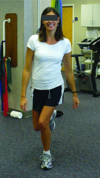
a 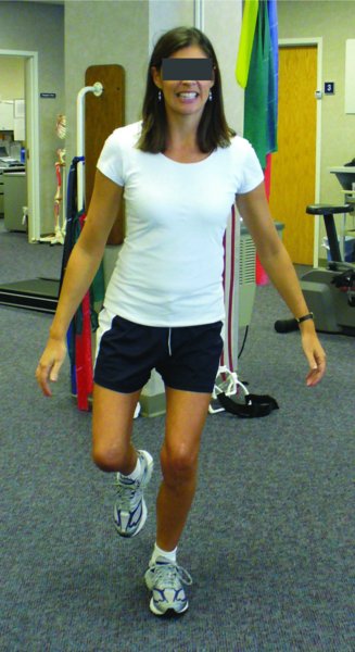
b
Stress fractures
Related posts:

Stay updated, free articles. Join our Telegram channel

Full access? Get Clinical Tree


