2.6 Training and Functional
 Functional Significance of the Pelvic Floor Musculature
The muscles of the pelvic floor form the dynamic closure of the abdominal cavity below. They interact with the muscles of the diaphragm above, the deep abdominal muscle anterolaterally, and the muscles of the spine posteriorly. Together, these muscle groups maintain a physiological pressure distribution, ensure the position of the abdominal organs, and provide for a stable but flexible lumbar and lower thoracic spine [Constantinou and Govan 1982, B⊘ and Stien 1994, Hodges and Richardson 1996, Sapsford et al. 1997a, 1997b, Heller 2002].
The pelvic floor muscles dynamically support and suspend the organs of the true pelvis. To maintain continence during the storage phase of urine and stool, the vesicourethral angle and anorectal flexure must remain stable [DeLancey 1988, Kuijpers 1990, Deindl et al. 1993].
The musculature of the pelvic floor includes the urethral and anal sphincters. These affect urethral and anal closure pressures and by this function can prevent or obstruct the elimination of stool and urine [Gosling et al. 1981, Lestar et al. 1989, Deindl et al. 1998].
The muscles of the superficial layer of the pelvic floor affect sexual functioning. They affect the ability to have penile and clitoral erections and also affect vaginal tone [DeLancey 1994, Van Kampen 2000].
The muscles of the pelvic floor form the active internal support of the pelvic girdle. Hence they affect the nutation of the sacrum, the stability of the sacroiliac joints, the symphysis pubis, and the lumbosacral joint [Lee 1999].
 Indications for Muscle Training of the Pelvic Floor
The various functions of the pelvic floor muscles indicate muscle training and functional exercises in the following dysfunctional conditions:
- Incoordination in the stabilizing system of the abdominal cavity
- Changes in the position of the pelvic organs: vaginal descent, cystocele, rectocele
- Disorders of continence: stress and urge incontinence (urinary and fecal)
- Disorders of emptying: constipation, urinary retention
- Sexual dysfunctions
- Loss of stabilization in the pelvic girdle: sacroiliac joints, lumbosacral joint
- Pelvic asymmetries: pelvic scoliosis
- Disorders of emptying: constipation, urinary retention
 Muscle Training—Therapeutic Exercise
 “Training is the systematic directed repetition of muscle contractions above threshold resulting in increased performance by morphologic and functional adaptation” [Weineck 2000, p. 16].
“Practice is the systematic directed repetition of movements to improve performance without demonstrable morphologic changes“ [Weineck 2000 p. 15].
“Therapeutic exercises are thought-out and planned series of movements or changes in activity designed to delimit or surround a defined deficit in habitual movements so that efforts to evade the dysfunctional movements are avoided” (Klein-Vogelbach 1992].
In the pelvic floor, as elsewhere, therapeutic exercises aim to develop or redevelop motor abilities. Hence, pelvic floor exercises follow the laws of movement training (see section 1.2). It is the task of the therapist during the initial examination to recognize the type and extent of injury to the pelvic floor muscles. Beyond that, deficits in the coordination of movements also have to be determined.
Patients may show muscular insufficiency in rapid contractions, strength, and strength endurance. Training needs to be designed to reestablish these muscular capabilities. On the other hand, good muscular strength may be present, but not functionally available. In such cases, the primary therapeutic goal is to establish the correct automatic movements by functionally coordinated exercises.
In working with the pelvic floor, as elsewhere, the principles of muscle training and movement education are therefore directed toward the treatment of individual functional deficits and the presenting pathology.
The Pelvic Floor and its Primary Stabilizers
The musculature of the pelvic floor is classified as a primary or local stabilizing group. It meets the criteria required for an actively stabilizing system of this type [Markwell and Sapsford 1995, Sapsford et al. 1997b]. The muscles are short, monarticular, or circular, surrounding a pelvic opening. The great majority consist of slow-twitch type I muscle fibers. Their normal function follows the principle of stabilization—i. e., early activation before increases in abdominal pressure (e.g., coughing, sneezing) [Constantinou and Govan 1982] or the continuation of the movements of adjoining segments of the body [B⊘ and Stien 1994]. The latter function represents a synergistic interaction with the stabilizing muscles of the lumbar spine and hip joints (multifidus, psoas muscles, and intrinsic rotators of the hip) [Sapsford and Hodges 2000, Sapsford 2004].
Exercises designed for muscle groups with comparable functions can be used for training of the pelvic floor. The treatment suggestions below are modeled on the corresponding recommendations for the training of the stabilizing muscles in the lumbar spine (transversus abdominis and multifidus muscles) [Richardson and Jull 1995, Cholewickij and McGill 1996, Hides and Richardson 1996, Hodges and Richardson 1996, Hamilton and Richardson 2000, O’Sullivan 2000].
Phases of Pelvic Floor Training
- Phase 1: Development of the ability to become aware of the region
- Phase 2: Specific localized training and relaxation
- Phase 3: Exercising functionally related muscle chains with graded load
- Phase 4: Integrating the movements into daily activities and making them automatic
Phase 1: Development of the Ability to Become Aware of the Region
Awareness, experiencing, observing, practicing. The ability to activate the muscles of the pelvic floor voluntarily and independently is a precondition for the beginning of localized training. The path to independent execution leads through education in bodily awareness to exercise.
The degree of the client’s awareness of the spine, pelvis, and shoulder girdle is an indication of the degree of dysfunction in dynamic stabilization [Comerford and Addison 2000]. Richardson and Jull [1995] point out the importance of improved awareness before localized exercises are initiated. In the course of normal everyday activities, people do not become aware of the work of their muscles [Klein-Vogelbach 2000]. They only become aware of demands on muscles when these become fatigued or cannot act economically and in coordination in the face of newly experienced movement patterns.
These considerations also apply to the muscles of the pelvic floor. We experience the processes of elimination and feel the genital and pelvic areas during sexual activity. These experiences and perceptions are not consciously perceived as muscular actions. Indeed, the muscles of the pelvic floor lie deep in the true pelvis, and access to them is difficult to achieve through visual and tactile awareness.
The pelvic floor is the site of our most intimate sexual feelings and experiences. Dealing with these either with shame or openness is determined by differences in education, different cultural and religious influences, and individual life events and experiences.
All training and educational information provided regarding the anatomy and functioning of the pelvic floor has to be transformed into a subjective experience through awareness training. Experiences in subjective awareness and the consequent emotional burden have to be borne in mind during examination and training instructions.
Practical procedure. Using illustrations, models of the pelvis, and brochures and in patient-friendly language, patients are educated in the anatomy and physiology of the pelvic floor and the organs of elimination and sex as they relate to their condition.
- Goal: Knowledge and understanding promotes insight into the treatment procedures and motivates self-directed practice.
Patients become aware of the bony pelvic girdle by touching and feeling their own body and “discover” it as the outer boundary of the pelvic floor musculature.
- Goal: Concentrate patients’ awareness on the pelvic floor. Teach them to distinguish the muscle groups lying in the true pelvis from those outside the pelvis and around the hip joint.
The muscle groups in the pelvis are described one by one, and patients can feel the direction of the movements they generate by visualizing and activating each in turn.
- Goal: Patients learn to become aware of the various parts and functions of the pelvic floor. They improve their ability to detect various weak areas and asymmetries.
Several publications dealing with local pelvic floor activation describe a number of aids to awareness and ways to explore the region [Heller 1998, pp. 185–213, Tanzberger 1998a, Carriere 2002, Tanzberger et al. 2004] (see also section 4.3). No comparisons of the effectiveness of the various procedures are available. It is thought that efforts to isolate and differentiate the ability to become aware of and activate the various parts of the pelvic floor are of decisive importance in treatment, but this concept remains a hypothesis. Studies by Deindl et al. [1998] describe differences in the effect of pelvic floor training on the voluntary external urethral sphincter and the pubococcygeus muscle.
Patients access sensory awareness at quite different levels. Emotionally stressful situations can impair sensory perception of the pelvic floor. The patient has to be motivated to undertake self-directed awareness work. The patient’s idea of the region may be imaginative, abstract, or concretely anatomical. An effective way of working in sensory awareness is to be nonjudgmental when dealing with such individual perceptions.
- Goal: Localized perception and activation of individual parts of the pelvic floor: ability to exercise.
Levator ani: simultaneous contraction of all parts of the muscle (Fig. 2.82)
- Goal: Stabilizing pelvic organs from below.
Starting positions: Supine side-lying, prone, sitting.
Primary movements: The perineum moves upward, the vagina and anal canal can be felt to move upward and forward.
Instructions:
- Tactile aids, providing direction: for the perineum, vaginal introitus, anus
- Image: lifting cotton balls or marbles with the vagina
- Encircling spiral
- Swinging the perineum upward
- Tactile checking with self-palpation. Manual exploration of the perineum, vaginal introitus, vagina.
- Visual checking with a hand-held mirror.
Aid to sensory awareness: A tampon is introduced into the vagina. If the exercise is performed correctly, an inward pull can be clearly felt when the string is pulled.
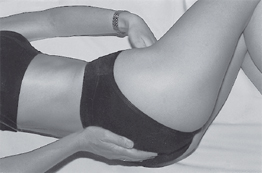
Puborectalis, pubococcygeus muscles (Fig. 2.83)
- Goal: Stabilizing the anorectal and vesicourethral angles.
Starting positions: Supine, side-lying, prone, sitting.
Primary movements: The pubis and coccyx move toward each other.
Instructions: The therapist’s hands on the pubis and coccyx provide tactile aid and direction.
Patient’s self-check: Tactile check: by palpating the coccyx, the patient can feel its slight but noticeable forward movement. At the same time, she senses constriction of the anal and vaginal openings.
Prerequisites: The lumbar spine and the hip joints do not move from their starting position.

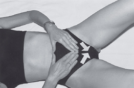
External urethral sphincter muscle (rhabdosphincter urethrae), deep and superficial transversus perinei muscles (Fig. 2.84)
- Goal: Raising pressure inside the urethra, stabilizing the urethra.
In anatomical textbooks, the transversus perinei muscle is shown as a muscular plate [Schünke 2000, p. 211] consisting of fibrous tissue in women (see section 1.1). Although it is not a real muscle, women can still visualize the direction of contraction of the transverse perineal muscle, as it is called. This teaches the patient to become aware of the anterior compartment of the pelvic floor in isolation. This exercise makes it possible to concentrate the action of the pelvic floor muscles on urethral closure (personal clinical experience).
Starting position: Sitting, leaning backward with the longitudinal axis of the body supported, side-lying, prone.
Primary movement: The rami of the pubis “move” toward each other. The urethra is constricted. The pressure in the symphysis pubis increases.
Instruction: The touch of the therapist’s hands indicates the direction at the symphysis or at the ischial tuberosities. Since this exercise does not result in perceptible bony movement, it is supported by imagery:
- “The vulva is closing.”
- “The symphysis is closing.”
- “The urethra is constricting.”
- “Little pellets or droplets are being lifted into the urethra (sucked inward).”
Self-checking: Tactile aids: the patient’s own tactile exploration helps her become aware of the active area.
Prerequisite: The hip joints and lumbar spine have to remain in their starting position.
External anal sphincter
- Goal: Increasing the pressure in the anus and lower anal canal.
Starting positions: Supine, side-lying, prone.
Primary movements: The external anal sphincter and the anal canal constrict. The perineum moves slightly upward.
Instructions: Tactile directions: finger on the perineum or the external anal ring.
Imagery: Marbles, cotton balls, holding small sponges in the anus, compressing, “kneading.”
Self-check: Tactile aids: the palpating finger rests on the anal ring.
Learning any type of exercise can be facilitated by linking it to expiration. As training progresses, the patient has to become able to hold a contraction independently of respiratory movements (kinetic control) [Comerford and Addison 2000]. The contraction of the deep muscles stabilizing the lumbar spine and hip joint is acceptable in all types of exercise.
Co-contraction of the superficial mobilizers can be observed with increasing force. This is acceptable as long as the primary activity originates in the muscles of the pelvic floor. As they train, patients can refine their awareness of the added activity of the superficial muscles, thereby learning the extent of the activity of the individual pelvic floor muscles.
Phase 2: Training and Relaxing Individual Muscles
To improve motor control and increase strength (maximal strength endurance). Coordination and strength performance is tested by vaginal or anal palpation (the “PERFECT” scheme—Laycock, see section 4.3). The aim is to improve the ability to recruit the pelvic floor muscles selectively. Muscle training primarily addresses the activity of the slow-twitch fibers of the pelvic floor musculature. In order to build the capacity for such recruitment, practice must address the ability to contract individual muscles to the extent possible voluntarily and at the appropriate time. The contraction of the muscles proceeds slowly. To facilitate the action, exercises are first practiced in neutral starting positions (sitting while leaning against support, side-lying, prone). Correct execution of the muscular activity is confirmed by the use of external feedback (electromyography, pressure feedback, palpation, mirror). Feedback by external means is gradually reduced until the patients have acquired the ability of intrinsic feedback (See section 1.2).
The increase in strength noted in the initial days and weeks is attributed to improvement in recruitment and frequency [Weineck 2000, Haas 2001, pp. 108–114].
Training variations
Improvement in intermuscular and intramuscular coordination and proprioception
- Contraction with minimal intensity (20–40 % of maximal strength), with 10–40 repetitions distributed through the day [Haas 2001, p. 119].
- The exercise is carried out in concentric and eccentric steps.
Enhancing strength and strength endurance
- Selective submaximal contractions are performed 8–10 times for 10s.
- Six to eight sustained contractions are combined with three brief maximal contractions [B⊘ et al. 1994].
- This series is practiced three to five times a day.
Training in the performance of rapid contractions with explosive strength, learning the ability to execute rapid contractions with variable intensity. The ability to execute rapid contractions can be tested by the procedure proposed by Laycock. The standard is 10 rapid contractions (see section 4.2). It is important to be able to develop a rapid dynamic response in the muscles of the pelvic floor by reacting rapidly and strongly to the development of sudden pressure (coughing, sneezing, jumping). Training in timely coordinated recruitment must be combined with the ability to develop considerable strength in a short time (training of the fasttwitch fibers in the pelvic floor musculature).
Training variations
Pelvic floor contractions are performed with brief tightening of individual muscle groups.
- Brief contractions are repeated rhythmically (series of 10 rapid contractions of submaximal intensity and brief pauses).
- Brief contractions are combined with sustained action [B⊘ et al. 1994].
- Timing and intensity of the contractions are varied within the series
- Brief contractions are combined with relevant acute everyday stresses (coughing, sneezing, jumping, etc.).
Learning to relax the pelvic floor muscles. The muscles of the pelvic floor form the boundary of the abdominal cavity and as such are exposed to constant variations in pressure. If the muscle tone is too low or too high, it can result in an imbalance in the response to these changing dynamic loads.
Involuntary losses of urine, gas, or stool, and uncontrollable urges or pain, as well as abuse, may result in an increase in the resting tone of the pelvic floor muscles. A pelvic floor muscle that is excessively tense loses dynamic elasticity and response preparedness.
While a reactive contraction such as that described above ensures continence, the ability to respond with relaxation and opening to downward pressure for the elimination of urine and stool, as well as for birth, must be preserved.
Micturition, defecation and the birth of an infant are the result of activation of smooth-muscle storage and bearing organs. Normal micturition by voluntary voiding does not require activation of abdominal muscles to increase abdominal straining. To initiate micturition and for uninterrupted voiding the muscles of the pelvic floor must be able to relax in a coordinated manner [Sapsford et al. 1998, p. 66].
If the voluntary muscles are habitually active during micturition and defecation, they interfere with normal emptying. Insufficient relaxation of the muscles of the pelvic floor delay voiding and can lead to incomplete emptying of the bladder [Sapsford et al. 1998, p. 66, B⊘ and Stien 1994].
Exercise variations
To develop the ability to contract and relax individualmuscle groups
- The four exercise patterns described on pp. 254–6 are performed with emphasis on the opening and relaxation phases.
- Starting positions without tightening are selected (prone, supported side-lying).
- The starting position is combined with cautious stretching of the pelvic floor (on all fours, knee–elbow position)
To become aware of respiratory movement in the pelvic cavity
Starting position: Side-lying.
- The patient’s hand explores the pelvic floor in the area of the perineum and anus.
- Expiration is prolonged. The ensuing inspiration is perceived as a downward movement at the point of contact with the hand.
Using phonation as an aid (humming vowels: ah, oh)
- The breathing exercises can be accompanied by relaxed humming of “M”, AH”, “OH.”
- The oropharynx is relaxed (awareness exercises for the oral cavity).
Changing the processes involved in voiding
- Sudden downward pressure toward the pelvic floor can result in reactive contraction of the pelvic floor muscles [Tanzberger 1998a].
- The mechanism of straining is replaced by visualization of a gradual push in the direction from anus to urethra.
Phase 3: Exercising Functional Muscle Sequences with Modulated Stress
Exercising functional muscle sequences. Working with functional muscle sequences presupposes an ability to contract and relax normally. Therapeutic exercises address as many variations of movement patterns requiring muscular coordination as possible. The closer the various selected exercises relate to the individual’s functional deficit, the more rapidly they will translate into everyday activities. Practicing in this way stimulates the patients’ own creativity and experimentation in applying the exercises to their daily life.
Exercise patterns
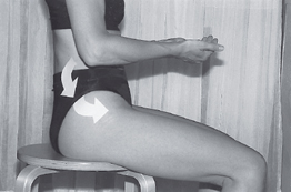
Activating movement sequences in muscles acting in the same direction
Starting position: Supine, side-lying, sitting, standing.
Basic movement: The coccyx moves forward, a little upward. The pelvis extends at the hip joints and the lumbar spine flexes. The posteroanteriorly aligned strap muscles of the pelvic floor are exercised in a movement sequence coinciding with the direction of movement of the coccyx (Fig. 2.85).
Functional effect of the exercise: Contraction of the muscle groups acting in alignment can result in radiation and reinforcement of contraction of the pelvic floor muscles. The patient must be able to contract the pelvic floor muscles with the lumbar spine flexed and the resulting increased pressure directed toward the true pelvis (e. g., coughing, lifting, sitting).
Using the body’s own synergies. Synergistic activation of the pelvic floor muscles by contraction of the transversus abdominis muscle was described by Sapsford et al. [1998] (Fig. 2.86). The author is not aware of any research indicating whether such synergic action can be relied on in patients with muscular deficits in the pelvic floor. In my own work with patients, this synergic effect is often present (measurement by superficial electromyography feedback and the perceptions of exercising patients).
Synergistic activation can also occur when the intrinsic rotators of the hip joint are contracted (personal clinical experience).
Effect on function: The stability of the lumbar and lumbosacral spine, the bony pelvis, and the hip joints is improved. Coordination of the stabilizing muscle groups of the abdominal muscle sheath is improved.
Activating movements counter to the dominant movement and reciprocal movements
Starting position: Supine, side-lying, sitting, standing.
Basic movement: The coccyx moves backward and a little upward. The pelvis is extended on the lumbar spine, and the hips are flexed. By contracting the lumbar extensors, the coccyx is impelled backward and upward (reciprocal effect). The contraction of the pelvic floor is inhibited or opposed (Fig. 2.87).
Related posts:

Stay updated, free articles. Join our Telegram channel

Full access? Get Clinical Tree


