Total Hip Arthroplasty in Patients with Protrusio
Michael E. Berend
Introduction
Acetabular protrusion or “protrusio” is where the femoral head has migrated medial to the medial wall or floor of the acetabulum (Fig. 75.1). The distorted hip anatomy with protrusion may be associated with early degenerative joint disease of the hip. It is a challenging condition for reconstruction of the hip in total hip arthroplasty (THA) for surgical exposure, hip dislocation, acetabular preparation and bone grafting, and restoration of leg length and offset (1,2,3,4,5,6,7,8,9,10,11,12,13,14,15,16,17). This chapter will discuss the clinical and radiographic evaluation, surgical treatment with THA, and clinical outcomes of THA in patients with protrusion of the hip.
History
Acetabular protrusio, which is also known as arthrokatadysis, or the “Otto Pelvis” is a condition more commonly seen in patients with inflammatory arthritis such as lupus, ankylosing spondylitis, and rheumatoid arthritis (Fig. 75.1). It is seen more often bilaterally than in unilateral hips. In young patients a rheumatologic evaluation may be warranted if no inflammatory condition has been ruled out. Relevant medications, especially newer biologic agents aimed at the treatment of inflammatory disease, should be assessed as they may increase the relative risk of infection during THA.
Clinical Examination
After a thorough patient history, a physical examination should be performed. Similar to the history, the examination should focus on the possibility of underlying systemic inflammatory disease that may affect the spine and other joints. These may commonly produce fixed flexion deformity of the hip and limited mobility of the spine. Gait assessment and limb length should be carefully assessed both standing and supine.
Range of motion of the hip should be carefully quantified. Importantly, hip flexion contractures and limited motion about the hip are more commonly seen in patients with bilateral inflammatory disease and protrusion deformities. Adduction contractures with limited passive and active abduction are common and must be noted and addressed intraoperatively. Supine and standing evaluation is helpful to complete the dynamic assessment of the severity and interaction of the deformities associated with protrusion of the hip. Often, limited internal rotation and flexion are noted. While performing a THA with severe adduction contracture, an adductor tenotomy may be required to improve abduction postoperatively.
Fixed spinal deformities should be examined as they may influence functional leg-length issues that may not be corrected or perhaps even exacerbated following THA if unrecognized and proper patient education should be entertained. Gait, single leg stance, and side-lying active abduction are valuable indications of abductor strength and integrity and
should be assessed. Often the influence of all of the deformities and contractures about the hip with resulting functional limb-length manifestations are best appreciated on gait examination.
should be assessed. Often the influence of all of the deformities and contractures about the hip with resulting functional limb-length manifestations are best appreciated on gait examination.
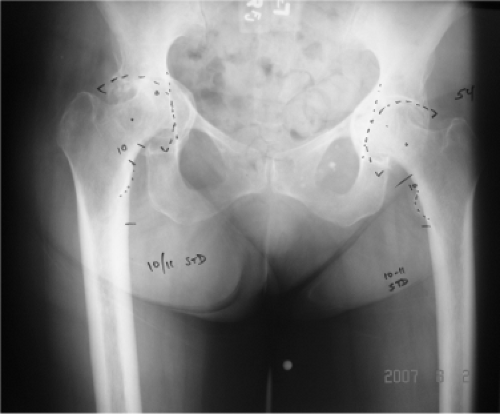 Figure 75.1. AP radiograph of bilateral acetabular protrusion in a patient with rheumatoid arthritis. Both femoral heads are medial to the ilioischial line. Preoperative templating demonstrating the goal of implanting an uncemented acetabular component at the anatomic position. |
Radiographic Evaluation
Anteroposterior (AP) radiographs should be evaluated to assess the position of the acetabulum. The routine femoral and acetabular landmarks of the hip and pelvis should be identified. The ilioischial or “Kohler’s line” should be outlined. This line begins in the sciatic notch and proceeds distally along the posterior column and onto the medial border of the ischium. If the medial aspect of the femoral head is at or medial to Kohler’s line then the hip is classified as “acetabular protrusio.” In severe cases the trochanteric region of the femur is adjacent to the pelvis (Fig. 75.1). Radiographic leg-length discrepancy is measured as well as the offset of the femur relative to the contralateral hip. Often little offset is present, which makes the surgical exposure more challenging, and the sciatic nerve closer to the surgical exposure of the acetabulum. Fixed pelvic obliquity should be recognized and the center of rotation of the femoral head should also be evaluated.
In cases of acetabular component loosening with resulting medial migration and protrusion of the implant, additional studies are often helpful. In addition to routine radiographs, a CT scan of the pelvis is quite helpful to further understand the osseous deformity about the pelvis. Three-dimensional (3D) reconstructions of the pelvis can be prepared to help better understand the deformity and aid in planning for surgical reconstruction in severe deformities (Fig. 75.2).
In cases where the medial wall is totally absent and the acetabular component has migrated significantly medially then an arteriogram may be performed to determine the position of the iliac vessels relative to the medialized component (Fig. 75.3). In cases of severe deformities, in which the vessels may be at risk, a retroperitoneal approach to the inner wall of the pelvis for isolation of the pelvis may more safely protect the vessels during acetabular revision and component extraction during revision THA.
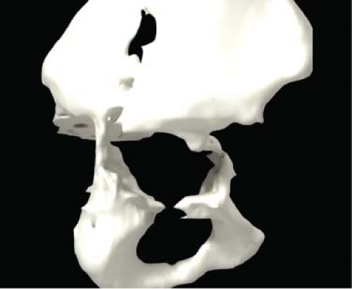 Figure 75.2. 3D reconstruction image/model of the right hemipelvis based on a CT scan of the pelvis in a case with loose acetabular component with severe acetabular protrusio and medial bone loss. |
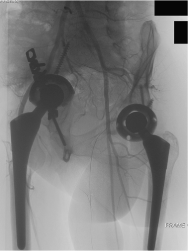 Figure 75.3. Arteriogram showing acetabular protrusion with medial cup migration and retroversion. The cup is displacing the iliac vessels as shown on the arteriogram. |
Preoperative Planning
Related posts:


Stay updated, free articles. Join our Telegram channel

Full access? Get Clinical Tree


