Total Elbow Arthroplasty for Primary Osteoarthritis
Emilie Cheung
Garet Comer
DEFINITION
Primary osteoarthritis (OA) of the elbow is a relatively rare condition that has an idiopathic etiology, although it is frequently associated with heavy use of the arm.
Unlike OA of other large joints, elbow OA is characterized by relatively preserved joint space and articular cartilage but with hypertrophic osteophyte formation and capsular contracture.
Primary OA at the elbow is characterized by pain and lost motion, strength, and, ultimately, function.
ANATOMY
The elbow is a modified hinge joint composed of the ulnotrochlear, radiocapitellar, and proximal radioulnar articulations.
The elbow has two planes of motion: flexion and extension in the sagittal plane and pronosupination in the axial plane.
Flexion-extension comes from motion at the ulnotrochlear articulation and has normal range of motion of 0 degree of extension to 145 degrees of flexion.
Pronosupination is a result of motion at the radiocapitellar and proximal radioulnar articulation and has a normal range of motion of 80 degrees of pronation and 80 degrees of supination.
Collateral ligamentous complexes are present on the medial and lateral sides of the elbow that—along with the inherent bony geometry—confer static stabilization to the elbow.
Lateral collateral ligamentous complex functions as a lateral stabilizer preventing posterolateral rotatory instability (PLRI) and is described as a Y-shaped confluence of three components.
Radial collateral ligament—extends from the lateral epicondyle of the humerus to the annular ligament
Lateral ulnar collateral ligament—extends from the lateral epicondyle of the humerus to the crista supinatoris of the ulna
Annular ligament—forms a ring around the radial neck, extending from the anterior sigmoid notch of the ulna to the crista supinatoris
Medial collateral ligament functions as the most important stabilizer to valgus stress and is composed of three components.
Anterior oblique ligament—extends from the medial epicondyle of the humerus to the sublime tubercle of the anteromedial facet of the coronoid
Posterior oblique ligament—extends from the medial epicondyle of the humerus and fans out to insert along the sigmoid notch
Transverse ligament—this thin band extends transversely from the posterior to anterior margin of the sigmoid notch
PATHOGENESIS
The cause of primary OA is likely multifactorial with hand dominance, history of heavy use, race, and other factors contributing.2
NATURAL HISTORY
Primary OA is a relatively rare condition that commonly affects the dominant arm of middle-aged males who have a history of heavy use due to sports or heavy equipment use.
The severity of the presentation is variable and dependent on disease progression.
Early OA is characterized by pain at terminal extension and flexion with patients frequently complaining of pain with carrying a heavy object with the elbow extended.
Radiographically, early disease is characterized by relatively preserved joint space and impinging osteophytes on the margins of anterior and posterior ulnotrochlear articulation as well as radiocapitellar osteophyte formation.
Late-stage OA presents with pain throughout the arc of motion with loss of articular cartilage and joint space narrowing.
PATIENT HISTORY AND PHYSICAL FINDINGS
Primary OA of the elbow is unique amongst OA of other joints, in that the disease severity and anatomic source of pathology allow for a variety of successful treatment options ranging from simple débridement to arthroplasty.
History
The classic presentation is a middle-aged male with a history of heavy use who presents with symptoms on his dominant side.
Symptoms may include pain, loss of motion, mechanical symptoms of catching or locking, or weakness.
When pain is the primary complaint, it is important to understand the degree of disability and impact on the patient’s function; identify the anatomic source of pain whether ulnotrochlear, radiocapitellar, or extra-articular; and identify whether pain occurs at the extremes of flexion and/or extension or throughout the arc of motion.
Patients with predominantly mechanical symptoms may need only arthroscopic débridement.
In patients who primarily complain of lost motion, it is important to understand the degree to which this affects the patient and whether the lost motion is in flexion, extension, or both.
Further history that is necessary to evaluate when considering arthroplasty include prior surgeries, history of trauma, history of infection, and the patient’s demand level and posttreatment expectations.
Physical Examination
The physical examination begins with inspection of the skin for prior surgical scars, other wounds, or evidence of infection.
Flexion and extension as well as pronosupination range of motion should be assessed.
It is essential to document at what point in the arc of motion pain is experienced as well as any catching, locking, or other mechanical symptoms.
Rotation through the radiocapitellar joint may not generate significant pain or stiffness despite a degenerative appearance radiographically.
Neurovascular examination specifically focused on evaluation for ulnar nerve compression at the elbow is essential to document.
IMAGING AND OTHER DIAGNOSTIC STUDIES
Anteroposterior (AP), lateral, and oblique radiographic views are adequate to make the diagnosis of OA.
Characteristic findings on plain films include osteophyte and/or loose body formation emanating from the olecranon and coronoid processes and extending into their fossae (FIG 1).
The radiocapitellar joint may also be affected and demonstrate osteophytes around the radial head.
Typically, radiographic changes in the ulnotrochlear joint precede those in the radiocapitellar or proximal radioulnar joints.
Computed tomography (CT) scan with three-dimensional reconstructions may be performed and is especially helpful for localizing osteophytes not well visualized on plain films.
Impinging shelf osteophytes within the olecranon, radial, and coronoid fossae are best appreciated on CT and may be missed on plain films.
Similarly, CT is helpful for identifying osteophytes in the medial gutter within close proximity to the ulnar nerve.
DIFFERENTIAL DIAGNOSIS
Posttraumatic arthritis of the elbow
Rheumatoid or other inflammatory arthropathy
Chronic septic arthritis
Crystalline arthropathy
Haemophilic arthropathy
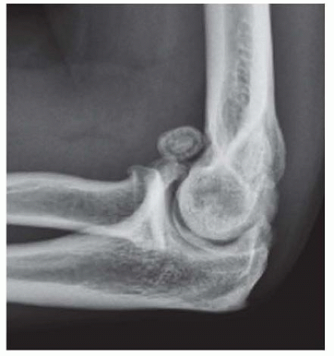 FIG 1 • Lateral x-ray demonstrating osteophyte formation, a loose body, and relatively preserved joint space. |
NONOPERATIVE MANAGEMENT
Nonoperative management is appropriate for the early stages of the disease, when the patient reports mild pain and motion loss of less than 15 degrees.
Nonoperative treatments include activity modification, nonsteroidal anti-inflammatory drugs, intra-articular corticosteroid injections, and physical therapy.
Physical therapy should focus on pain control, antiinflammatory modalities, and preserving range of motion and strength.
Intra-articular injections provide transient relief that is suitable for maintenance therapy.
SURGICAL MANAGEMENT
Total elbow arthroplasty (TEA) is considered in patients with the following:
Disabling elbow OA in patients older than 65 years
Mid-arc pain with activity resulting from cartilage loss of the ulnotrochlear joint
Willing to comply with low activity levels with their operative extremity
Prostheses come in several basic designs:
Linked prostheses
Linked devices have mechanically linked ulnar and humeral components that function as a hinge. Contemporary designs are semiconstrained implants, which about 7 degrees of varus/valgus and rotational laxity at the articulation.
Unlinked prostheses
Unlinked prostheses have no mechanical connection between the ulnar and humeral components and rely on the ulnar and humeral components’ congruence and the capsuloligamentous structures for stability. They have the theoretical benefit of lower bone-cement interface stress leading to less loosening, although clinical data has not yet demonstrated this.
Convertible prostheses
These implants allow for use in either a linked or unlinked fashion.
Preoperative Planning
Full-length x-rays of the arm and forearm must be obtained and scrutinized for deformity, prior hardware, or pathologic lesions.
Preoperative templates are available by many manufacturers and may be used as a guide for intraoperative component selection.
The condition of the soft tissue must be assessed preoperatively, including prior surgical or traumatic scars. We avoid creating skin bridges of less than 1 cm when there is a prior scar. When any doubt exists about the quality of closure, arrangements should be made preoperatively to have a wound vacuum-assisted closure device available.
Regional anesthetic infusion through an interscalene catheter is used throughout the perioperative period.Related posts:
 Open Reduction and Internal Fixation of Displaced Lateral Condyle Fractures of the Humerus
Open Reduction and Internal Fixation of Capitellum and Capitellar-Trochlear Shear Fractures
Corrective Osteotomy for Radius and Ulna Diaphyseal Malunions
Volar Plating of Distal Radius Fractures
Ligament Stabilization of the Unstable Thumb Carpometacarpal Joint
Dorsal Block Pinning of Proximal Interphalangeal Joint Fracture-Dislocations
Open Reduction and Internal Fixation of Displaced Lateral Condyle Fractures of the Humerus
Open Reduction and Internal Fixation of Capitellum and Capitellar-Trochlear Shear Fractures
Corrective Osteotomy for Radius and Ulna Diaphyseal Malunions
Volar Plating of Distal Radius Fractures
Ligament Stabilization of the Unstable Thumb Carpometacarpal Joint
Dorsal Block Pinning of Proximal Interphalangeal Joint Fracture-Dislocations

Stay updated, free articles. Join our Telegram channel

Full access? Get Clinical Tree


