Arthroscopic and Open Triangular Fibrocartilage Complex Repair
A. Lee Osterman
Emily Slate
DEFINITION
The triangular fibrocartilage complex (TFCC) is a complex anatomic structure located at the ulnar side of the wrist. It has several important biomechanical functions:
Extends the gliding surface of the radiocarpal joint
Cushions and stabilizes the ulnar carpus
Stabilizes the distal radioulnar joint (DRUJ)
Disorders of the TFCC are responsible for the ulnar-sided wrist symptoms of pain, weakness, and instability that affect the patient’s function.
The diagnosis and treatment of these injuries to the TFCC will restore stability, resulting in pain relief and a generally good prognosis for functional return.
ANATOMY
The TFCC is a cartilaginous and ligamentous structure interposed between the ulnar carpus and the distal ulna (FIG 1A). It arises from the distal aspect of the sigmoid notch of the radius and inserts into the base of the ulnar styloid.17
The TFCC attaches to the ulnar carpus via the ulnocarpal ligament complex (ulnolunate, ulnotriquetral, and ulnar collateral ligament) (FIG 1B).
The radioulnar ligaments stabilize the DRUJ, limiting rotation as well as axial migration.2
The dorsal and volar radioulnar ligaments are fibrous thickenings within the substance of the TFCC.
As a result of this anatomic configuration, they function as a unit rather than as independent ligaments.
The central, horizontal portion of the TFCC is the thinnest portion, composed of interwoven obliquely oriented sheets of collagen fibers for the resistance of multidirectional stress.
The vascularity of the TFCC has been carefully studied.4 The TFCC receives its blood supply from the ulnar artery through its radiocarpal branches and the dorsal and palmar branches of the anterior interosseous artery. These vessels supply the TFCC in a radial fashion (see Bednar et al4 for a good image of TFCC vascularity).
Histologic sections demonstrate that these vessels penetrate only the peripheral 10% to 40% of the TFCC. The central section and radial attachment are avascular.
This vascular anatomy supports the concept that peripheral injuries can heal if injured and treated appropriately, whereas tears of the central portion do not heal if sutured and are usually débrided.
Biomechanics
The TFCC has several important biomechanical functions: it transmits 20% of an axially applied load from the ulnar carpus to the distal ulna, it is the major stabilizer of the DRUJ, and it is a stabilizer of the ulna.1,6,16,18
The amount of the load transferred to the distal ulna varies with ulnar variance. A greater amount is transferred in positive ulnar variance than negative.
This results in a corresponding decreased thickness of the central portion of the TFCC in ulnar positive wrists.
There is a variable load placed on the TFCC with forearm rotation. Supination causes a negative ulnar variance due to the proximal migration of the ulna. This is reversed with pronation as the ulna moves distally, causing it to become ulnar positive.
The ulnar head also moves within the sigmoid notch in a dorsal direction with pronation and a volar direction with supination.
The dorsal and volar radioulnar ligaments, which form the peripheral portion of the TFCC, serve as major stabilizers to translation at the DRUJ during forearm rotation.
 FIG 1 • A. Anatomic coronal section demonstrating the triangular fibrocartilage (TFC) and its relation to the lunate (L), triquetrum (T), distal ulna (U), and radius (R). B. TFCC. |
PATHOGENESIS
Traumatic injuries of the TFCC result from either the application of an extension, pronation force to the axially loaded wrist, or a distraction force to the ulnar aspect of the wrist.
This will most commonly occur with a fall on the outstretched hand or a resisted torque force.
The lesions are more common with ulnar positive and neutral patients and are frequently found in patients with fractures of the distal radius.
Several authors have examined the incidence of intracarpal soft tissue injuries associated with distal radial fractures.
Geissler et al8 studied 60 patients, finding a TFCC injury in 26 (43%).
In Lindau et al’s12 series of 51 patients, 43 had a TFCC injury (84%): 24 had a peripheral tear, 10 had a central perforation, and 9 had a combined central and peripheral tear.
In a study of 180 wrist joints in 100 cadavers ranging in age from fetuses to 94 years, Mikić13 found that degeneration of the TFCC begins in the third decade of life.
This degeneration increases in frequency and severity as people age.
After the fifth decade of life, 100% of TFCCs appear abnormal.
However, these age-related TFCC lesions are often asymptomatic.6
NATURAL HISTORY
The classification system described by Palmer15 is the most useful for describing TFCC injuries, dividing them into traumatic and degenerative.
Traumatic lesions are classified according to the location of the tear within the TFCC. The traumatic class has been designated by Palmer15 as class 1, with subclasses of A, B, C, and D assigned to anatomic lesions within the TFCC (FIG 2A).
A class 1A lesion represents a tear in the horizontal or central portion of the TFCC. The tear is 2 to 3 mm medial to the radial attachment of the cartilage. It is usually oriented from dorsal to volar.
A class 1B lesion represents an avulsion of the peripheral aspect of the TFCC from its insertion onto the distal ulna. This can occur either with a fracture of the ulnar styloid or as a pure avulsion from its bony attachment. This type of injury disrupts the stabilizing effect of the TFCC on the DRUJ, resulting in clinical instability.
A class 1C lesion represents an avulsion of the TFCC attachment to the ulnar carpus by disruption of the ulnocarpal ligaments. These lesions result in ulnar carpal instability with volar translocation of the carpus.
A class 1D lesion represents an avulsion of the TFCC from its radial attachment. Isolated disc tears should be differentiated from disruption of the dorsal and volar radioulnar ligaments. Such global TFCC injury will result in DRUJ instability.
Degenerative type 2 lesions are age-related, nontraumatic lesions to the TFCC, typically characterized by central perforations and positive ulnar variance.6,22
The natural history of such degenerative lesions, when and if they become symptomatic, is a progressive cascade of degenerative changes, as reflected in Palmer’s type 2 classification (FIG 2B).
The deterioration proceeds from triangular fibrocartilage (TFC) wear through central perforation (type 2C) to lunatotriquetral ligament tear and arthritic changes of the lunate, triquetrum, and distal ulna (type 2D or E).
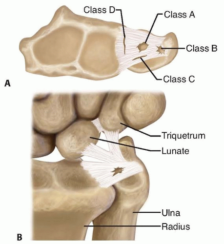
FIG 2 • A. Axial drawing looking at the radius platform and the TFCC. Dorsal is up and volar is down. Palmer classification for acute TFCC injuries. A class 1A lesion involves a tear in the central, horizontal portion of the TFCC. A class 1B lesion is a tear of the TFCC from the distal ulna with or without an ulnar styloid fracture. A class 1C lesion is a tear of the TFCC distal attachment to the lunate and triquetrum through the ulnolunate and ulnotriquetral ligaments. A class 1D lesion is a detachment of the TFCC from its insertion on to the radius at the distal sigmoid notch. B. Palmer classification for degenerative TFCC lesions, usually related to positive ulnar variance and ulnocarpal impaction syndrome. The degeneration occurs in a progressive cascade: types 2A and 2B, TFC wear; type 2C, fibrillated central TFC lesion; types 2D and 2E, arthritic chondral changes of the distal ulna and lunate and disruption of the lunatotriquetral ligament. (A: Modified From Palmer AK. Triangular fibrocartilage complex lesions: a classification. J Hand Surg Am 1989;14[4]:594-606.)
Treatment is based on the stage of involvement.
Degenerative and traumatic lesions can coexist, and injury can render a degenerative lesion symptomatic.
PATIENT HISTORY AND PHYSICAL FINDINGS
Symptoms consist of ulnar-sided wrist pain, frequently with clicking, that typically occurs after a fall.
The initial physical examination reveals swelling over the ulnar aspect of the wrist with inflammation of the tendon of the extensor carpi ulnaris (ECU).
Point tenderness is present over the TFCC and distal ulna. The more isolated the point of maximal tenderness, the more specific the diagnosis.
A fovea sign (point tenderness directly over the ulnar TFC origin) indicates a type 1A or 1B TFC injury or an ulnar extrinsic injury type 1C.
Ulnar deviation and axial loading of the wrist (TFCC compression test) will elicit a painful response and a click with forearm rotation.
The DRUJ must be assessed for instability. Instability is best assessed with the forearm in neutral rotation, but it is also checked in full supination and full pronation.
The examiner stabilizes the distal radius with one hand and applies a force to the distal ulna, moving it dorsal and
volar, looking for increased motion or subluxation of the distal ulna relative to the radius and comparing it with the opposite uninjured wrist.
Significant instability can present as laxity of the distal ulna with a positive “piano key” sign and dorsal prominence of the distal ulna. This may be due to a significant tear or detachment of the dorsal or volar radioulnar ligaments.
A click produced by ulnar deviation and supination over the ECU sheath at the distal ulna indicates ECU instability with subluxation out of its sixth extensor compartment.
A visual carpal supination deformity with ulnar prominence that can be passively corrected by a dorsally applied force to the pisiform indicates an ulnar extrinsic ligament tear.
TFCC injuries do not occur in isolation; they are often a component of a spectrum of injury to the ulnar side of the wrist. The examiner must therefore evaluate all of the commonly injured structures on the ulnar side of the wrist.
The lunatotriquetral joint must be assessed for instability due to a lunatotriquetral ligament tear. This would cause tenderness over the lunatotriquetral interval with a positive Shuck test (painful click as the lunate and triquetrum slide abnormally).
Point tenderness over the triquetrum may signify a triquetral avulsion fracture.
An audible clunk and visual subluxation of the carpus that occur with active ulnar deviation suggest that a midcarpal instability is present.
Crepitus and pain over the pisotriquetral joint on the shear test may indicate pisotriquetral arthritis.
The other soft tissue structures around the ulnar wrist should be examined, including the ulnar nerve, the dorsal ulnar sensory nerve branch, and the ulnar artery.
Grip strength measurements using a Jamar dynamometer, although subjective, are helpful in quantitating patient effort and as a parameter to follow therapeutic progress.
IMAGING AND OTHER DIAGNOSTIC STUDIES
The diagnostic workup should include plain radiographs and a neutral rotation posteroanterior and lateral view.
This will allow assessment for fracture, ligament instability resulting in carpal malalignment, and ulnar variance. It is important to determine ulnar variance because it will influence treatment options (FIG 3A).
The DRUJ must also be examined radiographically to determine if subluxation, arthritis, or ulnar styloid abnormalities such as an acute or chronic nonunited fracture fragment are present.
Magnetic resonance imaging (MRI) is useful in the diagnosis of TFCC tears, especially the class 1A and D lesions.9,11 T2-weighted images in the coronal plane are of the greatest diagnostic value (FIG 3B).
The TFCC has a homogenous low signal intensity. The synovial fluid of the joint appears as a bright image on T2 and will outline tears in the TFCC.
A gadolinium arthrogram enhances the visualization of TFCC tears.
The reported sensitivity and specificity of MRI in diagnosing injuries of the TFCC in the literature is variable.
Golimbu et al9 reported a 95% accuracy of MRI in the detection of TFCC tears. MRI findings were verified arthroscopically.
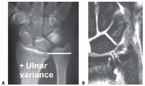
FIG 3 • A. Positive ulnar variance is often associated with degenerative TFC tears and ulnocarpal impaction syndrome. The radiograph should be taken in neutral rotation. B. Coronal T2 MRI wrist image. High signal of the joint fluid outlines the low signal substance of the TFC complex. Tears will show as high signal within the central region (arrow at right) or TFCC periphery. There is a normal clear area (arrow at left) at the insertion of the radial TFC into the medial articular cartilage of the radius.Related posts:
 Sauvé-Kapandji Procedure for Distal Radioulnar Joint Arthritis
Proximal Interphalangeal and Metacarpophalangeal Joint Silicone Implant Arthroplasty
Surgical Treatment of Acute and Chronic Paronychia and Felons
Skin Grafts and Skin Graft Substitutes in the Distal Upper Extremity
Treatment of Enchondroma, Bone Cyst, and Giant Cell Tumor of the Distal Upper Extremity
Clinodactyly
Sauvé-Kapandji Procedure for Distal Radioulnar Joint Arthritis
Proximal Interphalangeal and Metacarpophalangeal Joint Silicone Implant Arthroplasty
Surgical Treatment of Acute and Chronic Paronychia and Felons
Skin Grafts and Skin Graft Substitutes in the Distal Upper Extremity
Treatment of Enchondroma, Bone Cyst, and Giant Cell Tumor of the Distal Upper Extremity
Clinodactyly

Stay updated, free articles. Join our Telegram channel

Full access? Get Clinical Tree

 Get Clinical Tree app for offline access
