Thumb Carpometacarpal Arthrodesis
K. J. Hippensteel
Ryan Calfee
DEFINITION
The trapeziometacarpal joint of the thumb is frequently affected by osteoarthritis, second in frequency only to the distal interphalangeal joint, but often more disabling due to pain and weakness of grip and pinch strength.
The choice of surgical management for symptomatic thumb carpometacarpal (CMC) joint arthrosis varies according to patient age, medical comorbidities, functional demands, and radiographic staging.
Arthrodesis of the CMC joint was initially described by Muller13 in 1949. Despite the popularity of arthroplasty at this joint, arthrodesis can also provide excellent functional outcomes.
Arthrodesis is ideal for the younger patient with moderate to advanced thumb CMC degeneration (arthritic or posttraumatic) who anticipates higher load pinch and grip requirements.9
ANATOMY
The thumb CMC joint is a biconcave-convex (saddle) joint, permitting motion in three planes: flexion-extension, abduction-adduction, and pronation-supination. These multiplanar motions allow for power grip, power pinch, opposition, and delicate precision pinch.
Provided minimal osseous constraints, ligamentous structures are largely responsible for stabilizing the thumb CMC joint.
Sixteen ligaments have been described around the thumb CMC joint.
Seven are primary stabilizers of the thumb CMC joint:
Superficial anterior oblique ligament (sAOL) and deep anterior oblique ligament (dAOL)
Dorsoradial
Posterior oblique
Ulnar collateral
Intermetacarpal
Dorsal intermetacarpal
The remainder stabilize the trapezium, providing a stable foundation for the thumb.2
PATHOGENESIS
The pathogenesis of CMC joint arthrosis is multifactorial, involving biochemical,16 biomechanical, and genetic influences.
Osteoarthritis of the thumb CMC joint occurs more commonly in females compared to males.
Arthritic degeneration begins on the palmar aspect of the thumb metacarpal and trapezium. This may be secondary to compression in this area during pinch.
The dorsal ligament complex (dorsoradial and posterior oblique ligaments) is the thickest, strongest, and most important ligament stabilizing the thumb CMC joint. It prevents both the thumb metacarpal volar beak from disengaging from the volar recess in the trapezium as well as dorsal subluxation of the metacarpal base during power grip or pinch. Although the anterior (palmar) oblique ligament, or so-called beak ligament, was thought to be the most important stabilizing ligament of the thumb, multiple studies have suggested the dorsal ligament complex is the most critical stabilizer.3,19,22 The volar beak ligament is completely lax in opposition and taut only in the hitchhiker position.
Arthritis begins secondary to the compressive, rotational shear forces during power pinch and grip in the volar recess of the trapezium near to the volar beak of the metacarpal. After many years, the volar beak begins to wear down and instability develops during the screw-home-torque rotation seen in opposition.7 With progression of disease, osteophytes develop and eburnation progresses throughout the entire joint surface.
Osteoarthrosis can also develop from disruption of the articular cartilage. Any fracture involving the articular surfaces (most commonly the base of the thumb metacarpal) will predispose to, or accelerate the development of, arthrosis. This is the result of direct cartilage injury at the time of the accident or over time secondary to articular incongruity or articular surface irregularity.
Anatomic restoration of the joint surface can minimize this progression but not eliminate it completely.
NATURAL HISTORY
Arthrosis of the thumb CMC joint begins along the palmar aspect of the metacarpal secondary to the compressive rotational shear and dorsal subluxating forces during pinch and grip. These forces can approach 164 kg during power grasp at the CMC joint.4
The entire base of the metacarpal and the distal trapezium experience eburnation of the cartilage, which progresses to develop osteophytes.
As arthritis progresses, the thumb metacarpal subluxates dorsally and radially. The metacarpal adducts and flexes resulting in compensatory metacarpophalangeal (MCP) joint hyperextension. This hyperextension effectively brings the thumb pulp out of the palm to allow for grasp.
In fulminant arthritis, the entire surface of the trapezium becomes involved, resulting in degeneration between the proximal trapezium and the distal scaphoid.
PATIENT HISTORY AND PHYSICAL FINDINGS
Thumb CMC joint arthrosis will often present with pain at the base of the metacarpal.
The pain will be exacerbated with activities that load the thumb metacarpal base, such as turning a doorknob, twisting a lid off a jar, or turning a key.
Pain at rest may or may not be present.
This classic deep-seated pain at the base of the thenar muscles must be distinguished from de Quervain tendonitis, tendonitis of the flexor carpi radialis, radioscaphoid degeneration, and trigger thumb.
Symptoms do not always correlate with the clinical or radiographic appearance. A patient may have advanced clinical and radiographic disease with minimal symptoms. Conversely, a patient may have significant symptoms with minimal radiographic changes and no clinical deformity.
Physical examination of the patient with advanced disease reveals deformity.
The thumb metacarpal base subluxates in a dorsal direction and the metacarpal becomes fixed in adduction and flexion. This manifests as a metacarpal prominence at the CMC joint with decreased ability to abduct the thumb away from the palm.
In an effort to compensate for this limitation, the MCP joint will often hyperextend, creating a zigzag deformity.
Asking the patient to place one finger on the point that is most symptomatic helps localize the point of maximal tenderness to the CMC joint or another area.
CMC grind test: Reproduction of symptoms while axially loading the thumb and circumducting the thumb metacarpal confirms the CMC joint as a site of disease.
Finkelstein maneuver: Maximal tenderness over the radial styloid during ulnar deviation of the hand with a thumb in the fist suggests that de Quervain tendonitis may be a greater source of symptoms.
Phalen test: Reproduction of symptoms during wrist flexion indicates carpal tunnel syndrome as a more likely etiology.
Carpal tunnel compression test (Durkan test): Reproduction of symptoms with compression over the carpal tunnel indicates carpal tunnel syndrome as a more likely etiology of symptoms.
Trigger evaluation: Reproduction of pain, triggering, or locking of the thumb at the interphalangeal joint with activity flexion indicates trigger thumb as an etiology.
Allen test: The radial and ulnar arteries are compressed and the hand is exsanguinated. The ulnar artery is released and the circulation of the hand is assessed. The process is repeated, releasing the radial artery while the ulnar artery is occluded. Surgical procedures often involve mobilization of the radial artery in the snuffbox. Damage to this artery will require reconstruction if the ulnar artery cannot compensate.
IMAGING AND OTHER DIAGNOSTIC STUDIES
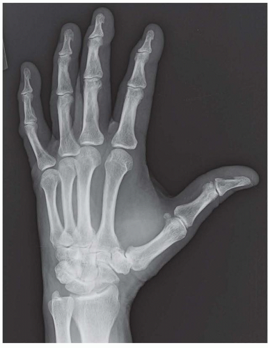
Plain radiographs are the imaging modality of choice for evaluation of thumb CMC joint arthrosis (FIG 1).
These include posteroanterior, pronated anteroposterior (AP) (Robert view), lateral, and Bett views.
Eaton and Littler6 have described a radiographic staging system that is commonly used.
Stage I: normal-appearing or widened joint space secondary to synovitis
Stage II: joint space narrowing and osteophyte formation smaller than 2 mm
Stage III: joint space narrowing with osteophytes larger than 2 mm
Stage IV: stage III appearance with the addition of narrowing or osteophytes in the scaphotrapezial joint
The scaphotrapezoid joint is not specifically addressed in this system and may be difficult to assess radiographically, but this joint should always be assessed at the time of surgery because it may be a source of continued pain.21
DIFFERENTIAL DIAGNOSIS
Thumb CMC arthrosis
De Quervain disease
Trigger thumb or stenosing tenosynovitis
Flexor carpi radialis tendonitis
Scaphoid pathology (fracture, nonunion, avascular necrosis)
Radioscaphoid arthrosis
Scaphotrapeziotrapezoid (STT) arthrosis
Carpal tunnel syndrome
Intramuscular (thenar) processes, such as vascular or tumor etiologies
NONOPERATIVE MANAGEMENT
Most patients with symptomatic thumb CMC joint arthrosis benefit from a trial of conservative therapy, which may include rest, oral anti-inflammatory medication, intra-articular corticosteroid injection, thenar isometric strengthening exercises, and splinting.1
Forty percent of patients can have significant and sustained relief of pain with steroid injection and splinting for
3 weeks regardless of radiographic staging. If separated by Eaton’s staging, over 80% with stage I can have sustained relief for over 18 months compared to approximately 33% with stage II or III or less than 25% with stage IV.5
Although nonoperative treatment does not eliminate the problem or alter the underlying disease process, it often reduces symptoms and may either negate the need for surgery or at least allow for delay of surgical intervention. During this time, patients are reassured that continued activity is not expected to change the disease course and that despite radiographic progression, the arthritic pain may at times improve over time.
SURGICAL MANAGEMENT
The indication for surgical intervention for symptomatic thumb basilar joint arthrosis is pain and weakness not sufficiently responsive to conservative treatments.
There are multiple procedures used to treat symptomatic CMC thumb arthritis, none of which have proven superiority.
The ideal patients for thumb CMC arthrodesis are younger, active patients who need to maintain power grip and pinch and regularly place high force demand on their thumb. These are typically younger manual laborers with stage II or III disease. However, arthrodesis can provide symptomatic relief in older patients with stage II or III disease.
Stay updated, free articles. Join our Telegram channel

Full access? Get Clinical Tree


