CHAPTER SEVEN THORACIC SPINE
INTRODUCTION
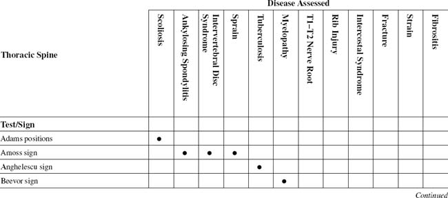
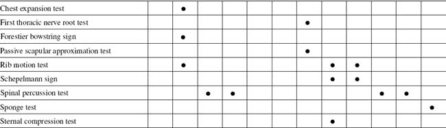
TABLE 7-2 THORACIC SPINE CROSS-REFERENCE TABLE BY SYNDROME OR TISSUE
| Ankylosing spondylitis | |
| Fibrositis | Sponge test |
| Fracture | Spinal percussion test |
| Intercostal syndrome | |
| Intervertebral disc syndrome | |
| Myelopathy | Beevor sign |
| Rib injury | |
| Scoliosis | Adams positions |
| Sprain | |
| Strain | Spinal percussion test |
| T1–T2 nerve root | |
| Tuberculosis | Anghelescu sign |
ORTHOPEDIC GAMUT 7-1 THORACIC AREA PAIN
Diagnostic keys for thoracic area pain include the following:
ORTHOPEDIC GAMUT 7-2 MECHANICAL SPINAL SYNDROME CLASSIFICATIONS
Adapted from Hefford C: McKenzie classification of mechanical spinal pain: profile of syndromes and directions of preference, Man Ther 13(1):75–81, 2008.
ORTHOPEDIC GAMUT 7-3 THORACIC SPINE
Stabilizing influences for the thoracic spine include the following:
ORTHOPEDIC GAMUT 7-4 THORACOLUMBAR SPINE
To assess range of motion of the thoracolumbar spine, the patient is directed to do the following:


ESSENTIAL MUSCLE FUNCTION ASSESSMENT
During the trunk extension test, the latissimus dorsi, quadratus lumborum, and trapezius assist back extensors. The head and neck extensor muscles and the hip extensors should be tested before the back extensors are tested (Fig. 7-7).
ESSENTIAL IMAGING
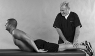
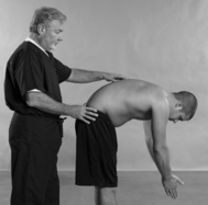
ADAMS POSITIONS
Assessment for Pathologic or Structural Scoliosis
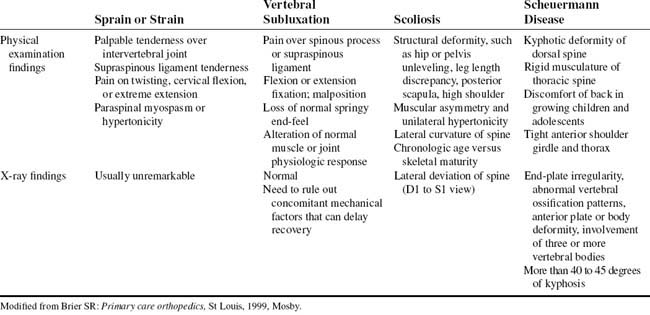
Comment
The spinal column centers the mass of the torso and head in a line along the vertical axis that falls through the pelvis. Disturbances of the spine, such as the curvatures associated with scoliosis, may significantly alter the normal balance and coordination of the spine (Table 7-3).
Scoliosis is also classified as either structural or nonstructural. Structural curves are fixed and nonflexible and fail to correct with side bending. Nonstructural curves, on the other hand, are flexible and readily correct with side bending. The Lenke Classification System helps examiners develop a more complete picture of the patient’s condition by understanding the scoliosis as multidimensional and considering it from more than one view. The Lenke classification method also gives more detailed shorthand for communicating about scoliosis in professional settings, using a widely understood set of criteria (Table 7-4).
TABLE 7-4 LENKE SCOLIOSIS CLASSIFICATION
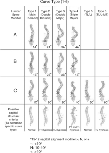 |
| The Lenke Classification System is simple, accurate, and easy to reproduce and communicate between health care providers. It relies on measurements taken from standard radiographs (X-rays). In this method, the examiner evaluates X-rays of the patient from the front, the side, and in bending positions. Each scoliosis curve is then classified in three steps by the region of the spine, the degree or angle of the curve, and the relationship of the side-to-side curve to the sagittal plane. For example, many scoliosis curves affect the presence or absence of kyphosis, which is the outward or convex curve normally found in the upper back. In addition, each aspect of the curve is evaluated for its relative stiffness or flexibility. |
From Dr. Lawrence Lenke, Washington University School of Medicine, St. Louis, Missouri. Available at: www.spinal-deformity-surgeon.com.
PROCEDURE
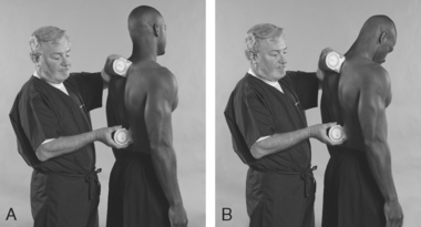
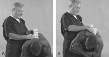
AMOSS SIGN
Assessment for Ankylosing Spondylitis, Severe Sprain, or Intervertebral Disc Syndrome
Comment
Ankylosing spondylitis (AS), an ascending disease, affects the thoracic region after the lumbar. Patients with this condition experience back pain, but the anterolateral chest pain and the limited chest expansion bother them the most. In some patients, these symptoms may occur rather early in the life of the disease, but they usually become bothersome after 6 years of illness. Chest pain, which usually occurs during inspiration, and limited chest expansion are caused primarily by involvement of costovertebral and manubriosternal joints, as well as the costochondral junctions and the clavicular joints. The girdle-like restriction may cause a sense of anxiety and dyspnea, particularly during exertion. However, respiratory problems are surprisingly uncommon, although restricted ventilatory volumes are detected by pulmonary function studies (Table 7-5). Of course, should concomitant disease result in impaired diaphragmatic breathing, a problem is likely to develop.
TABLE 7-5 MODIFIED NEW YORK CRITERIA FOR ANKYLOSING SPONDYLITIS DIAGNOSIS
| Clinical criteria | Low back pain and stiffness for more than 3 months that improves with exercise but is not relieved by rest |
| Radiological criterion | Sacroiliitis grade 2 bilaterally or sacroiliitis grade 3–4 unilaterally |
Adapted from Moll JMH: New criteria for the diagnosis of ankylosing spondylitis, Scand J Rheum 16(suppl 65):12-24, 1987.
PROCEDURE

Stay updated, free articles. Join our Telegram channel

Full access? Get Clinical Tree


