15 The upper arm and elbow
Apart from injury, disorders of the upper arm and elbow region are generally straightforward and present few special problems. They conform to the general descriptions of bone and joint diseases that were given in Part 2. Thus the humerus is subject to the ordinary infections of bone, and occasionally to bone tumours – especially metastatic tumours. The elbow is liable to every type of arthritis, none is particularly common though rheumatoid arthritis is commoner than osteoarthritis. After the knee, it is the joint most often affected by osteochondritis dissecans and loose body formation. The ulnar nerve lies in a vulnerable position at the back of the medial epicondyle, and the possibility of impairment of nerve function complicating disease or injury of the joint should always be remembered. Replacement arthroplasty of the elbow is undertaken increasingly, but compared with those of hip and knee arthroplasty the results still leave much to be desired, especially from the point of view of the longevity of the new joint.
SPECIAL POINTS IN THE INVESTIGATION OF UPPER ARM AND ELBOW SYMPTOMS
History
The interrogation follows the usual lines suggested in Chapter 1. It is important to ascertain the exact site and distribution of the pain, and its nature. Pain arising locally in the humerus is easily confused with pain arising in the shoulder, which characteristically radiates to a point about half-way down the outer aspect of the upper arm. Elbow pain is localised fairly precisely to the joint, though a diffuse aching pain is often felt also in the forearm. When the ulnar nerve is interfered with behind the elbow the symptoms are mainly in the hand.
Steps in routine examination
A suggested plan for the routine clinical examination of the upper arm and elbow is summarised in Table 15.1.
Table 15.1 Routine clinical examination in suspected disorders of the upper arm and elbow
| 1. LOCAL EXAMINATION OF THE ARM AND ELBOW | |
| Inspection | Power |
| Bone contours and alignment | Flexors |
| Soft-tissue contours | Extensors |
| Colour and texture of skin | Supinators |
| Scars or sinuses | Pronators |
| Palpation | Stability |
| Skin temperature | Lateral ligament |
| Bone contours | Medial ligament |
| Soft-tissue contours | The median nerve |
| Local tenderness | Sensory function |
| Movements (active and passive) | Motor function (opponens action) |
| Humero-ulnar joint: | Sweating |
| Flexion | The radial nerve |
| Extension | Sensory function |
| Radio-ulnar joint: | Motor function (extension of wrist, thumb, and fingers) |
| Supination | The ulnar nerve |
| Pronation | Sensory function |
| ? Pain on movement | Motor function |
| ? Crepitation on movement | Sweating |
| 2. EXAMINATION OF POTENTIAL EXTRINSIC SOURCES OF ARM PAIN | |
| 3. GENERAL EXAMINATION | |
| General survey of other parts of the body. The local symptoms may be only one manifestation of a widespread disease | |
Movements at the elbow
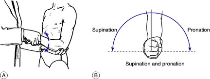
The elbow joint has two distinct components: the hinge joint between the humerus above and the ulna and radius below, allowing flexion–extension movement; and the pivot joint between the upper ends of the radius and ulna, allowing rotation of the forearm. It should be remembered that free rotation of the forearm is dependent not only upon an intact superior radio-ulnar joint: it demands also free mobility between the radius and ulna throughout their length, and distally at the inferior radio-ulnar joint. Flexion–extension: The normal range is from 0 to 155 ° (0 = anatomical position, with the arm straight). Supination–pronation: Rotation movements must be tested with the elbow flexed to a right angle, to eliminate rotation at the shoulder (Fig. 15.1A). The normal range is 90 ° of supination (palm up) and 90 ° of pronation (palm down) (Fig. 15.1B). If the range of rotation is restricted possible causes must be sought in the forearm and wrist as well as in the elbow.
The ulnar nerve
Because of the vulnerability of the ulnar nerve in its course behind the elbow, tests of ulnar nerve function should be carried out as part of the routine examination of the elbow. Examine for sensibility in the little finger and medial half of the ring finger, and test the ulnar-innervated small muscles of the hand for wasting or weakness: a simple test is to ask the patient to spread the fingers and to bring them together again forcibly, to prevent a card gripped between the middle and ring fingers from being withdrawn. Note whether the skin in the territory of the ulnar nerve sweats equally with the rest of the hand.
Extrinsic sources of pain in the upper arm
Pain in the upper arm is commonly referred from a lesion elsewhere—particularly from the shoulder, and from the neck when the brachial plexus or its roots are involved. Shoulder pain usually radiates from the tip of the acromion process to about the middle of the outer aspect of the arm, but it does not extend below the elbow. In contrast, nerve pain from interference with the brachial plexus often extends throughout the length of the arm and forearm into the hand and fingers; and frequently there is accompanying paraesthesiae in the form of tingling, numbness, or ‘pins and needles’.
DISORDERS OF THE UPPER ARM
ACUTE OSTEOMYELITIS (General description of acute osteomyelitis, p. 85)
Pathology. Except in time of war the humerus is seldom infected directly by organisms introduced from without, for compound fractures are rare. Infection is usually haematogenous, from a focus elsewhere in the body. This type of infection occurs mainly in children, and it usually begins in a metaphysis of the bone – more often the upper metaphysis than the lower. Since both the upper and the lower metaphyses are partly enclosed within the capsule of the shoulder and of the elbow respectively, a metaphysial infection is liable to spread directly to the adjacent joint, causing pyogenic arthritis (see Fig. 7.2).
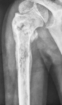
Imaging. Radiographs do not show any abnormality at first. After about two weeks there are often localised rarefaction and subperiosteal new bone formation (Fig. 15.2), but these changes may be slight at first. Radioisotope scanning shows increased uptake in the affected area.
Investigations and treatment are the same as for acute osteomyelitis elsewhere (p. 89).
TUMOURS OF BONE
Benign tumours (General description of benign bone tumours, p. 106)
Giant-cell tumour is uncommon in the humerus, but when it does occur the usual site is at the upper end, where it often extends close up to the articular surface. The tumour occurs chiefly in young adults, and its general characteristics are like those of giant-cell tumour of bone at other sites (p. 109).

Stay updated, free articles. Join our Telegram channel

Full access? Get Clinical Tree


