Fresh frozen allograft reconstruction has been used for a long time in massive bone loss in orthopedic surgery. Allografts have the advantage of being biologic reconstructions, which gives them durability. Despite a greater number of complications in the short term, after 5 years these stabilize with high rates of survival after 10 years. The rate of early complications and the need for careful management in the first years has led the orthopedic surgeon to the use of other options. However, the potential durability of this reconstruction makes this one of the best options for younger patients with high life expectancy.
Key points
- •
Fresh frozen allograft is a reconstructive biologic option for large-extremity osseous defects that, if there is not a major complication, are durable for many decades.
- •
Advantages of this reconstruction are that host ligaments and muscles could be attached to the graft, could be progressively incorporated by the host, and are available for all anatomic sites.
- •
Disadvantages include host-donor junction complications, deterioration of the joint, possible disease transmission, and that processing techniques, such as radiation, could affect the strength and elastic modulus of the graft.
- •
Outcome of massive allografts reconstruction could be improved with adequate anatomic matching, infection prevention, modern internal fixation, stable soft tissue reconstructions, and accelerated rehabilitation protocols.
Introduction
Massive bone loss due to tumor resection, high energy trauma, uncontrolled infections, or prosthetic revisions is a major problem in modern orthopedics centers. Although endoprosthetic reconstruction has improved in recent years, biologic reconstruction is a functional alternative option for large-extremity osseous defects.
Fresh frozen allograft reconstruction has a long history in orthopedics. Since Bauer, who in 1910 described transplantation of bones stored by refrigeration, in a hundred years there had been great advances in fresh frozen allografts. The aim of this article is to describe the outcome and surgical techniques in osteoarticular, intercalary, and allograft-prosthetic composite (APC).
Osteoarticular allograft
Total Condylar Allograft
This reconstruction is used to replace one side of the joint after major bone loss. This alternative does not sacrifice the contralateral side of the joint and allows the surgeon to reattach the host-donor soft tissues. Although, as with other allograft reconstruction, infection, fracture, and nonunion could be ensue, articular deterioration and joint instability are the 2 complications that are related directly with this type of reconstruction.
Infection is not only related to the allograft reconstruction; others factors such as extensive bone resection with soft tissue loss, duration of surgery, and adjuvant treatments such as chemotherapy and radiotherapy influence the rate of infection. This complication is seen not only in bone allograft reconstruction but also in endoprosthetic reconstructions, with an infection rate of 8.4% in large series. The most extensive series of allograft evaluated for infection reports an incidence of 7.9% of primary infections in 945 patients. In the same series, fracture rates (18%) and nonunion rates (16%) were higher than infection rates. However, an infection complication usually leads to an allograft removal so it is related with a high failure rate.
Fractures of the allografts can be avoided if the allograft is protected with internal fixation for its entire extent. Although in the 1990s the use of plates and screws was reported as increasing the rate of fracture, in recent reports it is the use of intramedullary nails that was related with higher fracture rates and nonunion rates. The use of rigid fixation with plates and screws for allograft reconstruction is recommended in most publications. The addition of vascularized fibular graft had been advocated as useful; however, in a recent study they do not show significant differences comparing allograft with and without this. Protecting the allograft with intramedullary cement should be avoided in osteoarticular graft due to the possibility of resurfacing the joint with prosthesis in the future.
Nonunion could be avoided if host-donor osteotomy is stabilized with an adequate internal fixation, which is mostly obtained with rigid fixation with compression plates and screws. Chemotherapy had a deleterious effect on allograft reconstruction ; however, the effect of chemotherapy seems to be a reversible process. When nonunion occurs, it is usually solved with autologous bone graft.
Articular deterioration and joint instability are complications related directly with osteoarticular allografts. Clinicopathologic studies performed in retrieved human allografts found earlier and more advanced degenerative articular cartilage changes in specimens retrieved from patients with an unstable joint than in those from patients with a stable joint. Poor anatomic matching of both size and shape between the host defect and the graft can significantly alter joint kinematics and load distribution, leading to bone resorption or joint degeneration. To have a more accurate size match between the donor and the host, preoperative planning platforms, based on three-dimensional (3D) computed tomography (CT)–derived bone models to choose the best allograft using a virtual bone bank system ( [CR] ), have been described. Bone measures in a 3D virtual environment determined the best match from a bone bank, providing more information to the surgeons for allograft selection before surgery. Longevity of these grafts is also related with the joint stability obtained in surgery with reconstruction of ligaments, tendons, and joint capsule. Deterioration of the joint is accelerated with malalignment of the limb.
Surgical Technique
The surgical technique begins with the proper selection of the graft in preoperative planning (see [CR] ). The selected allograft is taken out of its packaging and placed directly in warm solution. After being thawed, the donor bone is cut to the proper length with the use of a navigation system, and soft tissue structures such as cruciate ligaments, collateral ligaments, and posterior capsule are prepared for implantation. With the navigation system the adequate rotation is marked on the graft.
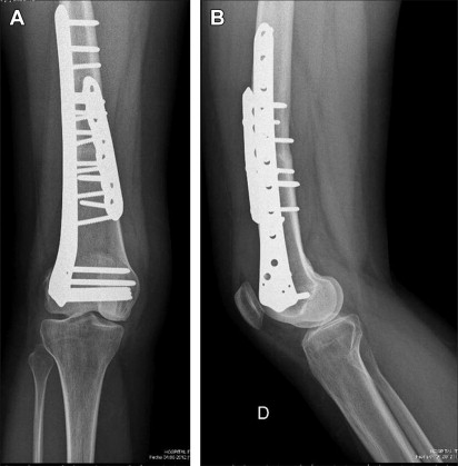
We use an extended anterior-medial approach to the knee, and the extensor mechanism is released when a proximal tibial osteoarticular allograft is performed. The bone tumor is resected with an adequate margin of normal tissue according to preoperative staging studies. All resections are intra-articular and intracompartmental. A transverse osteotomy guided by navigation is used in every case, and the rotation is marked to correlate with one of the allografts. A short anterior six-hole locking compression plate (LCP) is used to reduce the osteotomy. We first apply compression to the osteotomy with 2 cortical screws and then secure the plate with locking screws in the remaining holes. To obtain rigid fixation, a locking compression condylar plate is used on the lateral side of the reconstruction with the goal of obtaining a solid allograft construct by spanning the entire length of the allograft ( Fig. 1 ). Soft tissues from the allograft are attached to corresponding host tissues to obtain the best stability. This reconstruction includes repair of the posterior capsule, anterior and posterior cruciate ligaments, medial and lateral collateral ligaments, and, in tibial allografts, the patellar ligament.
Results
It is difficult to analyze results in an anatomic location with a specific type of reconstruction (such as osteoarticular allograft of the proximal tibia). Usually, the series included multiple locations with the same type of reconstruction (osteoarticular allograft of all locations) or multiple reconstructions of the same location (reconstruction of the distal femur with osteoarticular, intercalary, and composite allografts). Ogilvie and colleagues analyzed 20 osteoarticular allografts (8 distal femur, 6 proximal tibia, 4 proximal humerus, 1 distal radius, and 1 proximal ulna) with a minimum follow-up of 10 years. They found a fracture rate of 45%, degenerative joint disease of 25%, nonunion rate of 20%, and infection of 10%, with a 10-year allograft survival of 45%. Campanacci and colleagues reported 20 osteoarticular allografts (10 distal femur and 10 proximal tibia) in patients younger than 14 years, with a minimum follow-up of 7 years. The allograft survival at 10 years was 58% in distal femur and 20% in proximal tibia; the failures were related to graft fractures. It is interesting that both reports show allograft fractures as the primary cause of failure. The low allograft survival in this small series could be related because these 2 series included long-term follow-up patients so they are those patients of the beginning of a long learning curve; an extensive series is needed to improve the outcome.
With respect to specific anatomic locations, we have reported on 80 osteoarticular distal femur allografts with a minimum follow-up of 5 years. There were 6 infections and 4 fractures with an allograft survival of 78% at 5 and 10 years of age and the rate of allograft survival without the need of a subsequent knee prosthesis resurfacing was 71% at 5 and 10 years. Toy and colleagues reported 26 osteoarticular distal femur allografts with a minimum follow-up of 8 years. There were 4 infections and 5 fractures with a 5- and 10-year allograft survival rates of 69% and 63%, and 5- and 10-year survival rates of the joint surface of 79% and 65%.
Sixteen osteoarticular proximal tibia allograft were reported by Clohisy and colleagues, with a survival rate of 56% and an average of 90° of active motion of the knee with a good or excellent Musculoskeletal Society Tumor score in 9 patients. Five patients had extension strength in the involved knee equivalent to that in the contralateral knee, 6 had strength that was greater antigravity but less than normal, and 3 had only antigravity strength. Brien and colleagues evaluated 17 proximal tibia reconstructions; they remarked that mild instability and extensor weakness was the most common problem. Hornicek and colleagues reported 38 patients with a proximal tibial osteoarticular allograft with a survival rate of 66%. They did not find significant difference between patients who received chemotherapy and those who did not. Muscolo and colleagues reported 58 proximal tibial osteoarticular allografts with a minimum follow-up of 6 years. Infection was the principal cause of failure (13 patients), with an allograft survival rate of 65% and joint survival rate of 57%. Seventy-two percent of the retained allografts (23 of 32) had some articular deterioration in this series.
Regarding proximal humeral osteoarticular allograft, Gebhardt and colleagues reported 60% of satisfactory results in 20 patients with this reconstruction. Getty and Peabody reported 68% of allograft survival rate in 16 patients but they abandon the routine use of osteoarticular allografts to replace the proximal aspect of the humerus due to high complication rate. DeGroot and colleagues reported 31 patients, of whom 23 were filled with cement, with a survival of 78% at 5 years. van de Sande and colleagues found that osteoarticular allograft reconstruction of the proximal humerus had higher complication rates (62%) than endoprostheses (21%) and APC (40%). The authors analyzed their series in a recent article, and we found high articular deterioration and fracture rate in proximal humerus osteoarticular allograft.
Osteoarticular allografts of the distal part of the radius replace an articular surface for which prostheses are not readily available. Kocher and colleagues analyzed 24 patients with osteoarticular allografts of the distal radius. The most frequent cause of revision was fracture (4 patients) or wrist pain (2 patients). There were 4 other complications necessitating additional operative management. Of the 16 patients in whom the graft survived, 3 had no functional limitation, 9 reported limitation only with strenuous activities, and 4 had limitations in the ability to perform moderate activities. Bianchi and colleagues reported 12 patients with distal radius osteoarticular allograft. Distal radioulnar joint instability was observed in 8 cases, and subchondral bone alterations and joint narrowing were present in all cases but were painful in only 1 patient. Scoccianti and colleagues reported 17 patients with only one failure due to fracture of the graft. Although no patient reported more than modest nondisabling pain and 6 reported no pain at all, radiographs showed early degenerative changes at the radiocarpal joint in every patient.
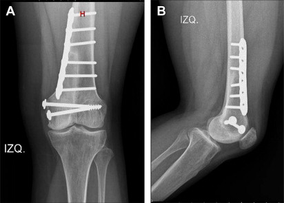
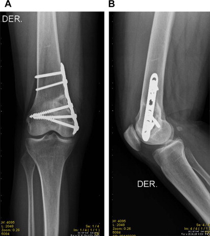
Unicondylar allografts
Unicondylar osteoarticular allografts ( Fig. 2 ) are used after bone tumor resection or trauma bone loss. The surgical technique is demanding, but this type of reconstruction is clearly improved with the use of intraoperative navigation assistance. Fracture and nonunion rates are insignificant in this reconstruction due to the metaphyseal location of the reconstruction. The major problem with this reconstruction is degenerative joint disease and instability. These problems could be prevented with appropriate preoperative selection of the allograft in a virtual bone bank and the consequent allograft cut with the use of intraoperative navigation assistance. Although the authors select donors younger than 35 years, the accuracy in size matching between the donor and the host is the most important issue in this allograft. For appropriate selection, the allografts and the host are measured with CT scans. The maximum total width and anteroposterior dimension of the medial and lateral condyles are the more important measures for allograft selection.
Surgical Technique
After proper selection of the allograft, the surgical procedure begins with resection of the lesion, including biopsy scars with appropriate bone and soft tissue margins. The authors use the same exposure described for total condylar osteoarticular allograft to have an adequate exposure of the knee joint. To perform the unicondylar resection 2 perpendicular osteotomies are done. The use of navigation systems clearly improved the accuracy of these osteotomies. It is crucial that the donor bone is cut to the proper length to avoid distal or proximal malposition offset with a consequent varus or valgus deformity of the knee. After the unicondylar allograft is cut to the proper length, it is fixed with 2 cancellous screws with washers. Then an LCP is used to obtain a more rigid fixation and to avoid any fracture in the diaphyseal area. Soft tissues from the allograft are attached to corresponding host tissues to obtain the best stability. This reconstruction includes repair of the posterior capsule in all cases, in lateral unicondylar allografts the anterior cruciate and lateral collateral ligaments and in medial unicondylar allografts the posterior cruciate and medial collateral ligaments.
Results
Muscolo and colleagues reported 40 unicondylar osteoarticular allografts of the knee (29 distal femur and 11 proximal tibia) followed for a mean of 11 years, with a survival rate at 5 years of 85%. There were 6 failures due to 2 local recurrences, 2 infections, 1 fracture, and 1 massive resorption. Bianchi and colleagues reported 12 unicondylar osteoarticular allograft (9 distal femur and 3 proximal tibia) with a minimum follow-up of 4 years. They had 2 failures, and the remaining patients had mild or severe degenerative changes.
Intercalary allografts
Allograft Arthrodesis
Massive allograft arthrodesis is a salvage procedure reserved in cases when a great loss of soft tissue is present such as after extra-articular tumor resection or failed previous reconstruction with complete loss of muscle function involved in the joint movement.
Benevenia and colleagues reported on 25 patients, 11 of whom had nononcological complications following intercalary allograft arthrodesis of the knee. Weiner and colleagues reported 18% of nonunion in 39 patients who had an allograft arthrodesis after resection of a tumor around the knee. Mankin and colleagues reported that only 3% had an excellent result and 51% had a good result, according to their scoring system after reviewing 71 patients who had an allograft arthrodesis. Donati and colleagues reported a two-center study of 92 patients with knee arthrodesis. They found that complications were greater (infection 20%, fracture 25%, nonunion 44%) and outcome less successful, in comparison with other allograft reconstructions. This is due to the greater soft-tissue resection after extra-articular resection. In addition, the allografts implanted in replacement of the knee may not be strong enough to resist high rotational forces ordinarily acting on the knee.
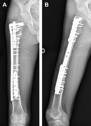
Intercalary Segmental Allografts
Intercalary segmental allograft avoids complications associated with osteoarticular allograft, such as degenerative joint disease or instability. The functional results are better than osteoarticular or allograft composite, with lower infection rate (6%). The main complications in this type of reconstruction are mechanical such as fracture and nonunion. These 2 complications could be avoided with a rigid internal fixation with plates and screws. Frisoni and colleagues analyzed factors that affected outcome in intercalary segmental allograft and found that the use of intramedullary nails had an adverse relationship with fracture and nonunion. Although this reconstruction combined with a vascularized fibular graft diminished the rate of nonunion, they found no differences in fracture rates. The authors found similar results in intercalary femoral allograft with a fracture rate of 17%, leading to replacement of the graft in all cases except one. In the tibia, however, we found that the fracture rate was lower (11%) with intercalary allograft, and all these fractures were treated with autologous graft without failure of the original allograft. These differences between femur and tibia allografts could be explained by the presence of the fibula, which could help to transmit load to the ankle joint. Although Ortiz-Cruz and colleagues reported that they did not find any differences in nonunion in diaphyseal or metaphyseal bone, or with different internal fixation, Cara and colleagues and Muscolo and colleagues reported nonunion mainly in diaphyseal osteotomies. In the last study, the nonunion rate for diaphyseal junctions was higher (15%) than the rate for metaphyseal ones (2%).
Surgical Technique
The surgical procedure begins with resection of the lesion, including biopsy scars with appropriate bone and soft tissue margins ( [CR] ). The surgical resection could be performed more accurately assisted by computer navigation. After being thawed in a warm solution a fresh deep-frozen allograft segment, sized to fit the bone defect, is cut to the proper length. All allograft-host junctions are made with a transverse osteotomy. Each osteotomy is first stabilized with a short LCP plate; this simplifies the reduction of each osteotomy. After compression of the host donor junction with at least 2 cortical screws, we placed locking screws. When both osteotomies are secured the authors placed a third long plate to achieve a more rigid fixation and protect the whole graft ( Fig. 3 ).
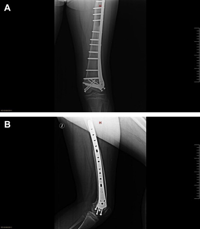
The use of intercalary allografts in the last 2 decades has increased with advances in imaging techniques that allow for more accurate transepiphyseal osteotomies with epiphyseal preservation. The epiphysis can then be fixed to intercalary segmental allografts, maintaining the articular cartilage and joint ligaments of the patient. These osteotomies usually heal without complication, allowing limb stability and normal joints motion. In recent years, the use of navigation was reported to perform these osteotomies more accurately.
The surgical technique after transepiphyseal osteotomies differs in that only cancellous screws are used for fixation at the epiphyseal host-donor junctions when a thin epiphyseal segment is saved ( [CR] and [CR] ). However, the authors recommend placing at least 1 or 2 screws of the long plate in the host epiphysis to protect the construct when possible ( Fig. 4 ). They found that although this junction healed quickly, the metaphyseal area is the one that suffers most of the allograft fractures.
Hemicylindrical Intercalary Allografts
Hemicylindrical intercalary allografts were originally described after resection of low-grade surface tumors ( Fig. 5 ) or to reconstruct the cortical window after intralesional curettage of a benign lesion ( [CR] ). In recent years, this type of reconstruction had been described after multiplanar osteotomies to save the joint with or without the assistance of intraoperative navigation.
Related posts:
 Soft Tissue Balance in Total Knee Arthroplasty with a Force Sensor
The Role of Acromioplasty for Rotator Cuff Problems
Soft Tissue Balance in Total Knee Arthroplasty with a Force Sensor
Trauma
Evaluation and Medical Management of Fragility Fractures of the Upper Extremity
The Principles and Applications of Fresh Frozen Allografts to Bone and Joint Reconstruction
Soft Tissue Balance in Total Knee Arthroplasty with a Force Sensor
The Role of Acromioplasty for Rotator Cuff Problems
Soft Tissue Balance in Total Knee Arthroplasty with a Force Sensor
Trauma
Evaluation and Medical Management of Fragility Fractures of the Upper Extremity
The Principles and Applications of Fresh Frozen Allografts to Bone and Joint Reconstruction
Stay updated, free articles. Join our Telegram channel

Full access? Get Clinical Tree


