11 The liver and spleen
 The Liver
Portal hypertension
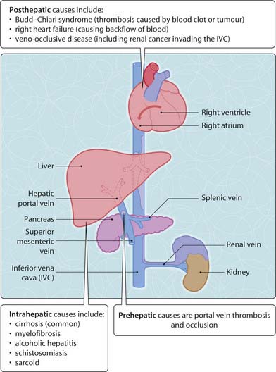
Portal hypertension (Fig. 3.11.1) occurs when the outflow to the hepatic portal vein is blocked, causing backflow. The portal system has five sites of anastomoses with the systemic circulation (portal systemic anastomoses; Table 3.11.1). When portal hypertension occurs, portal blood flow through these sites increases and causes formation of varices. The most important site is the lower oesophagus, as these oesophageal varices may cause massive upper gastrointestinal haemorrhage and possibly death (Ch. 8). Portal hypertension can also cause ascites (below).
Table 3.11.1 Portosystemic Anastomoses
| Area | Portal veins | Systemic veins |
|---|---|---|
| A Lower oesophagus | Left gastric | Azygos (via oesophageal veins) |
| B Rectum | Superior rectal into inferior mesenteric | Middle/inferior rectal into pudendal/internal iliac |
| C Bare area of liver | Hepatic | Inferior phrenic |
| D Periumbilical | Paraumbilical vein | Anterior abdominal wall |
| E Retroperitoneum | Colic and splenic | Lumbar |
Management
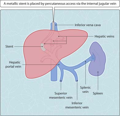
The transjugular intrahepatic portosystemic shunt (TIPS; Fig. 3.11.2) may be used for long-term management. It is an interventional radiological procedure. A metallic stent creates a communication between the hepatic vein (low pressure) and the portal venous system (high pressure), allowing blood to flow into the inferior vena cava without passing through the liver.
< div class='tao-gold-member'>
Stay updated, free articles. Join our Telegram channel

Full access? Get Clinical Tree


