43 Examination of the neck


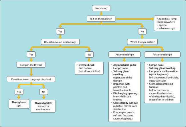
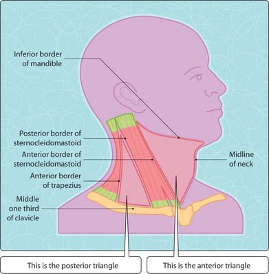
The scheme shown in Fig. 3.43.1 contains the steps needed to differentiate neck lumps. These can be considered as midline or non-midline (Ch. 42) and whether they are in the anterior or posterior triangle of the neck (Fig. 3.43.2). The anterior triangle is bordered by the midline of the neck (anterior border), the anterior border of sternocleidomastoid (posterior border) and the inferior border of the mandible (superior border). The posterior triangle is bordered by the posterior border of sternocleidomastoid (anterior border), the anterior border of the trapezius (posterior border) and the superior border of the clavicle (inferior border).
The most effective investigations for an unknown lump are CT, MRI and fine needle aspirate cytology.


