The fascial distortion model

7.15


The fascial distortion model


Introduction: connective tissue as mechanosensory system


In spite of the fact that connective tissue is in general envisioned as a scaffold with minimal metabolism and blood supply, it serves as a transportation network for all adjacent tissues. All structural changes in the connective tissue, whether caused by trauma or disease, lead to an alteration of the metabolism of all other tissues involved. This close relationship enables the connective tissue to be a very sensitive indicator for virtually any dysfunction in all areas of the body. Proprioception and nociception – the senses for position, touch and pain – are located in the connective tissue.


Wherever disease, tumor, or trauma cause damage in the body, pain occurs as soon as fascia is involved. This phenomenon, although well known in the intestines and the brain, is often not applied to the musculoskeletal system. The signaling function is not exclusively responsible for pain and touch, and muscle energy (with or without movement) is observed and measured by the surrounding connective tissue. Due to the close connection between fascia and nerves – every nerve consists mainly of connective tissue – information is precisely conducted. Overall, the fascial net is a complex and precise mechanosensory system. Not only are the positions of the limbs observed, but also all traumatic or metabolic dysfunctions.


The patient as expert – the Typaldos model


Mainstream medicine takes little advantage of this most complex information network. Physicians increasingly ignore it and instead use technological investigation devices. The patient gets the impression that his perception and description of pain or discomfort disturbs, rather than supports, the diagnostic process. Accordingly, magnetic resonance imaging (MRI) scans are used to locate the disorder even though key studies suggest that MRI scans are, in spite of their spectacular resolution, questionable tools to locate the source of pain (Jensen 1994; Chou 2009).


In most contemporary medical healing approaches, it is the expert practitioner who makes a diagnosis due to his extensive training and skills. The patient in general is unable to be involved in these deliberations, due to the lack of a common language and the impression that their knowledge is inferior.


This frustrating experience was the origin of Stephen Typaldos’ fascial distortion model (FDM) (Typaldos 2002). Typaldos, an osteopath and emergency physician, found it a frustrating experience to learn how little medical and osteopathic treatments could help patients suffering from so-called soft tissue injury. Conditions such as sprained ankles, lower back pain, and neck pain seemed to be almost unresponsive to the treatments he could offer within an emergency department. Improving his manipulation skills or increasing the diagnostic effort (MRI, ultrasound, nerve conduction, etc.) did not improve the outcome.


Typaldos instead started to ask the patients what kind of treatment they themselves thought might bring relief, and, to the patients’ surprise, he applied the treatments they proposed; for example, pressing on a particular spot in a patient’s back, which she was unable to reach herself, or pulling on the arm or on the skin of a patient as they suggested. In many cases these procedures had better results than any other therapy these patients had undergone previously in their career. This again was a big surprise to Dr. Typaldos. This method of diagnosis and treatment planning was unknown in both mainstream medicine and in the kind of osteopathy he had learned. On the other hand, the symptoms and reported improvement could not be explained by conventional diagnoses.


Typaldos’ impression was that the patients had subconscious understanding of the nature of their condition and of possible solutions. Studying the patients’ stories and gestures, he found recognizable entities for which he saw fascia as the common denominator. Typaldos postulated three-dimensional alterations of fascia that could apparently be repaired by the maneuvers and techniques he had successfully performed under the guidance of his patients. Typaldos discovered six “fascial distortions”, as he called these new diagnoses (Table 7.15.1).


The fascial distortions


The six distortions are listed in the order they were discovered by Typaldos.


Herniated triggerpoint (HTP)


All compartments of the human body are separated by fascia. The main purpose of this kind of fascia is to seal the cavities and prevent enclosed organs from protruding out of the cavity. In general (apart from the thorax) the pressure gradient makes tissue and fluids prone to protrude towards the surface as soon as a gap in the sealing fascia opens (a hernia). Some of these hernias (e.g., inguinal hernia) are well explored and there are successful (mainly surgical) treatments available. Others are less well known and the complaints are not understood as a hernia. Only the concerted occurrence of an opening of the canal, a pressure peak, and the protrusion of tissue out of the compartment can be identified as pathologies. Each component occurring on its own is physiological.


Once a hernia is formed it is maintained by the pressure gradient. Spontaneous healing is not expected but reduction leads to repair. A closure of the aperture (which is physiological) is not strictly necessary but might help avoid a relapse.


In the FDM, hernias within the musculoskeletal system play a major role in explaining patients’ complaints, and entirely new treatment options utilizing the concept of reduction, which would be deemed impossible in other models. Other medical models that concern these complaints are interpreted very differently.


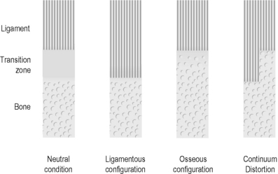
Continuum distortion (CD) (see Fig 7.15.1)


To understand CD, it is necessary to look into the nature of bone and ligament in a fundamental way. In the traditional model the point where bone and ligament come together is called “insertion”. A junction of two obviously different structures is postulated. This is contradictory to the enormous stability of this junction. In the continuum theory, as part of the FDM, bone and ligament are envisioned as one structure. Ligament is accordingly considered to be demineralized bone and a bone calcified ligament. Single bones are not considered to be components of “in-vivo anatomy” and only come into existence after collagen biodegradation, in mechanical injury, or in medical dissection studies. The same principle applies for ligaments.


That the majority of fibrous pathways are within bone suggests an important role for collagen fibers in bone stability. Fascial fibers can be considered as the highly tensile component of this compound, analogous to the steel in ferroconcrete. A purely mineral-based material can never be resistant to bending forces: it is only resistant to compression forces. The FDM postulates that the tissue in the transition zone has the ability to shift between two states (i.e., between bone and ligament). Depending on the demands, it can become ligamentous or osseous by shifting calcium matrix out of or into the bone. Since the calcium concentration is high in the bone and low in the ligament, the soluble mineral is prone to spread into the ligamentous zone driven by the force of entropy. Physical activity forces the calcium back into the bone by osteoblast activity in the bone and mechanical stress on calcium that has shifted into the ligament. The opposite is a common finding in immobilized patients with a lack of physical activity in intensive care units. Osteoporosis in the bones and exostosis in the ligament are the manifestations of this.


According to the FDM, this phenomenon occurs several times a day on a small scale when physical activity leads to a small shift of the transition zone towards its ligamentous configuration, and inactivity leads to a small shift towards osseous configuration. This shift is only possible if synchronized in the entire “insertion”. As soon as one part of the transition zone shifts in the osseous configuration while the rest shifts into the ligamentous configuration continuum distortion occurs, leading to immediate loss of functional ability due to disturbed proprioception. CD is envisioned as a pathological step in the transition zone between bone and ligament. To envision bones and ligaments as one structure (the continuum) with calcified and decalcified zones allows an entirely new perspective on injury and complaints located in or adjacent to “insertions”.

Stay updated, free articles. Join our Telegram channel

Aug 24, 2016 | Posted by in ORTHOPEDIC | Comments Off on The fascial distortion model

Full access? Get Clinical Tree

Get Clinical Tree app for offline access