The deeper fasciae of the neck and ventral torso

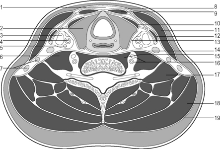
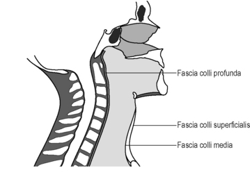
1.7


The deeper fasciae of the neck and ventral torso


Introduction


In early embryogenesis the originally unitary body cavity is divided by the development of the transverse septum, the future diaphragm, and the pleuropericardial septum secondarily into the thoracic cavity and the peritoneal cavity. Both structures arise from the mesenchymal concentrations of connective tissue in the neck (see below).


The two pleural cavities and the pericardium – pericardial cavity – are formed within the thoracic cavity. These cavities are completely filled with their respective contents (lungs, heart, abdominal organs) and a small quantity of serous fluid.


Both pleural cavities are lined with the parietal pleura; this continues along the hilum of the lungs into the visceral pleura. At the top, the parietal pleura forms the two pleural domes, which rise up on both sides over the upper thoracic aperture and reach the posterior head of the first rib. The parietal pleura folds in on itself in a caudal direction deep into the costodiaphragmatic recess. In this area the pleural and peritoneal cavities are separated from each other by the thin muscular plate of the diaphragm. The domes of the diaphragm, which rise up well into the thoracic space, are joined to each other by the central tendon.


Medially, the two pleural cavities border the mediastinum, which is closed at the bottom by the diaphragm, but in a cranial direction is in direct connection with the connective tissue-filled crevices of the neck.


The anterior mediastinum contains the pericardium with the heart and its arterial and venous vessels; posterior from here lie the trachea with the origins of the bronchial tubes, the aorta, the esophagus with the branches of the vagus nerve, and the thoracic duct.


The cranial border of the abdominal cavity is the diaphragm, the lateral border is the abdominal wall, and the caudal border is formed by the pelvis and the pelvic floor. With the parietal peritoneum as internal lining, it encloses a serous cavity, which contains the digestive organs. Posteriorly, the parietal peritoneum borders a connective tissue space, the retroperitoneal space, which contains the retroperitoneal vessels and organs.


Neck fascia


A visceral cord lies in front of the cervical spine consisting of nerve and vascular bundles and cervical organs. A transverse section at the level of the first tracheal cartilage shows that the visceral cord is fairly central and the posterior border is directly on the cervical spine. It is surrounded by a connective tissue muscular coating composed of three fascia and pretracheal and prevertebral muscles and a dense covering (Fig. 1.7.1).


The visceral cord in the neck is therefore mobile against its covering and is lifted and lowered by the act of swallowing and can follow any movement in the cervical spine. Furthermore, the vascular nerve bundle formed by the carotid sheath is arranged with the internal jugular vein, the common carotid artery, and the vagus nerve so that it remains as protected as possible from the changes in position and shape of the cervical spine and the viscera of the neck. The junction of the carotid sheath with the omohyoid muscle further ensures that the reflux venous blood flow runs from the brain independently of position.


Arrangement of the three neck fascia


Fascia colli superficialis (lamina superficialis fasciae cervicalis)


Lying directly below the skin and platysma, the superficial sheet of the cervical fascia stretches over a wide area. As continuation of the masseteric fascia it runs from the mandible and the floor of the mouth over the interposition of the hyoid bone in a caudal direction, after which it is attached to the clavicle and the manubrium sterni and continues into the pectoral fascia. It shifts significantly during respiration because of its attachment to the sternum. It forms a lateral sheath around the sternocleidomastoid muscle, which glides freely within this covering. At the back it covers the corpus adiposum of the lateral cervical triangle and continues to the trapezius muscle. Upwards in a posterior direction it traverses the mastoid process and extends from there as far as the superior nuchal line.


The superficial neck fascia is structured differently in different regions; in the area of the digastricus muscle, the anterior part of the trapezius, and the lower third of the sternocleidomastoideus it is delicate. In the cranial third, the superficial fascia is very strong and is almost immobile against the subcutis. In the lower part of the lateral cervical triangle, it is like a sieve because of the passage of the supraclavicular nerve and its accompanying vessels. Because of its many connections to neighboring structures it is under permanent tension in a living being (Fig. 1.7.2).


Fascia colli media (lamina pretrachealis fasciae cervicalis)


The middle neck fascia is composed of the fascia coverings of the infrahyoid muscles and forms a firm triangular skirt in front of the cervical viscera. Cranially, it grows fixed to the hyoid bone; caudally it runs with the coverings of the sternohyoidei and sternothyroidei through the upper thoracic aperture behind the sternum and inserts at the manubrium sterni. Laterally, it is fixed to the posterior surface of the clavicle and is bordered posterolaterally by the fascia coat of the omohyoideus, which arises from the medial angle of the scapula and runs in an arc to the hyoid. The two omohyoid muscles span the middle neck fascia and are firmly held in place by their tendons. As the middle neck fascia is involved in the formation of the carotid sheath, the lumen of the internal jugular vein is held open by its traction. This principle also applies to the superficial veins of the neck and the deep veins, which come from the area of the neck and shoulder girdle.


Above the sternum is a space between the middle neck fascia and the superficial neck fascia filled with fatty tissue and veins, the suprasternal space. It extends laterally as far as the sternocleidomastoideus and cranially as far as the level of the ring of cartilage around the larynx. Both fasciae are welded together to the hyoid bone.


Fascia colli profunda (lamina pretrachealis fasciae cervicalis)


The deep neck fascia is fixed to the anterior longitudinal ligament of the cervical spine and provides a connective tissue cover for the prevertebral muscles (Mm. longi colli) and the lateral neck muscles (Mm. scaleni). These coverings are therefore considerably involved in the construction of the posterior wall of the inside space of the neck. Cranially, the deep neck fascia is fixed to the base of the skull. Laterally, it is connected to the fascia of the levator scapulae, the nuchal fascia and the superficial sheet of the neck fascia. From the scalenus medius it reaches the clavicle, and together with the scalenus anterior it reaches the outer surface of the thorax. Over this area it covers the nerve and vessel bundle to the upper extremities – the brachial plexus and the subclavian artery.


Caudally, the deep neck fascia travels into the endothoracic fascia.


Connective tissue tracts radiating from the deep neck fascia of the neck into the suprapleural membrane contribute to the fixation of the pleural dome (see below).

Stay updated, free articles. Join our Telegram channel

Aug 24, 2016 | Posted by in ORTHOPEDIC | Comments Off on The deeper fasciae of the neck and ventral torso

Full access? Get Clinical Tree

Get Clinical Tree app for offline access