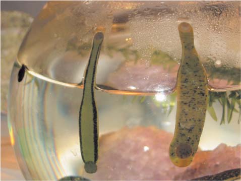
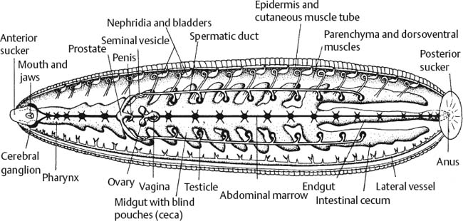
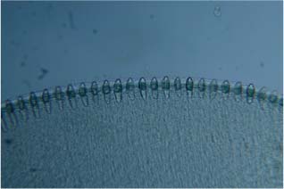
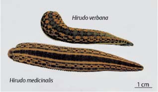
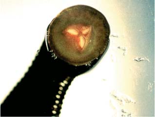
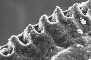
3 The Biology of Leeches Roughly 600 leech species have been identified to date, but only about 15 are used in medicine. Leeches classified as “medicinal leeches” in the narrower sense have been used to treat patients around the world, especially in Europe and the United States, for centuries. Because of its excellent therapeutic properties, the medicinal leech described here is considered to be the favorite species. In the past, the medicinal leech was generally assumed to be a single species with regional color differences. Based on this logic, the two phenotypes with the most distinctive coloring were originally named Hirudo medicinalis medicinalis and Hirudo medicinalis officinalis (Fig. 3.1a, b). The latter, which is also known as the “Hungarian leech,” was later renamed Hirudo officinalis after it was given species status. According to current scientific understanding, the different patterns on their body surfaces indicate that the two presumed phenotypes are indeed two different species: Hirudo medicinalis Linnaeus, 1758 and Hirudo verbana Carena, 1820. The species concept was recently proved by DNA analysis [26]. In leech therapy, the existence of two species seems to be irrelevant because both have been used as equivalents throughout history. As far as can be determined, no differences in their spectrum of activity are known or have ever been investigated. The composition of their saliva is also identical [24]. Since the natural supply of Hirudo medicinalis became scarce due to the intensive exploitation of leeches in the 19th century, Hirudo verbana has been virtually the only leech species used around the world for decades. Because the two species were originally assumed to be color variants of a single species, most authors (e.g., [39]) referred to both as “Hirudo medicinalis,” without differentiating between the two types. In this book, however, the terms “leech,” “medicinal leech,” and “Hirudo” will therefore be used synonymously to refer to both Hirudo medicinalis and Hirudo verbana. Fig. 3.1a There are no known differences in the activity spectrums of the close relatives Hirudo medicinalis and Hirudo verbana (Carena 1820). The two medicinal leech species are used as equivalents. Because of overharvesting of Hirudo medicinalis in the 19th century, Hirudo verbana is now predominately used in medicine around the world. Hirudo medicinalis typically has a drop-shaped pattern on the lateral aspect of its body (from [26]) Fig. 3.1b Hirudo verbana (left) can best be distinguished from Hirudo medicinalis (right) based on differences in color patterns on their ventral body surfaces. (Photo by M. Roth) While on the subject of terminology, it should also be noted that the leech is not a worm. The term “worm” (vermis) is no longer used in the zoological nomenclature because the animals formerly grouped under the heading of “vermis” are wormlike members of completely different groups, as is now known today. Nonetheless, the leech is currently classified in the nomenclature as an annelid or “ringed” worm, making the earthworm its close relative. The term “worm” has therefore prevailed, although there is now a distinction. The term is a graphic description as well because, according to a leading German dictionary, the Duden [10], “worm” also has the meaning “wriggler.” The fact that many people suffer from misconceptions about leeches needs no further explanation. In German, leeches are called “Blutegel,” which is reminiscent of the word “Ekel” (disgust), but has completely different etymological roots. “Egel” derives from the Greek “echis” which, by the way, is related to “Igel” (hedgehog) and is the source of the Old High German word “Blutigel.” “Igel” means “not a snake”—which is a good thing or we would otherwise be dealing with “blood snakes”! Though zoologically untenable, this might perhaps improve the image of the leech because, phonetically, it is not as closely related to “Ekel” (= “disgust,” see above). The Swedish name is similar, namely “igle.” The leech’s role as a bloodsucker is emphasized in various other languages. For example, it is called “sangsue” in French, “sanguijuela” in Spanish, “sanguisuga” in Italian, “bloedzuiger” in Dutch, and “sanguisugolam” in Latvian (a very melodic name, indeed). In English, leech is derived from the Old English word “læce,” which also meant “physician” in the Middle Ages. The aim of this chapter is to convey knowledge of the biological characteristics of leeches from which information relevant to clinical practice can be derived and, secondly, to abolish misconceptions about leeches. Both patients and physicians suffer from misconceptions that can sometimes hamper treatment. The opposite is also the case, in that some experienced leech therapists make use of the unusual and archaic nature of this form of treatment to back up the merits of its unspecific active component. This mindset turns the leech into a kind of “performance artist” in the physician–patient relationship. This chapter will therefore elucidate leech biology from a treatment perspective. Leeches were presumably “applied” very much earlier than the documented records of medicine show—although not by humans. In the Cambrian explosion of life form development roughly 400–500 million years ago, prehistoric annelids evolved as segmented invertebrates from the first multicellular organisms. These “arch-annelids” ultimately evolved into leeches. Since they were among the first hunters in the earth’s history, they were one of the factors that guided selection and drove the pace of evolution. The prehistoric annelid was also an ancestor of humans. Segmentation is a consequential biological trait shared by the two species. Surprisingly enough, the blood of leeches and humans also has similarities, suggesting a common ancestry: Hemoglobin serves as the carrier of oxygen in medical leeches as well as in humans, but is dissolved in the respiratory fluid of the one (leech) and is stored in blood platelets/erythrocytes in the other (human). Since the leech is a skeletonless invertebrate, confirmed fossil finds of Hirudinea are rare. The only two confirmed finds date back to the Jurassic period. Based on fossil evidence, we know that the general structure of the Jurassic leech was very similar to that of the modern leech [22]. Therefore, the basic model for Hirudo was already “set” at a very early stage of history, and the remaining time was available for “fine-tuning.” If one assumes that evolution follows a goal-oriented course, it might seem that Nature is intentionally making refinements that facilitate the development of cooperation between the “perpetrator” (leech) and the “victim” (mammals, patients, etc.).1 The medical significance of the biochemistry of the leech clarifies this relation. The saliva of leeches is a highly efficient and highly complex trait acquired over the course of the evolution of the species. The composition of the saliva was apparently a “main area of evolutionary development”: When the leech bites its victim, the effect is a combination of at least 30 individual chemicals. However, only eight constituents of leech saliva have so far been characterized in terms of chemical structure and mechanism [2] (see p. 131 and Fig. 11.1). In some cases, the functional value of the compound (e.g., hirudin) to the leech is immediately apparent. Hirudin enables the blood to flow for some time without it clotting. This is the fundamental fact that makes the process possible at all. This anticoagulant protein serves to make the victim’s blood flow more readily while the leech is sucking it and was therefore favored by evolution. Calin (see p. 134 and Fig. 11.1) is another constituent of leech saliva. The main function of this protein is to induce secondary bleeding, which can last up to 12 hours. At first glance, you might wonder why the leech would need to produce a substance that makes the wound bleed for a relatively long time. After all, the leech detaches itself an hour or less after biting its victim and is no longer connected to the victim’s blood supply. Consequently, the leech has no direct benefits to gain from the prolonged blood flow. Nature normally does not allow itself the luxury of producing a protein for no apparent reason. The answer to this puzzle may be that the secondary bleeding is designed to attract other leeches, which would function as a means of preserving the population. However, water movement alone is a well-known signal that serves this purpose, so it still would not be necessary to produce a protein with the same function. The most likely answer is that the induction of prolonged bleeding is designed to help the host cleanse the wound and thus serves as a simple yet effective means of disinfection. It reduces the risk of infectious diseases and sepsis, which the prey could contract through infection, for example with the leech-borne bacterium Aeromonas (see p. 143 ff), or through secondary contamination. The production of calin can therefore be interpreted as a form of “resource management.” In other words, if the leech’s prey dies of sepsis, it cannot return to the place where it was bitten, and if it develops a painful infection, it may not be able to come back, but a “satisfied” host is more likely to return. The inclusion of calin in leech saliva therefore functions to preserve the population of the leech’s ecological niche (for the biochemistry of leech secretions, see Chapter 11, p. 131 ff). Leech saliva has other generally beneficial effects. Therefore, encounters between the leech and its victims within the ecological niche are not entirely coincidental because the victim is not unhappy about returning. This behavior establishes the basis for joint evolution characterized by species cooperation. The prey or host animals, including humans, had millions of years’ time to become accustomed to the leech. Though not systematically investigated, it is probable that animals ranging from lower species to mammals learned to “appreciate” the leech as a “therapist.” This hypothesis is based on various evidence, for example, the fact that cattle with joint diseases apparently like going to leech-infested waters. According to reports that I have received from people in Greece and India, among other places, cattle seem to deliberately—as far as one can judge a cow’s intentions–expose themselves to leech bites (i.e., “get treatment”) for extended periods in order to leave the site more “light-footed” than before. Leech therapy has also been successfully introduced in modern veterinary medicine. Most dogs are said to be unusually quiet while the leech is “docking” onto them, as if they instinctively knew it was going to help them [32]. One can therefore assume that even Pithecanthropus, his ancestors and indeed Homo sapiens, also recognized and exploited the healing effects of the leech. In the Stone Age, people employed different techniques of venesection with and without leeches. They interpreted leeching as a simple method for “removal of evil spirits” [17]. Likewise, some para-medical therapists today refer to this as the “removal of the evil.” After prehistoric times, Hirudinea entered the written history of medicine and has since passed through it in waves of popularity. The infor mation in ancient writings does not always allow one to clearly identify which of roughly 600 existing leech species was used. According to Arndt [1], 15 species are used for medicinal leech therapy. The natural leech supply was never seriously endangered before the 19th century, during which, however, entire leech populations were then devastated. Mainly supported by the teachings of the French physician Broussais, the practice of leeching grew explosively in the 19th century, which ultimately led to the outbreak of true “leech mania” or “vampirism” in Europe (see Chapter 2, p. 9). In those days, leeches were returned to nearby ponds after use. There was no fear of transferring diseases through microorganisms since people did not know that microbes existed. However, other practices led to a drastic reduction in the domestic leech supply. One such practice was the unrestrained removal of leeches from their natural habitats in order to ship them by the ton, for example from Hamburg to France, America, Australia, and England.2 It is also safe to assume that many of the leeches died after the treatments and, thus, were never returned to the cycle of nature.3 German businessmen proposed the foundation of a prestigious leech trading company, the “Actiengesellschaft Hirudinea” in 1863 [31]. This seemed like a very promising business opportunity, but their idea came too late. In the age of Koch, Pasteur, and Virchow, leeching disappeared from the medical repertoire toward the end of the 19th century. The once endangered leech populations had a relatively long time to recover during the 20th century, but regained the justifiable attention of physicians in the second third of the century. Now that the mechanism of action of leeching was relatively well known, leeches were applied very selectively and usually in small numbers. This hiatus in the 20th century did not suffice for regeneration of European leech populations, because the required wetlands had meanwhile been lost through drainage or had otherwise become unfit for leech survival. A large portion of natural leech biotopes (e.g., shallow ponds in meadow-lands) had been destroyed, and the remaining leeches no longer had access to the prey they needed for reproduction—namely mammals. Furthermore, environmental toxins have made it even more difficult for these sensitive organisms to survive in Europe. The number of natural leech habitats existing in Europe today is very small indeed. Most leeches now used in Central Europe for medicinal purposes are imported (mainly from Turkey) and are seldom grown domestically. Because the active constituents in leech saliva are effective ingredients in ointments and other topical products, the Turkish leech populations are very tightly monitored because leeches are very much in demand, alive or dead. Several tons of leeches, both fresh and frozen, are imported to Europe each year. Conservationists had hoped that recombinant hirudin manu factured from genetically modified bacteria and yeasts would soon be able to reduce the pressure on leech populations, but this hope has yet to be fulfilled. Because of the variety of constituents and complex mechanisms of action of leech saliva, live leeches are more effective than pure hirudin in a number of cases. The number of live leeches used in Germany alone is probably around 300 000–400 000 each year. Assuming each leech weighs around 3 g, this equates to roughly 1.2 tons of leeches per year. On July 24, 2004, the American Food and Drug Administration (FDA) officially approved the leech as a “medical device” [37]) based on scientific evidence of the efficacy of leeching in diseases such as osteoarthritis of the knee [30]. This may spark a new round of pressure on the species. Although the approval of leech therapy may be good news for humans, it is not so great for the leech—at least not yet. Ironically, the evolutionary feat of adapting to its prey ultimately resulted in more harm than good for the leech. Considering the beneficial effects of the leech, a discussion has arisen as to whether it is more biologically appropriate to classify the leech as “symbiotic” (a much more congenial and just description) rather than “parasitic” or “ectoparasitic.” The roles of the protagonists have changed: Leeches are now being parasitized by humans. As mentioned above, the usefulness of the leech for humans is based on a survival strategy developed by the leech over the course of evolution. Considering that leeches have repeatedly been on the brink of extinction, it would be smart to develop new and efficient strategies to ensure their continued well-being for a change. For these reasons, Hirudo medicinalis was included in Appendix II of the Washington Endangered Species Act, which was designed to impose harvesting quotas and control leech trade. Under this agreement, it is mandatory for anyone buying or selling leeches to submit a CITES report form.4 In other words, written permission to conduct the transaction must be documented by means of a CITES form whenever leeches are imported or exported. In Germany, the corresponding legislation is supervised and executed by the Federal Agency for Nature Conservation (BfN): “A person who goes to the zoo, eats herring filets, keeps a caged parrot, or purchases a leech extract ointment to improve the blood flow rarely ever sees how these events are related. In countries around the world, including Germany, wild animals and wild plants are being removed from their natural environments for trade and manufacturing purposes. Many animals around the world are currently endangered because they are being used as foodstuffs, traditional remedies, or live commodities. On the initiative of the Federal Ministry for the Environment, Nature Conservation and Nuclear Safety and the Federal Agency for Nature Conservation (BfN), criteria specifying ways in which the use of wild fauna and flora can be conducted in harmony with Nature have now been proposed.“[6] In order to understand the structural concept of leech anatomy from a biological perspective, we will take a look at how the animal behaves in freshwater ponds. Its natural behavior is characterized by the following cycle of recurring events: a) Quietly searching for food and observing potential prey while swimming slowly and floating near the water surface for extended periods; this can last months to years; b) Quickly attaching to prey and feeding in a rapid series of movements; feeding is usually finished within a few minutes; c) Submersion to a hiding place in the depths of the water body to rest and digest the meal; this resting period may last several months to years. There is frequently a smooth transition from (c) to (a). The leech can ingest relatively large amounts of blood (up to 10 times its body weight) each time it feeds. This allows the leech to survive for up to two years between feeding. This thrifty feeding behavior is partially due to the limited supply of mammal hosts and is reflected in the simple but very advantageous body structure of the leech (Fig. 3.2). With respect to organ size, Hirudo consists almost entirely of a double-walled “digestive tube.” The foregut, midgut (stomach), and hindgut occupy the largest volume of organs and tissues in both the filled and empty states. The foregut, comprising the mouth, pharynx, and esophagus, makes up roughly two tenths of the entire length of the animal. The midgut (stomach), which consists of 10 pairs of blind pouches, makes up five tenths of the total length. The three-part hind-gut makes up roughly three tenths of the total length. Only one bacterium species is equipped for long-term survival in the leech stomach (cf. p. 142 ff). This species happens to be the symbiont the leech needs to digest its food: Aeromonas biovar sobria veronii (formerly called Pseudomonas hirudinis and many other names [14, 15]). This amazing coincidence is very helpful for medicinal leeching. The leech stomach (crop) is an enormous “storage chamber” that enables the leech to survive for years without food. It is connected to the hindgut, the site where low-energy digestion takes place (anaerobic digestion can also be performed as needed). Even after the stomach contents have been fully depleted, the leech can subsist for months on its own body substance. The leech has a small anterior sucker which surrounds its pointed apical mouth. The anus opens on the dorsal surface above the large rear sucker. The leech also has two other types of excretory organs: the segmental nephridia and hermaphrodite (male and female) reproductive organs. Respiration takes place through the body wall. Hemoglobin dissolved in the blood transports oxygen to all parts of the body via a contractile capillary network. As an invertebrate animal, the leech has a soft body that lacks a skeleton. The body structure of Hirudinea resembles that of their phylogenetically close relatives, the earthworms (Annelida, Oligochaeta), in that it also displays segmentation. The body of the medicinal leech is composed of 34 segments. Segments 9–11 form the clitellum, the organ responsible for cocoon secretion, which is only visible in the summer months. The last seven segments form the large rear sucker. Fig. 3.2 Anatomical structure of the medicinal leech (ventral surface). Secondary annulation is seen as folding of the body wall (adapted from Storer & Usinger, in [41]) Secondary annulations, consisting of 105 external rings (annuli), obscure the internal segments. Each segment is covered by up to five annuli. The annuli can stretch like an accordion. They provide skin reserves that expand to accommodate the large volumes of blood ingested during feeding. The annuli also assist in locomotion. Externally, the only sign of internal segmentation is the recurrent reddish orange/olive/black striped pattern on the back of Hirudo verbana and Hirudo medicinalis (Fig. 3.1a). No two patterns are exactly identical. Internally, segmentation is reflected by the metameric arrangement of the 32 ganglia, excretory bladders, nephridia, and seminal vesicles (Fig. 3.2). As simple as the gross anatomy of the leech may initially appear, the individual structures are surprisingly complex. Its central nervous system, which is crucial for successful hunting, localization of enemies, and general coordination, is very highly developed and specialized (see p. 28 ff and 33 ff). The cephalic region of the leech fulfills attachment, biting, secretory, sucking, and locomotor functions, contains gustatory receptors, temperature receptors, and eyes, and is connected to the lower pharyngeal ganglion. The leech head was an early climax in increasingly specialized head development over the course of evolution. The body wall of the leech must also fulfill numerous tasks. The strong muscles of the body wall permit prolonged swimming, crawling, and respiratory movements and they help the leech to remain attached to its prey and other structures (see p. 31 ff). The oral anatomy and function of medicinal leeches plays a central role in leech therapy. To find the right spot to bite, the leech must probe the host’s skin. It uses highly sensitive chemical, heat, and touch receptors distributed most densely in the upper lip region to test the skin for the desired characteristics. If the leech tastes blood, glucose, or sweat, senses that the temperature is between 35°C and 40°C, and feels pulsating movements indicating the proximity of an artery, then it knows it has found a good feeding site. The primary oral cavity (oral sucker) of the medicinal leech is separated by a velum from the secondary oral cavity (mouth) containing the jaws (Fig. 3.3a). Both can be retracted and extended. Before feeding, the leech starts to pump air into and out of the cavities and positions its jaws on its victim. While feeding, the leech slides its many-toothed jaws back and forth to slice into the victim’s skin in a sawing motion, while the cavities (including the pharynx) contract and dilate rhythmically. The pumping motion creates a vacuum, which results in the suction of blood. This can readily be seen by the outside observer, especially in the final stages of feeding. The oral sucker spreads dorsally to form an upper lip (prostomium), which contains taste receptors. An adhesive substance (mucus) secreted by the oral sucker and the pharyngeal pump mechanism help the front and rear suckers achieve powerful suction pressure (roughly 0.2 atm). As a result, the leech can attach itself to almost any surface: Sandpaper or glasspaper of any grain size, Velcro, coarse or fine wire, or plastic wickerwork, and fabrics or even vertically positioned surfaces coated with Vaseline are no major problem for a leech to climb. Fig. 3.3a Cross section of the oral sucker of the leech The mouth of the medicinal leech is located in the anterior sucker. Just within the oral cavity are three jaws, the three sides of which form 120° angles, resembling the Mercedes-Benz symbol (Fig. 3.3a). Figure 3.3b provides a view of—or better, into—an oral sucker of a leech attached to a glass plate. The yellowish orange lenticular regions are the lips, which contain the hardened masses of muscle comprising the jaws. The bladelike jaws have 60–100 small teeth along their edges, as is seen from a dorsolateral view in Figure 3.3c. Under a scanning electron microscope, interdental pores through which leech saliva is secreted can be identified (Fig. 3.3d) [31]. Leech saliva is produced in discrete, scattered salivary glandular cells that do not connect to form a proper gland. The external openings distributed across the dental ridge between each pair of teeth are the endpores of a central canal, that is, the endings of the secretory ducts of the glandular cells. Antagonistic muscles at the base of the jaw work in concert to move the jaw back and forth in a semicircular motion similar to that of a circular saw. Through this muscle activity, the relatively small contact area of the curved, semicircular jaw penetrates deeper and deeper into the skin. Successive penetration has two distinct advantages: First of all, it requires less force than, for example, a jaw with a linear tooth configuration. Secondly, the victim perceives less discomfort and is less likely to notice the bite. Thus, the chances are greater that the uninvited guest will go unnoticed. Leech saliva may also contain a substance with anesthetic properties that enhance this effect (for other chemicals in leech saliva, see Chapter 11, p. 131 ff). The leech secretes saliva into the wound through the interdental pores while sawing its teeth deeper and deeper into the host’s skin. Mechanically speaking, the anatomical structures work like extremely efficient micropipettes that inject pharmaceutically active substances into the wound. No microsurgical tool can perform such complex functions with such high precision, not to mention the perfect coordination of the “interlocking” spectrum of action of the active constituents in leech saliva. Fig. 3.3b Oral sucker attached to a prewarmed glass plate. The pharynx (striped) is contracted. The leech has expelled all the air between the sucker and the glass. The pharyngeal muscles begin to contract rhythmically, creating the suction pressure required to withdraw blood. At the same time, the sawlike blades of the jaws of the leech (yellow) penetrate deeper and deeper into the host’s skin while emitting pharmaceutically active substances. In the middle of the throat, at the imaginary point of intersection of the jaws, the pharynx now opens. (Photo by C. Morkel) Fig. 3.3c Segment (approximately one-third to one-quarter of the total jaw length) of the leech jaw, which can have up to 80 small, calcified teeth. The lower edge of the picture is equivalent to roughly 1.5 mm. The external openings (nephridiopores) of the salivary gland cells located roughly 5–10 mm away can be seen as discrete stripes in the hardened muscle tissue at the lower edge of the photograph. This anatomical configuration can be likened to a microsurgical/biochemical precision tool. (Photo by E. Schulte) Fig. 3.3d Segment of the leech jaw showing the interdental pores through which leech saliva and the substances contained in it are secreted. (Photo by C. Morkel) The pharynx (throat) connects the oral cavity with the esophagus (gullet). The esophagus is a muscular coat consisting of annular, longitudinal, and radial muscles (Fig. 3.3a). It has the highest concentration of different muscle types in the leech body. These muscle groups interact to produce the rhythmic pharyngeal muscle activity that occurs while the leech is feeding. In the first phase of feeding (suction), radial muscles lead to active dilatation of the pharynx. In the second phase (transport), annular muscles are used for active pharyngeal contraction. These peristaltic movements of the pharynx start at a frequency of roughly 2.4 Hz and slow to approximately 1.2 Hz or less toward the end of feeding [18]. The body wall of the leech has some remarkable features. As the interface between the leech and outside world, it must cope with the special demands of leech life. Therefore: • It is extremely elastic; • It performs respiratory functions; • It serves as a barrier against harmful substances and infections; • It permits osmoregulation and regulates the exchange of chemicals and gases; • It functions like a single complex sensory organ; • It can resist mechanical attacks; • It can shed its outer layer (cuticula); • It is pigmented for camouflage purposes; • It serves as a means of species recognition;
Introduction
The Evolutionary History of Leeches
Anatomy and Function
Oral Anatomy and Function
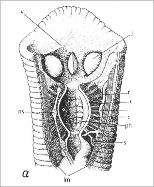
(from [19]).
Key to symbols:
j jaw
r radial muscles
c annular muscles
l lacunae
ph pharynx
s salivary ducts
lm longitudinal muscles
ns nervous systems
v velum
Skin, Muscles, Nerves, and Senses
Related posts:

Stay updated, free articles. Join our Telegram channel

Full access? Get Clinical Tree


