Sutures and glenoid anchors for instability

CHAPTER 5 Sutures and glenoid anchors for instability


Introduction


Shoulder instability is a common problem especially in the young and athletic population. This instability can be unidirectional (anterior or posterior) or multidirectional. In addition, the instability can manifest as a complete dislocation or lesser subluxations. The glenoid is a shallow concave socket, and its depth is increased at least 50% by the attached glenoid labrum. Providing glenohumeral stability to augment the labrum are the capsuloligamentous structures. Anteriorly static stability is provided by three glenohumeral ligaments. The superior, middle, and inferior glenohumeral ligaments resist anterior and inferior dislocation of the humeral head. The inferior glenohumeral ligament has anterior and posterior bands with an intervening sling and is the primary stabilizer to anterior humeral translation at 90 degrees abduction. The anterior band reciprocally tightens in abduction and external rotation, and the posterior band tightens with forward elevation and internal rotation.


When the glenohumeral joint dislocates, damage may occur to the bone, labrum, and the ligamentous attachments. The extent of this damage and the physiologic characteristics of the individual sustaining the dislocation influence the prognosis and have a bearing upon the appropriate treatment. A significant bone fragment may require reattachment of replacement. An engaging Hill-Sachs lesion may require a posterior humeral anchor to attach the infraspinatus tendon to fill the defect in the humeral head. Anchoring techniques may be required to reattach the fracture fragment. Concurrent with bony, labral, and ligamentous disruption, significant capsular stretching is a real possibility. To maximize the chances of success, the surgical treatment for shoulder instability must repair not only the torn labrum and bone but also address any stretched capsular tissue.


Arthroscopic stabilization techniques have advanced as a result of two major developments: suture anchors and ultra-high molecular weight polyethylene (UHMWPE) suture material. Although these anchors and sutures are now routinely used for both open and arthroscopic procedures, there can be no question that the innovation targeted arthroscopic procedures. Over time biomechanical studies on the properties of suture anchor fixation have lead to improvements in these anchors. The obvious benefits of stronger sutures have led to their wide acceptance.


Product innovation is not the only reason arthroscopic techniques have become the new “gold standard” for shoulder instability surgery; technique changes also play a major role. Thumbtacklike devices that reattach the torn capsuloligamentous structures medially on the glenoid neck have been abandoned because of the recognition that they are creating an anterior labral periosteal sleeve avulsion (ALPSA) lesion instead of reestablishing normal anatomic relationships. The recognition of the need for at least three anterior fixation points, locating the anchor sites on top of the glenoid cartilage (rather than medially on the neck), considering rotator interval closure to address inferior subluxation, adding posterior inferior sutures when appropriate, including a capsular plication stitch to address a patulous capsule, and better knot-tying techniques have all resulted in clinical outcomes consistent with those of open techniques.


Suture anchor designs are procedure specific. Some designs are best suited for holding multiple sutures in osteoporotic bone (rotator cuff repairs), or they allow static sutures to slide through the anchor eyelet with the option for independent tensioning after anchor insertion (lateral row cuff anchors). The focus of this chapter is on the anchors and sutures best suited for use in the glenoid labrum. The common features that make anchors well suited for a shoulder instability application include smaller size, reduced requirement for multiple sutures, and shorter lengths to avoid penetrating through the inferior glenoid at the 6 o’clock position.


Although not an absolute requirement but certainly preferable, biodegradable material is very attractive in a suture anchor, especially for one destined to be placed in the glenoid. While metal anchors were commonly used in the past, biodegradable anchors have comparable pull out strength; do not create problems in revision surgery or postoperative imaging;1 and, with the introduction of biocomposite materials, offer the prospect of osteoconductive behavior leading to their replacement with bone.


This chapter covers the current state of sutures and suture anchors appropriate for glenohumeral instability, including the materials and their properties, various knot configurations, biocomposite materials, and suture anchor characteristics. Each subject area concentrates on areas orthopaedic surgeons should be familiar with in order to make informed decisions when selecting these implants for patient use.


Suture material


Desirable material properties for an arthroscopic suture are similar to those for any surgical suture and include good handling characteristics, biocompatibility, adequate strength, and good loop and knot security. What distinguishes a superior arthroscopic suture is greater strength for a standard size and a low friction surface conducive to tying in the arthroscopic environment.


Many sutures are available and in addition to material and size differences can be monofilament, braided, blended, and absorbable or nonabsorbable. Each of these features has advantages and disadvantages of which the surgeon should be aware. A common biodegradable monofilament suture (polydioxanone or PDS) is adaptable to the arthroscopic environment and frequently used in shoulder instability surgery. PDS is often used as a shuttling suture for passing braided sutures through the glenoid capsuloligamentous tissue. It is often used for rotator interval closure stitches because it can be inserted directly with common suture hook devices (Spectrum system, Linvatec, Largo, FL; Ideal suture hook, Depuy Mitek, Rayham, MA) without the need for a shuttling device or suture. Because polydioxanone suture material degrades in a relatively short interval, its choice for suturing the rotator interval is based on the likelihood that should tightening be excessive, the degradation of the suture will allow the overconstrained tissue to stretch out with therapy. PDS degrades steadily after insertion, losing both mass and strength. At 2 weeks in vivo, PDS sutures retained 60% of the original strength and by 6 weeks 40%. The suture was almost completely reabsorbed by 9 weeks. Although PDS is easy to use and has relatively good strength, the stiffness associated with it results in PDS having a “memory,” with the tendency for knots tied with it to unravel if an insufficient number of backup half hitches are not tied. Nonetheless, PDS is a commonly used suture in glenohumeral instability surgery.


Until recently, braided polyester suture was the most commonly used suture in shoulder surgery. Although different brands were available, Ethibond (Ethicon Inc., Somerville, NJ) was the “gold standard” because it was easier to handle, more pliable, and passed easily through tissues because of a polybutylate coating. This coating is not present on Mersilene (Ethicon Inc., Somerville, NJ) and other braided polyester sutures. This changed when Arthrex (Naples, FL) introduced FiberWire suture, which has a braided polyester coat around a central core of multiple small strands of ultra-high molecular weight polyethylene (UHMWPE).


By way of background, the United States Pharmacopeia (USP) is an official public standards–setting authority. The USP standard correlated suture size and suture strength and set standard ranges for both. Following the USP standard, all number 2 size sutures were to have a breaking strength within a specific range. From the standpoint of the arthroscopic surgeon, this presented a problem. Unless considerable finesse was used during arthroscopic knot tying, the leverage applied to the suture by a knot pusher could break the suture. Additionally, braided polyester sutures were inclined to fray against the eyelet of a metal suture anchor or the insertion instrumentation, further diminishing the suture’s resistance to breakage. The introduction of FiberWire redefined suture performance. Although initially ignored by suture manufacturers, other arthroscopic instrumentation companies realized the advantages of the concept of “super strength” suture materials.


Dyneema fiber is an ultra-high-strength polyethylene fiber that offers a maximum strength combined with a minimum weight. It is up to 15 times stronger than quality steel and up to 40% stronger than aramid (aromatic polyamide) fibers on a weight-for-weight basis. This fiber was made available to many companies with various braid designs and was introduced into the field of arthroscopic surgery under several different brand names. These brand names include Hi-Fi (ConMed Linvatec, Largo, FL), Force Fiber (Stryker Endoscopy, San Jose, CA), Ultrabraid (Smith & Nephew, Andover, MA), Magnum Wire, (Axya Medical, Beverly, MA, and ArthroCare, Sunnyvale, CA) and MaxBraid PE (Arthrotek, Warsaw, IN) (Table 5-1). The newer UHMWPE-containing sutures have been shown to have 2 to 2.5 times the ultimate strength of traditional braided polyester suture and a 500-fold increase in resistance to fraying.2


Table 5-1 Common Ultra-High Molecular Weight Polyethylene–Containing Sutures


Suture Composition
FiberWire (Arthrex: Naples, FL) Braided polyester outer covering, UHMWPE fiber core
Orthocord (DePuy Mitek, Raynham, MA) UHMWPE and PDS (polydioxanone)
Hi-Fi (ConMed Linvatec, Largo, FL) Braided UHMWPE
Ultrabraid (Smith & Nephew, Andover, MA) Braided UHMWPE
Force Fiber (Stryker Endoscopy, San Jose, CA) Braided UHMWPE
MaxBraid PE (BioMet Sports Medicine, Warsaw, IN) Braided UHMWPE
MagnumWire (ArthroCare, Sunnyvale, CA) Braided UHMWPE

Subsequent to the release of the Dyneema family of braided sutures, Ethicon released Orthocord suture, which is used in many of the DePuy Mitek suture anchors. Orthocord is unique in that it combines UHMWPE with a degradable material. The size No. 2 consists of a combination of UHMWPE (32%) and polydioxanone (PDS) (68%) and is coated with polyglactin 910. The Orthocord design comprises a PDS core with a UHMWPE sleeve.3 This configuration is designed to leave a lower profile suture after the PDS has dissolved while retaining strength from the outer sleeve. It should be noted that the percentage of PDS and UHMWPE differs slightly with the different sizes of Orthocord suture.4


Although these UHMWPE-containing sutures have distinct advantages in arthroscopic shoulder instability surgery, concerns about mechanical irritation, articular cartilage erosion, tissue abrasion while running the suture through tissue, and impingement persist. As yet, no completely absorbable ultra-high-strength suture exists, although it is a desirable goal.


Suture performance and knots


Knot security and loop security are the two basic principles in arthroscopic knot tying. Loop security is defined as the ability of the suture loop to hold the tissue and to maintain a tight suture loop as the knot is tied. A knot with good loop security will not allow the backsliding of the knot once tied, which ensures continued proper tension between the repaired tissues. Loop security is influenced by the suture’s mechanical properties and by the tension the surgeon applies while tightening the knot. Knot security is the ability of the knot to resist slippage when a load is applied. Both knot and loop security vary with the different suture materials.


Arthroscopically tied knots consist of an initial slip knot that removes any slack at the tissue and a locking mechanism, usually a series of half hitches. When a surgeon ties knots by hand, he or she is able to apply square throws. In contrast, arthroscopic half hitches result from asymmetric tension being applied to the two strands. This feature may result in a less secure knot and may explain why arthroscopic knots fail at a greater rate than hand-tied square knots. To counter this tendency of half hitches to slide, more complex sliding locking knots have been developed. These knots have more internal resistance and result in greater knot and loop security.


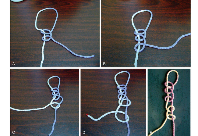
For arthroscopic shoulder instability surgery (as with any arthroscopic knot tying), the surgeon should master two types of knots: nonsliding and sliding knots. Nonsliding knots are used in cases when the suture is caught, frayed, or otherwise cannot slide freely. These consist of a series of half hitches that vary the direction of the throw and alternate the post around which the half hitch is thrown. The most common nonsliding knot (Fig. 5-1), the Revo knot, has been shown to test well compared with sliding knots.5,6 The first two half hitches of the Revo knot are throwing in the same direction on the same post. The third half hitch reverses that direction but uses the same post. These three half hitches are tensioned at this point using a “past pointing” technique with a single lumen knot pusher. Two additional half hitches are then thrown using the other suture limb as the post and alternating the direction of the throw.


When the suture slides freely through the suture anchor eyelet and soft tissue, sliding knots can be used. A major advantage of a sliding joint is that it is assembled entirely outside the cannula. When tying a sliding knot, the post strand is shortened to less than half the length of the loop strand. This is done because as the knot is delivered down the cannula and into the joint, the post limb is pulled out of the joint, lengthening it, and the loop limb slides into the joint, shortening it. Once the main knot is delivered to the fixation site, it is reinforced with alternating half hitches on alternating posts. Traction must be maintained on the post of a sliding knot while the half hitches are thrown and locked. This is not needed for a sliding locking knot.


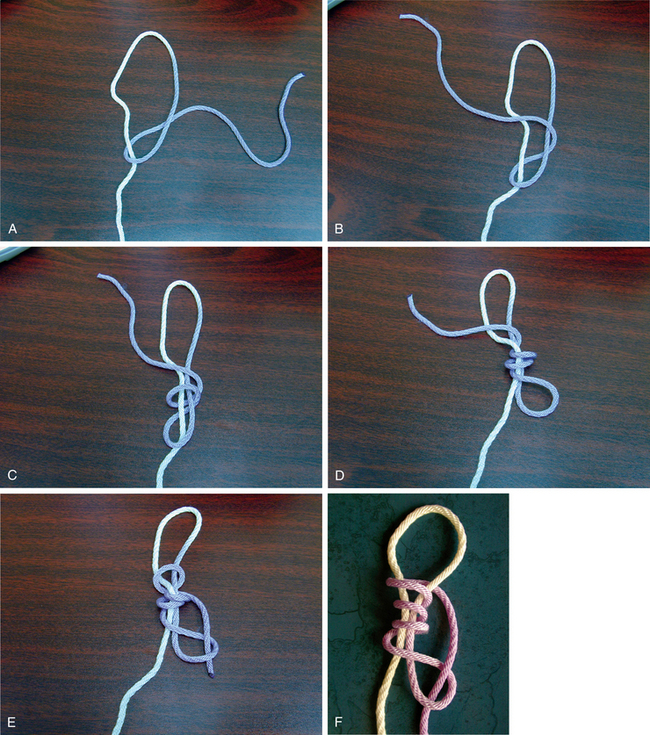
Sliding knots can be subdivided into locking and nonlocking knots. The Duncan knot is a sliding nonlocking knot. To tie a Duncan loop (Fig. 5-2), the sutures are grasped between the thumb and index finger and a loop is created by passing the loop strand over the post. The loop strand continues in the same direction to place four subsequent throws around the post limb. The free end of the loop limb is the passed through the original loop created, and the knot is tightened to remove the slack from the knot configuration. Once the knot is “policed,” the post strand is pulled and the knot advanced. At least three reversed half hitches should be used to reinforce the knot.


Stay updated, free articles. Join our Telegram channel

Jan 21, 2017 | Posted by in ORTHOPEDIC | Comments Off on Sutures and glenoid anchors for instability

Full access? Get Clinical Tree

Get Clinical Tree app for offline access