CHAPTER 37 Surgical Approach
All revision shoulder arthroplasty in our practice is performed through a deltopectoral approach. The utilitarian nature of this approach makes it ideal for use in revision shoulder arthroplasty. The deltopectoral approach is easily extended into an anterolateral approach to the humeral diaphysis should it be necessary for extraction of a humeral component or for fixation of a periprosthetic fracture.
TECHNIQUE FOR THE DELTOPECTORAL APPROACH
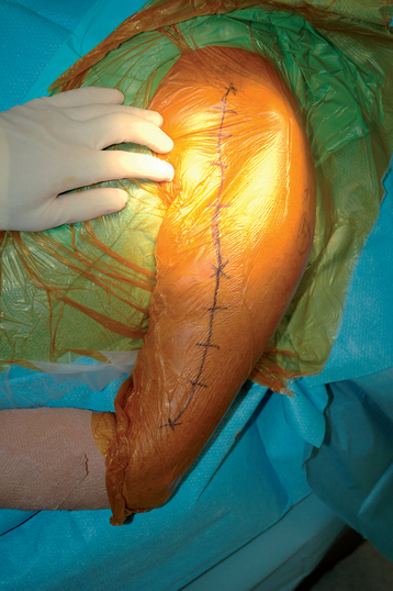
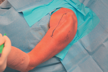
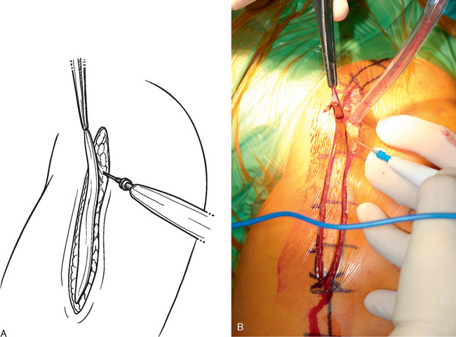
The deltopectoral approach used for revision shoulder arthroplasty follows the same technical principles described for primary shoulder arthroplasty (see Chapter 8). Patient positioning is the same as for primary shoulder arthroplasty. Draping differs in revision arthroplasty in that the stockinet is placed only up to the elbow to allow distal extension of the surgical approach if necessary (Fig. 37-1). Any previous skin incisions are delineated with a sterile surgical marking pen (Fig. 37-2). This makes these incisional scars more visible after skin preparation and placement of the occlusive drape. The previous skin incision is used if possible. If the previous incision is topographically positioned within 5 cm of our standard deltopectoral skin incision as described in Chapter 8, we will use the previous incision site (Fig. 37-3). If the previous incision deviates substantially from our standard deltopectoral incision, we will make a new incision altogether. If the previous incision has resulted in a hypertrophic scar, the scar is elliptically excised with a no. 10 scalpel blade (Fig. 37-4). The incision extends for 10 to 15 cm, depending on the size of the patient. Occasionally, the original incision is longer than what we consider necessary. In that situation we use only a portion of the original incision. To minimize hemorrhage, we use a needle tip electrocautery for subcutaneous dissection and for most of the deep dissection throughout the procedure. Medium-size skin rakes are used for retraction during this portion of the approach.
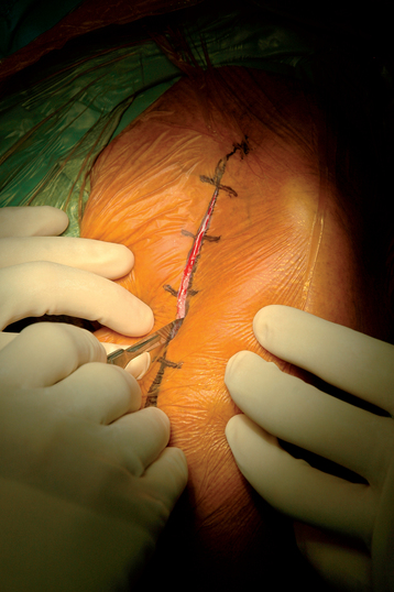
Figure 37-3 Use of the previous incision for the deltopectoral approach during revision shoulder arthroplasty.
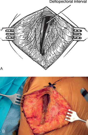
The cephalic vein, when present, is located to identify the interval between the deltoid and pectoralis major. In many cases the cephalic vein is not identified during revision shoulder arthroplasty. If such is the case, the deltopectoral interval can be readily located proximally by identifying a small triangular area devoid of muscle tissue between the proximal portions of the deltoid and pectoralis major muscles (Fig. 37-5). If located, the cephalic vein is dissected free of the pectoralis major muscle with Metzenbaum scissors. We prefer to retract the cephalic vein laterally with the deltoid because most of the branches of the cephalic vein are deltoid based. Medial retraction of the cephalic vein with the pectoralis major disrupts these deltoid branches and introduces unwanted hemorrhage.
Stay updated, free articles. Join our Telegram channel

Full access? Get Clinical Tree


