Sporotrichosis
Michael R. McGinnis
Michael B. Smith
Sporotrichosis is a chronic fungal infection typically limited to cutaneous and subcutaneous tissues. Dissemination of the infection is unusual. The causative agent is the dimorphic fungus Sporothrix schenckii. Sporotrichosis occurs worldwide but is found primarily in warm, temperate, and tropical regions. With few exceptions, the fungus gains entry into the body through trauma to the skin. In children, the infection often occurs as a solitary lesion on the face (Fig. 214.1A) or neck. Subsequent discrete nodular lesions tend to develop and spread along the lymphangitic system (Fig. 214.1B). Most cases of sporotrichosis occur in adults after intradermal inoculation of the fungus on contaminated thorns or splinters, reeds, or grasses. In many instances there is no memory of localized trauma. Floral nursery workers, gardeners, tree farmers, and others are at highest risk, although epidemic forms have been recorded in adults and children. In some endemic regions of Latin America approximately 60% of those infected are children under 15 years of age. Risk factors include playing in fields of crops and owning a cat. Cats have been associated as a risk factor by several investigators. Rare cases of disseminated sporotrichosis have been reported in patients with human immunodeficiency virus (HIV) infection. The pulmonary and extracutaneous forms are caused by inhalation of the fungus, which initially causes primary pneumonia. Secondary spread to joints, bone, muscle, and other organs is rare but may occur in the immunosuppressed host.
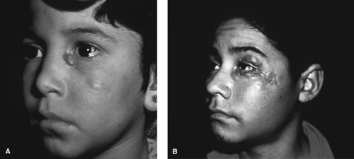 FIGURE 214.1. A: Sporotrichosis in its classic appearance as a solitary lesion on the face of a child. B: Sporotrichosis spread. |
ETIOLOGY
S. schenckii is a dimorphic fungus that exists in a mycelial form in culture at 25°C. The invasive, yeast-like form is grown easily on enriched media, such as brain–heart infusion agar, at 37°C. The yeast is nearly round to oval and may form one or more daughter blastoconidia. Cigar-shaped blastoconidia are produced also from the parent yeast cells, most frequently in disseminated infections. The histologic picture in sporotrichosis is that of a mixed suppurative and granulomatous tissue reaction, with fibrosis and microabscess formation (Fig. 214.2). This tissue reaction is seen in both localized and disseminated infections. Typically, demonstration of fungi in tissue is very difficult. Culture of pus and biopsy material should be performed because the fungus is isolated easily in culture.
Related posts:

Stay updated, free articles. Join our Telegram channel

Full access? Get Clinical Tree


