5
SPINE
Lisa Huynh, Patricia Z. Zheng, and David J. Kennedy
INTRODUCTION
Low back pain (LBP) is a widely prevalent condition, with documented lifetime prevalence rate as high as 90% (1). Most cases of acute LBP are self-limiting, although recurrence may occur in 60% to 73% of people (2). Not only is LBP problematic for the general population, it can be debilitating or even career ending for a professional athlete. During sports, athletes have considerable loads placed on their spines throughout a dynamic range of motion (ROM), which may predispose them to spine injuries. Therefore, it is not surprising that LBP is the most common reason that athletes miss a game (3), with up to 20% of all sports-related injuries involving the spine (4). Sports with high prevalence of LBP include gymnastics, weightlifting, swimming, tennis, volleyball, and football (5–7).
Studies on sex differences as related to the spine remain largely inconclusive. Frequently it is difficult to separate the sex influences on spine pathology from the influences of specific sports or activities that have a gender preference. An example would be gymnastics, in which males and females compete in different activities. Therefore, the results of studies evaluating rates of spine pathology associated with specific sporting events could be due to the event or the sex differences. This example epitomizes how determining the exact contribution of gender differences in injury rates can be challenging. However, there are many other sports such as basketball that are played by both sexes and help facilitate comparisons between genders.
Despite these difficulties, some studies have found distinctions between the sexes. Female athletes are more likely to suffer from LBP as compared to males (4,5,7). According to NCAA Injury Surveillance Data from 1997 to 1998, female athletes were twice as likely to sustain low back injuries compared to males (7). Low back injury was the most common injury in women’s volleyball and gymnastics, second most common in women’s soccer, and third most common in women’s basketball (7). This chapter reviews supported data on sex differences as they relate to anatomical and pathological conditions and offers potential insights where data are limited. We start by reviewing anatomic and alignment differences between the sexes, then offer insights and data on gender differences in spine pathology among several common spine disorders.
SPINE ANATOMY
The spinal column has multiple functions. It must offer osseous protection of the contained spinal cord and nerve roots. It must also offer significant stability to allow load transfer between the upper and lower limbs. In addition to these stabilizing features, the spine has a segmental multiplanar motion that is crucial for mobility. This combination of stability and segmental mobility results in unique demands on the spinal column. As such, the spine, especially the lumbar spine, is predisposed to great stress, putting it at risk of acute injury and repetitive overuse injuries. In assessing pathology in the spine, it is important to note that distinct gender differences exist in the spine anatomy secondary to sexual dimorphism. These differences are likely due to the load-bearing adaptations necessary for the accommodation of bipedal pregnancy.
Bony Anatomy
Although some minor variations in numbering exist, the vertebral column typically consists of 7 cervical vertebrae, 12 thoracic vertebrae, and 5 lumbar vertebrae along with the fused sacrum and coccyx. Vertebrates consist of a vertebral body anteriorly and laminae, spinous process and transverse processes posteriorly connected by two pedicles. The superior articular process (SAP) and inferior articular process (IAP) arise from the junction of the pedicle and lamina to join with the vertebrae above and below in the form of zygapophyseal joints (z-joints) as shown in Figure 5.1.

FIGURE 5.1: Lumbar zygapophyseal joint.
IAP, inferior articular process; SAP, superior articular process.
Source: Adapted and modified from Ref. (8). Peh W. Image-guided facet joint injection. Biomed Imaging Interv J. 2011;7(1):e4.
There are regional differences in the vertebrae. The cervical vertebrae have foramen in each transverse process to house the vertebral artery. The first two cervical vertebrae are further specialized to allow rotation with the first level (C1 or atlas) supporting the head and the second (C2 or axis) forming a tooth-like dens that serves as a pivot to allow lateral rotation. The C0-1 joint provides the most amount of flexion extension at approximately 25° while the C1-2 joint is responsible for 80° of axial rotation (9). The thoracic vertebrae have costal facets on the antero-lateral ends of the transverse processes for articulation with the ribs. The lumbar vertebrae are broader and heavier to help facilitate load transfers from the lower limbs through the pelvis to the upper body (10).
Studies have shown that vertebral width and disc-facet depth of male cervical vertebrates are greater, yielding more stable intervertebral coupling (11). Such differences in stability may partially explain the higher susceptibility to trauma-related neck pain that is found in females (12,13). Lumbar vertebral bodies in females also have reduced cross-sectional areas (CSA) and volumes (14). This is true even when matched for age, weight, vertebral bone density, and vertebral body height (14). This difference confers an additional risk for vertebral fractures in females, as the smaller female vertebrates confer a 30% to 40% higher mechanical stress for equivalent applied loads even in the setting of comparable vertebral bone densities (15).
Alignment
Besides inherent differences in bony structure, there are also gender differences in alignment. Janssen and colleagues showed that the T1-L5 segments of the female spine are more dorsally inclined in the sagittal plane (16). Based on animal models, this dorsally directed shear load could predispose the female spine to reduced rotational stability (17). A comparative study of men and women with chronic LBP demonstrated that men exhibited earlier and greater lumbopelvic rotation ROM during hip medial rotation (18). Additionally, the study found a greater proportion of men reported an increase in LBP symptoms with hip medial rotation compared to women, suggesting that movement of the hip medial rotation may be more problematic for men than women with LBP. This could potentially increase risk of developing LBP in men who participate in rotational sports such as golf or tennis.
Pelvic incidence, defined as the angle between the line joining the hip axis and the center of the S1 end plate and the line orthogonal to the S1 end plate, is also higher in women. It has been shown that an abnormally high pelvic incidence upsets the spinal balance and is associated with higher rates of spondylolisthesis and increased likelihood of disease progression (19,20) and severity of scoliosis in the elderly population (21).
Lumbar lordosis is an important adaption for bipedalism. However, since pregnancy shifts the center of mass anteriorly, the female spine must have increased lumbar curvature and reinforcement to compensate for the increased bipedal obstetric load (22), as shown in Figure 5.2. Additionally breasts have been theorized to affect spinal alignment. Radiographic studies suggest that breast size can affect the thoracic kyphosis and lumbar lordosis angles (23), with larger breast sizes being associated with both increased thoracic kyphosis and lumbar lordosis, as shown by the difference in lordosis when comparing male to female vertebral columns in Figure 5.3. However, a study has shown that reduction mammoplasty does not seem to change the measurable thoracic kyphosis and lumbar lordosis angles (24).

FIGURE 5.2: With pregnancy, human females have to adapt with increased lumbar lordosis to accommodate the anterior-translated center of mass. (A) Nongravidad female. (B) Gravidad female without lumbar lordotic compensation. (C) Gravidad female with lumbar lordotic compensation.

FIGURE 5.3: Male vs. female lumbar spine.
Joints, Ligaments, and Musculature
Spine mobility is maintained through the segmental intervertebral discs and a gliding movement of the paired synovial zygapophyseal joints. The cartilaginous nature of these joints may also provide shock absorption throughout the spine. Studies have shown that z-joint cartilage thickness is lower in females and the gap in the dorsal region is greater (25). Cervical z-joint shear and distraction motions in females were higher than that of males, providing a mechanism to explain why women may be more susceptible to whiplash injury (26). Studies have shown that disc height in women is higher than in men (27). Disc space narrowing is more frequent in women than men (28) while degenerative changes were observed more frequently in males (29).
Although spinous ligaments provide stability, the musculature is felt to be the primary stabilizer of the spine. In fact, it has been demonstrated that a cadaver with bones and ligaments intact but muscles removed will buckle under about 20 pounds (30). The anterior longitudinal and posterior longitudinal ligaments help to support the intervertebral discs. The intrinsic muscles of the back act primarily as extensors and rotators and help stabilize the spine. While gender variations in ligament and muscle activation have been documented in other joints and parts of the body (31–33), few available studies have described gender differences in spine ligament and musculature.
PATHOLOGY-SPECIFIC GENDER DIFFERENCES
Scoliosis
Scoliosis denotes pathological lateral curvature of the spine in the coronal plane. The four major types include congenital, neuromuscular, degenerative, and idiopathic scoliosis. Congenital scoliosis is associated with vertebral malformations; neuromuscular scoliosis is found in patients with trunk weakness associated with spina bifida, cerebral palsy, or neuromuscular disorders; and degenerative scoliosis results from traumatic or degenerative bone collapse secondary to osteoporosis. The prevalence of congenital, neuromuscular, and degenerative scoliosis shows little gender predilection apart from that associated with the primary condition.
Idiopathic scoliosis is however the most common etiology, accounting for 65% of structural scoliosis, and it affects adolescents during the growth spurt years (34). The exact pathophysiology is uncertain but it has been hypothesized that there is a complex interplay of defect in central control of growth as well as the spine’s inherent susceptibility to deformation. Researchers are looking into genetic, hormonal, collagen, and platelet-related causes (35). Interestingly, some studies report that athletes of certain sports have a higher incidence of scoliosis, most noticeably among female rhythmic gymnastic trainees (36,37), though it is not clear whether this is because of the demands of the sport or the particular gender predilections of the participants.
Regardless of exact pathophysiology, idiopathic scoliosis is much more common in females. The female-to-male ratio ranges from 1.5:1 to 3:1, with a 7.2:1 predilection in curves greater than 40° (38). While the reasons are still unclear, females may be more predisposed given that they go through relatively shorter and more rapid adolescent growth of the spine. Thus, they may be more predisposed to any defect in control of growth affecting the spine.
Timing of screening for scoliosis depends on age of adolescence. The American Academy of Orthopaedic Surgeons (AAOS), the Scoliosis Research Society (SRS), the Pediatric Orthopaedic Society of North America (POSNA), and the American Academy of Pediatrics (AAP) recommend that girls should be screened twice, at age 10 and 12, and boys once, at age 13 or 14, by the Adam’s forward bending test. The Adam’s test is a visual scoliosis screening tool performed with the patient bending forward at the waist with feet together, knees straight, and arms hanging freely. The examiner observes the patient from behind to look for asymmetric rise of one side of the rib cage next to the vertebral column. The peak rise of the convex side can be measured more objectively using a scoliometer, a specialized tool with a bubble level and angle measure to help determine the degree of curvature, as demonstrated in Figure 5.4 (39).
Given the predisposition of scoliosis in females, research from Norway has demonstrated that screening is cost saving when performed in girls only (40). Referral to a spinal deformity expert and x-rays should be considered if the Adam’s test is positive. Bracing should be initiated in patients with curves above 20 ± 5° Cobb who are still growing or demonstrating progression of deformity (41). Interestingly, males are less likely to respond to brace treatment and have been theorized to have more rigid spines (42).
Surgical intervention is usually considered when curves exceed 50°, are causing loss of pulmonary function, or are progressive (43). Fortunately, despite males having possibly more rigid spines, sex does not seem to affect outcomes of surgery for adolescent idiopathic scoliosis (44). Most experts advise a return to physical activity at the same level after surgery (36). A small study also showed that sports activity is not more restricted after surgical intervention than after nonoperative treatment; however, surgical and nonsurgical groups with scoliosis had reduced sport activity secondary to back pain as compared to matched controls without spinal disorder (45).
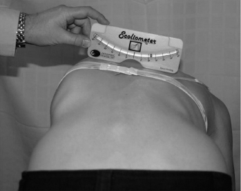
FIGURE 5.4: Scoliometer.
Source: From Ref. (39). Izatt MT, Bateman GR, Adam CJ. Evaluation of the iPhone with an acrylic sleeve versus the scoliometer for rib hump measurement in scoliosis. Scoliosis. 2012;7(1):14.
Kyphosis
The sagittal alignment of the thoracic spine displays a dynamic range and usually kyphosis naturally increases throughout life. Females exhibit significantly higher kyphosis after the age of 40 (46). This is often accompanied by decreased lumbar lordosis, and together the syndrome is termed lumbar degenerative kyphosis (47). In Takemitsu’s epidemiological study, 90% of subjects with the syndrome report significant LBP. There have been studies associating increases in thoracic kyphosis with higher spinal loads and trunk muscle forces (48). The resultant flat back has been associated with anomalous pelvic tilt (49). Higher thoracic kyphosis, as determined by occiput wall distance, has also been associated with shoulder pain and subacromial impingement (50). Treatment of excessive thoracic kyphosis includes physical therapy focusing on posterior kinetic chain strengthening and even surgical fixation in some cases (51).
A distinct entity of thoracic kyphosis is Scheuermann’s kyphosis, which is defined as the basis of anterior wedging of 5° or more of at least three adjacent vertebral bodies and typically occurs during adolescence (52). The prevalence of Scheuermann’s kyphosis ranges from 0.4% to 8.3% (53). While historically it was thought to be more prevalent in males (54), more recent studies have shown no gender predilection (35,55,56). Mild cases are treated symptomatically for back pain with exercise and anti-inflammatory medications while kyphosis greater than 45° requires bracing before progressing to surgery for curves greater than 75° (52). In regard to sports, some experts recommend more extension-biased sports such as gymnastics, swimming, and basketball while avoiding sports associated with repetitive flexion and heavy loading (57). Most authors recommend a rehabilitation program focusing on maintaining flexibility, strengthening the extensor muscles of the spine, and correcting any postural component of kyphosis (58).
Spondylolysis and Spondylolisthesis
Spondylolysis is a unilateral or bilateral defect in the pars interarticularis of the vertebra, most commonly found at the L5 level (59), as illustrated in Figure 5.5. The exact pathogenesis of lumbar spondylolysis remains controversial and is likely multifactorial. The most accepted mechanism is biomechanical stresses due to chronic low-grade trauma from repetitive flexion, extension, or rotation of the lumbar spine on a congenitally weak or dysplastic pars interarticularis (60,61). There are two main mechanisms of how the stress fracture occurs. The first is the “nutcracker” mechanism in which there is direct compression of the IAP of the cranial vertebra on the pars interarticularis of the caudal vertebra when the lumbar spine is in extension (62–65). The second mechanism is when the pars interarticularis fails in tension through a traction mechanism (62,63,66,67).

FIGURE 5.5: Radiographic findings in a patient with L5 pars interarticularis fracture and mild L5 on S1 spondylolisthesis: anteroposterior (A) and lateral (B) images. (White arrow points to the fracture and black arrow points to spondylolisthesis of L5 on S1.)
LBP, low back pain.
Source: From Ref. (68). Zukotynski K, Curtis C, Grant FD, et al. The value of SPECT in the detection of stress injury to the pars interarticularis in patients with low back pain. J Orthop Surg Res. 2010;5:13.
The incidence of asymptomatic spondylolysis has been estimated to be approximately 6% of the general population (59,66,69). Some studies have shown the positive association between spondylolysis and other diseases including spina bifida occulta (66,69–71), Scheuermann’s disease (72,73), and scoliosis (74). Numerous studies have reported a high incidence of lumbar spondylolysis among family members (up to 69%), which suggests a genetic predisposition to this condition (69,75–77).
The incidence is higher in athletes than in the nonathletic population (78–80). Most studies report 10% to 15% of adolescent athletes have been reported to have spondylolysis (59,81,82), especially those involved in sports with repetitive flexion/extension such as gymnastics, weight lifting, diving, and rowing (59). Most cases of spondylolysis are asymptomatic. In children and adolescent athletes, however, it is the most identifiable cause of back pain (59,67), usually manifesting as axial LBP that is exacerbated with lumbar hyperextension.
Spondylolysis is also more frequently found in males, with an incidence ratio of 2:1 (66,67,69), although spondylolisthesis affects females two to three times more frequently (59,67,83). Some have suggested that the increased incidence of males compared to females, as previously reported, might be a result of a time when females were not as active in sports as males. Fredrickson et al. refutes this claim; he reported an incidence of 2:1 ratio of boys to girls at age 6, at which activity levels of both sexes are similar (69). The etiology of this difference between males and females remains unknown, although the progression to spondylolisthesis may be due to differences in pelvic incidence between males and females.
Bilateral pars defects can result in the development of spondylolisthesis. Patients with known spondylolisthesis, bilateral pars defects, or possibly those presenting with unilateral pars defects at a young age, should be screened for the development or progression of a spondylolisthesis by intermittent standing spine films from a lateral view. This is especially critical during the adolescent growth spurt, as this condition can develop without any changes in symptomology.
Adolescent athletes with extension-biased pain and high suspicion of stress fracture of the pars interarticularis should be treated presumptively as spondylolysis. Treatment includes relative rest until pain free, with a gradual return to sports after correction of biomechanical abnormalities through physical therapy. These injuries typically require several months of rest and therapy before a full return to activities. Nonsteroidal anti-inflammatory medications are typically avoided as these medications may delay bone healing. Bracing is controversial; while it may limit activity it may also increase movement through the affected segment. Practitioners may also consider screening for vitamin D deficiency as a contributor to the pathology. However, there are no studies supporting sex-specific workup or management algorithm of spondylolysis in athletes.
Intervertebral Disc Pathology
Lumbar intervertebral discs degenerate not only with normal aging but also as a consequence of intrinsic and extrinsic factors. Although the exact cause and mechanism of disc degeneration is still under investigation, genetic link (84–86), obesity (87,88), smoking (89,90), and physical loading related to occupation and sports are associated with disc degeneration (91–94). Clinical manifestation of discogenic pain includes LBP typically associated with activities that increase intradiscal pressures, such as sitting, bending forward, coughing, and sneezing.
Some studies report sex differences in disc degeneration. Miller et al. analyzed 600 intervertebral discs excised from 273 cadavers and found male discs degenerated earlier than female discs and to a greater extent, most significantly in the second decade (95). In this study, nearly 40% of males had evidence of Grade II degeneration, as compared to no degeneration found in females. While the exact mechanism explaining the gender difference remains unclear, the author proposes several contributory factors including increased mechanical load on the male lumbar spine, greater CSA of male discs resulting in longer nutrient pathways, and differences in biochemical composition of the discs between males and females.
Kjaer et al. performed a cross-sectional cohort study on 439 thirteen-year-olds and found patterns of differences when results were stratified by gender (96). In boys, there was a statistically significant association in upper lumbar disc findings as compared to girls, with association found in the lowest lumbar spine segments. Additionally, there was a strong association between “seeking care” and disc protrusion/high intensity zone in girls, but not in boys. Despite this literature on gender differences, there are no known implications for participation in sports.
Ankylosing Spondylitis
Ankylosing spondylitis (AS) is a progressive chronic inflammatory disease that affects the axial skeleton, with variable involvement of entheses, peripheral joints, and, rarely, internal organs. Clinical features of AS include low back or buttock pain, peripheral joint pain, enthesitis, dactylitis, and/or progressively limited spinal mobility. Extra-articular comorbidities include acute anterior uveitis, psoriasis, inflammatory bowel disease, cardiovascular disease, and pulmonary disease.
Historically, it was thought that AS was found predominantly in males with sex ratios quoted as 10:1 (97). Recent studies, however, indicate the male-to-female ratio is closer to 2:1 (98). A suggested explanation for underestimation of female prevalence includes milder and less extensive clinical manifestations with less disabling symptomatology among females (99–101). Other possible explanations include slower development of radiographic findings in women, more peripheral arthritis in women leading to alternative diagnoses, and traditional regard of AS being a disease of men (101). Clinically, women with AS tend to have more neck and peripheral joint pain (98).
Radiographic differences between men and women have been studied, although no conclusive findings have been found. Some studies have found higher frequency of cervical spine involvement in females (102,103) and higher lumbar spine involvement in males. Other studies refute this claim showing no sex differences in cervical involvement despite higher frequency of cervical pain (104). Radiographic progression is more severe in males with AS than females (98,105). It has been suggested that radiographic severity was associated with males due to earlier diagnosis and therefore longer duration of disease. However, a study by Lee et al. correcting for age and duration of disease showed that males were still more likely than females to have increased levels of radiographic damage (98).
Special consideration should be given to athletes with AS. Over time, development of osteoporosis and ossification of the axial skeleton and ligamentous structures result in progressive rigidity, loss of lumbar and cervical lordosis, and altered biomechanical properties of the spine. Limitations in mobility and altered biomechanics may result in increased risk of spinal fractures, particularly in the cervical spine, even from low energy impact (106,107). While there are no set guidelines, Harper and Reveille recommend cautioning patients with AS to avoid sports at high risk of spinal injury, including football, ice hockey, wrestling, diving, skiing, snowboarding, rugby, cheerleading, or baseball (108).
Related posts:

Stay updated, free articles. Join our Telegram channel

Full access? Get Clinical Tree


