Spinal Cord Stimulation for Chronic Pain Management Implantation Techniques

CHAPTER 30 Spinal Cord Stimulation for Chronic Pain Management Implantation Techniques


INTRODUCTION


The fact that electricity might beneficially affect painful conditions has been known since antiquity.14


Spinal cord stimulation (SCS) was introduced by Shealey in 1967.5 Initially, the electrodes were placed over the dorsal columns in the subarachnoid space through a laminectomy. Subsequently, the electrodes were implanted, always through a laminectomy, between the two layers of the dura or epidurally. Some authors demonstrated efficacy of the procedure even with electrodes implanted ventrally to the spinal cord.68 This did not prove to be a practical way of conducting spinal cord stimulation and was subsequently abandoned. In 1975, Dooley described percutaneous implantation of electrodes in the dorsal epidural space. Manufacturers involved in the early stages of SCS included Medtronic, Avery, Cordis, Clinical Technology Corporation. Initially, the stimulating systems were only radiofrequency (RF)-driven passive receivers. In the mi-1970s, Cordis introduced the first pulse generator powered by a lithium battery. This was then followed by the Itrel pulse generator manufactured by Medtronic. In the first stages, stimulation was delivered through a unipolar electrode. Subsequently, bipolar arrays were made available. Many different types of percutaneous and plate-type arrays were developed. In all of them, however, the contact combinations were hardwired, and could no be reprogrammed after the pulse generator was implanted. A very important advance stemmed by the collaboration of Joseph Waltz and the Neuromed company in the early 1980s; they produced the first percutaneous quadripolar electrode with contact combinations that could be reprogrammed noninvasively through the external transmitter.9,10


In the late 1970s, there was a surge of enthusiasm for spinal cord stimulation among neurosurgeons in Europe and in the US. Thousand of patients with almost any type of painful condition were subjected to SCS. Poor patient selection, technical problems with the implanted equipment, and implantation by surgeons with minimal experience and commitment, resulted in a large number of patients with poor results; in the early 1980s the procedure fell into disrepute and was viewed with skepticism. Only a few dedicated neurosurgeons continued to apply this procedure for pain management. Gradually, in the past 10 years, the procedure has regained acceptance in the management of chronic nonmalignant pain; its role is currently firmly established in the armamentarium of the pain specialist as proven by several reports in the literature. The procedure has been acquired by other specialties, first among them anesthesiologists specializing in pain management. Other specialties have also demonstrated interest in the procedure, although to a lesser extent, such as rehabilitation medicine and orthopedic surgery. Interestingly, a large percentage of the SCS implants currently performed in Europe (particularly in Italy and Spain) are performed by vascular surgeons for the management of peripheral vascular disease.


Because of the emerging complexity of the structures being involved by the stimulation, the term ‘dorsal column stimulation,’ which was originally applied to this procedure, has been in general replaced by the term ‘spinal cord stimulation.’


Spinal cord stimulation, even though not considered an extremely technically demanding surgical procedure, commands extreme care in the details of the planning and of the execution. One has to be extremely fastidious about the correct positioning of the electrode(s), both in the longitudinal and transverse direction in the spine, about the position of the pulse generator, about the location of the subcutaneous wires and about the hook-up of the whole system. If only one of the various factors is not optimal, the effectiveness of the whole procedure might be negated.


This chapter will discuss the various available hardware solutions and some of the technical details of surgical implantation.


CURRENTLY AVAILABLE EQUIPMENT


Currently available equipment for SCS consists of electrodes, pulse generators, radio receivers, and transmitters.


Electrodes


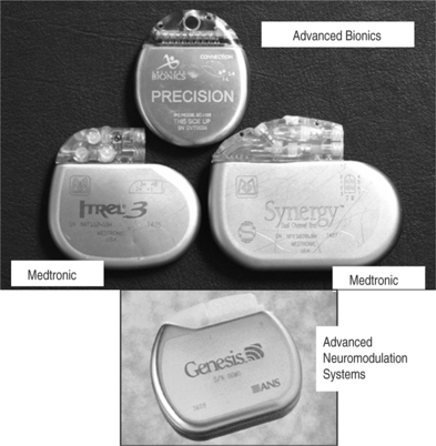
There are two main types of electrodes, the catheter-type (otherwise commonly referred to as ‘percutaneous’ leads) and the plate-type (otherwise commonly referred to as ‘laminotomy’ or ‘surgical’ leads) (Fig. 30.1). Percutaneous electrodes are commonly used both for trial stimulation or for permanent implantation. The most commonly used electrodes are either quadri- or octopolar. The general trend is to utilize one or two quadripolar electrodes for limb pain, and one or two octopolar electrodes for axial pain. A percutaneous electrode recently introduced by Advanced Bionics (Sylmar, CA) has 16 electrical contacts. The electrodes might be connected directly to the pulse generator/receiver, or they can be connected to an intermediate subcutaneous extension which, in turn, interfaces with the pulse generator/receiver.


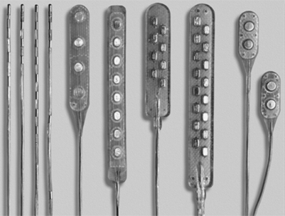
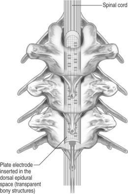
Plate-type electrodes require surgical implantation under direct vision (Fig. 30.2). The amount of actual bony removal varies and is often limited to a small portion of the lamina and spinous process. The simplest quadripolar plate electrode is the Medtronic Resume and Resume-TL, and the Advanced Neuromedulation Systems Lamitrode 4 (Medtronic Inc., Minneapolis, MN; Advanced Neuromedulation Systems, Plano, TX), with all four contacts arranged linearly in one paddle. The Medtronic Specify and the ANS Lamitrode 44 have eight contacts arranged in two parallel columns. Another electrode (ANS Peritrode) consists of two smaller paddles, each with two contacts; this configuration allows the surgeon to place the paddles in two different locations or with two different orientations and therefore offers a greater degree of flexibility. Plate electrodes with one or two columns of eight contacts are also available (ANS Lamitrode 88 and Lamitrode 8).


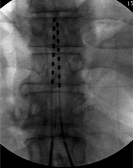
With modern technology, both types of electrodes are safe and effective ways of delivering electrical stimulation to the spinal cord. The percutaneous technique is appealing because it allows one to insert the electrode without muscle dissection and bony removal (Fig. 30.3). This is a substantial advantage when one wants to perform a trial stimulation to assess candidacy for a permanent implant. Percutaneously placed electrodes can also be advanced over several segments in the epidural space, thus allowing testing of several spinal cord levels. By inserting multiple parallel electrodes, various configuration matrices can be constructed that allow creating extremely focused electrical fields. Placement of percutaneous electrodes must be performed under fluoroscopic guidance (Fig. 30.4). This requires wearing heavy shielded garments and potentially exposes the implanting physician to non-negligible levels of radiation. The plate electrodes require open surgical intervention. Bony removal can be very limited. In the thoracic area the lower two-thirds of the spinous process and a small portion of the lamina usually have to be removed. In the cervical area, bony removal is often not necessary, and this is particularly true when placing electrodes at the C1–2 level. Most ‘laminotomy’ implants can be done through a small (1–1.5) skin incision. By advancing the electrode in a cephalad or caudal direction, one can explore at least three spinal levels in the thoracic and 4–5 in the cervical spine. (Fig. 30.4) Multiple arrays or different electrode configurations can be also constructed by utilizing more than one plate electrode. In the author’s experience, the main advantage of plate electrodes resides in their greater inherent stability in the dorsal epidural space and lesser propensity to migrate. Plate electrodes are the only option in case of previous spine surgery at the planned implant levels. The pattern of stimulation–induced paresthesiae provided by plate electrodes might be superior to the ones produced by the percutaneous electrodes. In a randomized, prospective study, North et al. proved that the performance of plate electrodes significantly exceeded that of percutaneous electrodes.10 Concordance of stimulation paresthesiae with pain was statistically better for plate electrodes. Plate electrodes are electrically more efficient. This is due to the fact that all the current is directed toward the dura instead of being dispersed circumferentially, as in the percutaneous electrodes. Plate electrodes therefore have a lower current requirement. Another advantage of plate electrodes with two columns of contacts lies in the fact that, unlike two parallel percutaneous electrodes, the relation among the electrical contacts is fixed and completely predictable. Some situations clearly command one of the two methods (i.e. a percutaneous system in the case of an outpatient percutaneous trial, or a plate electrode in the case of prior spine surgery). In most other situations, the choice is usually dictated by individual preferences and patterns of practice. A skilled implanter can usually achieve a similar stimulation matrix with either a plate or a percutaneous electrode. The differences between plate and percutaneous electrodes is likely to become more blurred with the development of miniaturized plate electrodes that can be introduced epidurally through a percutaneous device
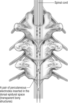


Pulse generators/receivers


See Figure 30.5


Stimulation consists of rectangular pulses delivered to the epidural space through the electrodes. Two types of systems are currently available. The totally implantable pulse generators contain a lithium battery in the pulse generator. They are activated and controlled by outside transcutaneous telemetry and, once activated, do not require any patient input to function. They can also be turned on and off through a small magnet. Lifespan of the battery greatly varies with usage and with the utilized parameters (voltage, rate, pulse width, etc.). Most patients can expect, under average use, the battery to last 2.5–4.5 years. Available lithium-powered pulse generators allow stimulation to be given in increments of 0.1 V. and with rates up to 130 Hz. The Precision pulse generator, manufactured by Advanced Bionics, contains a rechargeable lithium battery. The battery is recharged by wearing an outside recharger while the stimulator is in use. The interval between charges varies with usage time and power requirements and can be expected to be in the order of at least several days under normal utilization. Lithium-powered pulse generators make up the majority of implanted spinal cord stimulator.


Radiofrequency-driven systems, instead, consist of a passive receiver, implanted under the skin, and the transmitter which is worn outside of the body. An antenna, which is applied to the skin in correspondence of the receiver and connected to the transmitter, transmits the stimulation signals transcutaneously. In order for the system to function, the transmitter has to contain charged alkaline batteries and the antenna must make adequate contact with the receiver. This requires the patient to wear the outside system in order to receive the stimulation. RF-driven systems can deliver stimulation with a rate up to 1400 Hz, and can be customized to deliver more power than the corresponding lithium-powered systems.


Both systems have advantages and disadvantages. The main disadvantage of the RF systems is the inconvenience of having to wear the antenna and the radio receiver. The problem might go beyond pure inconvenience in individuals who have handicapped motor function in the upper extremities and cannot properly go through all the steps required to make the external unit function properly. Other patients, particularly individuals affected by a complex regional pain syndrome type 1, might not tolerate the antenna taped to the skin. The equipment cannot be worn while swimming or showering, and severe perspiration might make proper contact of the antenna problematic. Besides these considerations, the patient has to worry about changing batteries on a regular basis and making sure that the proper coupling exists between the antenna and the receiver at all times. These inconveniences are obviated by a lithium-powered system that runs automatically without any patient intervention. The RF system is usually reserved for patients who have greater power requirements and who would have to undergo replacement of the lithium-powered pulse generator with an unacceptably high frequency. The advent of the rechargeable lithium battery might redefine this requirement and the indications for an RF system.


The distribution of the electrical fields within the intraspinal structures is affected by the position of the electrode array as well as the polarity of the individual contacts. In order to generate an electrical field, one must have at least one negative contact activated (cathode) and one positive contact activated (anode). With the ANS and Medtronic systems, each contact can be either on or off. The Advanced Bionics System, instead, allows each electrical contact to be activated in fractional increments, thus allowing an almost seamless change in the distribution of the electrical field.


WHAT STRUCTURES ARE BEING STIMULATED


The spinal canal contains several nervous and non-nervous structures that, when stimulated electrically, give rise to a variety of responses. The electrical properties of the intraspinal contents can be characterized as the ones of an nonhomogeneous conductor (Fig. 30.6) Knowledge of the different type of responses and their correlation with the underlying anatomical substrate is extremely important in implementing strategies for spinal cord stimulation.11,12
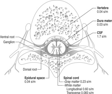


The width of the cerebrospinal (CSF) space is the most important factor in determining the stimulation parameters, particularly the perception and discomfort thresholds (Fig. 30.7). Dorsal root fibers in general have a lower stimulation threshold than dorsal column fibers and this is particularly evident with increasing thickness of the CSF layer. This is due in large part to the fact that dorsal root fibers have a very high conductivity at their entry into the spinal cord.


Stimulation of the large myelinated afferent fibers at the intraspinal level can occur in four different areas: the dorsal root, the dorsal root entry zone, the dorsal horn, and the dorsal columns. Electrical activation of these structures elicits tingling paresthesiae that are always ipsilateral to the stimulating electrode. If the stimulation voltage is increased, discomfort and pain occur. Clinically, it is extremely important to differentiate activation of the segmentary large myelinated afferents (dorsal root/entry zone/dorsal horn) versus activation of the ascending long tracts in the dorsal columns. Activation of the segmentary afferents causes paresthesiae located in the radicular dermatome at the level of the electrode. For electrodes in the thoracic area, this usually means paresthesiae along the anterior chest wall. With electrodes placed at T12 or L1 the usual pattern is paresthesiae along the anterior aspect of the thigh or in the inguinal area. In the cervical spine, paresthesiae will be elicited in various segments of the upper extremity.


The stimulation threshold for the segmentary system is lower than the one for the dorsal columns, and usually ranges 0.1–0.5 volts Differentiating between stimulation of the dorsal root, dorsal root entry zone, or dorsal horn can be exceedingly difficult. One can assume dorsal root stimulation can be expected if the electrode is placed laterally in the spinal canal. On the other hand, stimulation of the dorsal root entry zone and/or dorsal horn is more likely if the electrode is placed near midline, and segmentary paresthesiae are rapidly followed by activation of the dorsal columns with a small voltage increment. Stimulation of the longitudinal fibers of the dorsal columns is characterized by ipsilateral paresthesiae occurring in areas of the body caudal to the level of the electrode


The exact distribution of the paresthesiae varies with the level of the electrode and with some degree of interindividual variability. In some individuals, activation of the dorsal columns gives rise to a smooth tingling sensation which is uniformly distributed in all the dermatomes caudal to the implanted electrode. This is usually the hallmark of upcoming good therapeutic effects. Other individuals, fortunately a minority, perceive the stimulation with a patchy distribution, affecting separate body segments that are not connected by paresthesiae. Individuals that exhibit this pattern of distribution of the paresthesiae, unfortunately, almost never experience any meaningful pain relief.


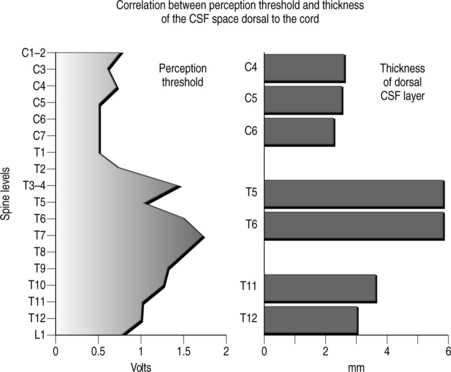
Activation of the dorsal columns usually occurs at a threshold that is at least 0.5–1.0 volt higher than the segmentary pathway. A thicker dorsal CSF space usually favors more selective activation of the segmentary sensory system as opposed to the dorsal column fibers. It is common to observe that, initially, systems are simultaneously activated, but after a few weeks the stimulation pattern is confined to a segmentary band. For this reason electrode placement in the upper thoracic spine (where the CSF space is the widest) seldom results in satisfactory long-term stimulation of the dorsal lemniscal pathway.


When stimulating the intraspinal structures epidurally, most commonly one observes a mixture of dorsal column, dorsal root entry/zone, dorsal root stimulation. This is true particularly with electrodes placed in the low thoracic–upper lumbar area (T11–12, L1) where the spinal cord tapers into the conus medullaris and the cauda equina nerve roots are a prominent component of the intraspinal structures.


Stimulation of the motor structures results in muscle contractions. Activation of the segmentary motor system (ventral root/motor neurons) results in muscular contractions in the somatic distribution of the stimulated segment. Activation of the descending corticospinal pathways, instead, causes muscle contractions in segments caudal to the level of the electrode. With laterally placed electrodes, stimulation of the segmentary motor system can occur at a threshold equal to or lesser than the one for large myelinated afferents. This invariably results in unpleasant contractions that can completely mitigate the benefits of the stimulation. In the vast majority of patients, stimulation over the dorsal columns does not result in activation of the motor system unless the stimulation is increased to a much higher voltage. The author, however, has occasionally seen patients in whom, even with a perfectly placed midline electrode, activation of the motor system occurred simultaneously with the sensory system. In these instances, selective stimulation of the dorsal column cannot be successfully performed.

Stay updated, free articles. Join our Telegram channel

Sep 8, 2016 | Posted by in PHYSICAL MEDICINE & REHABILITATION | Comments Off on Spinal Cord Stimulation for Chronic Pain Management Implantation Techniques

Full access? Get Clinical Tree

Get Clinical Tree app for offline access