Soft Tissue Closure of Severe Diabetic Foot Infections: A Combination of Biologics, Negative Pressure Wound Therapy, and Skin Grafting

Current Concepts and Techniques in Foot and Ankle Surgery


Surgical Soft Tissue Closure of Severe Diabetic Foot Infections: A Combination of Biologics, Negative Pressure Wound Therapy, and Skin Grafting


Crystal L. Ramanujam, DPM, MSc, Thomas Zgonis, DPM *


Division of Podiatric Medicine and Surgery, Department of Orthopaedic Surgery, University of Texas Health Science Center at San Antonio, 7703 Floyd Curl Drive-MSC 7776, San Antonio, TX 78229, USA


* Corresponding author.


E-mail address: zgonis@uthscsa.edu


Keywords


• Diabetic foot • Wounds • Neuropathy • Biologics • Negative pressure wound therapy • Amputation


Foot infections and associated wounds are the leading cause of hospitalization of diabetics.1 Expedited closure of diabetic foot wounds can reduce the risk for major limb amputation and decrease costs associated with prolonged wound care.2 Well-established surgical techniques such as primary closure, flaps, and skin grafting are not always suitable for early closure of extensive diabetic foot and ankle wounds. Recent advances in wound care modalities, including tissue-engineered products such as bilayer matrix scaffolds, can accelerate healing in these patients.3 The Integra Bilayer Matrix Wound Dressing (Integra Life Sciences, Plainsboro, NJ, USA) is a collagen-based synthetic graft that facilitates cellular invasion and capillary growth into the wound, and its outer layer is made from silicone. The adjunctive use of negative pressure wound therapy (NPWT) can augment the healing process and prepare for definitive closure through skin grafting. This article presents a stepwise approach detailing the combination of these surgical techniques for the closure of complicated diabetic foot wounds.


Case report


A 53-year-old diabetic man presented to the emergency room for treatment of left foot pain, redness, and swelling. A rock had fallen on it 3 days before. He initially self-treated the injury by soaking the foot in hot salt water, but noticed worsening redness of the entire foot and ankle. He denied constitutional symptoms on presentation. His past medical history was positive for uncontrolled diabetes mellitus and hypertension and he was noncompliant with outpatient medications.


General physical examination revealed a well-nourished, Spanish-speaking man in no acute distress. Vital signs showed an increased blood pressure but absence of fever or tachycardia. Foot and ankle examination showed palpable pedal pulses with pitting edema and cellulitis extending from the level of the toes to the ankle. Full-thickness ulceration was located at the medial aspect of the fourth digit with probing to deep soft tissues, serosanguinous drainage, and severe sloughing of necrotic skin on the entire toe. Small superficial ulcerations were found at the medial aspect of the fifth digit and lateral third digit. The patient also had leukocytosis and severe hyperglycemia. Radiographs of the foot and ankle revealed subcutaneous emphysema at the fourth digit. Based on the clinical and radiographic findings, the patient was admitted for urgent surgical intervention. He was medically optimized and cleared by the medicine team, and consented for left foot debridement to the level of an open partial fourth ray amputation. The initial surgical procedure consisted of aggressive debridement, cultures of bone and soft tissue, and open partial fourth ray amputation. He was maintained on intravenous broad-spectrum antibiotics with revisional surgery 4 days later for soft tissue debridement and NPWT device placement because of the depth of the remaining surgical wound defect. The patient was discharged to home on culture-specific oral antibiotics following normalization of laboratory values and no further evidence of clinical infection.


Five weeks later, after undergoing local wound care 3 times weekly for NPWT dressing changes, the wound depth had significantly decreased without recurrence of infection. Because of the large length and width of the surgical wound, the patient was brought back to the operating room for surgical application of Integra Bilayer Matrix Wound Dressing to promote further epithelialization. Surgical wound bed preparation to stimulate healthy bleeding tissue was performed via hydrosurgical debridement. The Integra Bilayer Matrix Wound Dressing was moistened in sterile saline and meshed in a 1:1 fashion to allow wound drainage and prevent fluid accumulation beneath the graft. The graft was then secured to the wound with staples, with the silicone layer facing away from the wound bed and with the graft well adhered to the wound surface with minimal tension. NPWT (VAC, Kinetic Concepts Inc., San Antonio, TX, USA) was also applied over the biologic wound bilayer to enhance incorporation during the early healing phase. Subsequent intraoperative wound cultures at this time were negative and the patient had removal of the NPWT device in the outpatient setting within the following 8 days. The patient was then transitioned to a non–weight-bearing lower extremity posterior splint for an additional 3 weeks and subsequent staple removal.


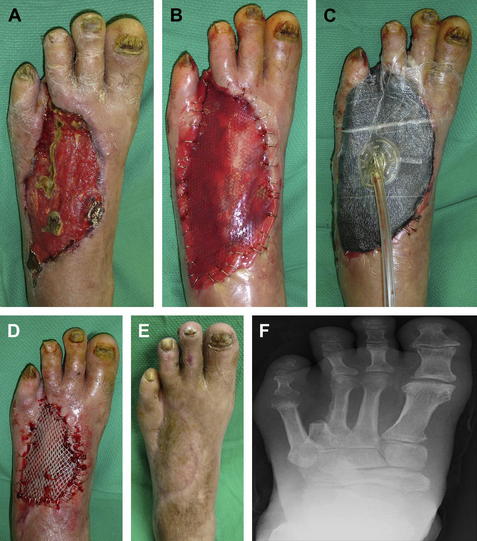
Eight weeks later, and after local wound care with moist to dry dressings, the patient returned to the operating room for definitive wound closure through split thickness skin grafting (STSG). The recipient wound bed was first prepared through hydrosurgical debridement. The donor site at the lower lateral aspect of the ipsilateral leg was used for the skin harvesting. An electric dermatome was used to carefully harvest the appropriately sized skin graft, which was meshed in a 1:1.5 ratio. The harvested STSG was anchored to the recipient bed by staples followed by application of a bolster dressing and a well-padded lower extremity posterior splint. After 3 weeks of non–weight bearing to the operative foot, the dressing and staples were removed revealing great healing signs at both the recipient and donor sites. The patient was then transitioned to full weight-bearing status in a postoperative shoe and eventually progressed into extra depth shoes. At the patient’s latest clinical visit at 18 weeks, he had no recurrence of wound or infection and no difficulty with ambulation (Fig. 1).


Stay updated, free articles. Join our Telegram channel

Mar 20, 2017 | Posted by in MANUAL THERAPIST | Comments Off on Soft Tissue Closure of Severe Diabetic Foot Infections: A Combination of Biologics, Negative Pressure Wound Therapy, and Skin Grafting

Full access? Get Clinical Tree

Get Clinical Tree app for offline access