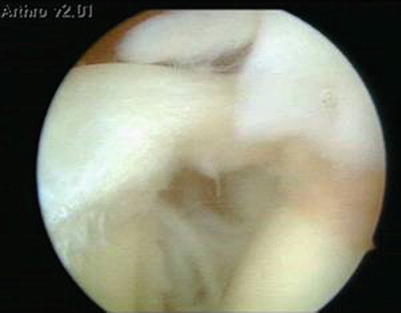
Fig. 20.1
Hyperangulation entails a greater loading phase of the throw with evident increase in force (like a catapult) but also a delicate balance: the stabilisation complex is, in fact, repeatedly taxed by numerous attempts to withstand also greater tensile stresses in external rotation, predisposing the capsule to anterior laxity. Tm teres minor, Is infraspinatus, Sup supraspinatus, Sub subscapularis, LHB long head biceps, SGHL superior gleno humeral ligament, MGHL medium gleno humeral ligament, IGHL inferior gleno humeral ligament
This mechanism, known as “peel back”, may lead to a potential detachment of the posterior biceps anchor (posterior SLAP II lesion) or the extension of a previous injury.
This SLAP lesion may also occur during the deceleration phase of the throw [11].
Consequences
Cadaver studies have reported lengthening of the anterior band of the inferior glenohumeral ligament [12] and a consequent increase in anteroinferior translation capability [13] as well as (further) external rotation capability, with a decline in performance and the onset of pain.
A SLAP lesion also leads to a posterosuperior instability which, combined with repetitive movement in abduction-external rotation, results in a progressive posterior insertional stress of the supraspinatus and its progressive surrendering.
The greater external rotation and the more posterior position of the humeral head increase the likelihood of the overhead motion causing pinching of the cuff between the posterior glenoid rim and the greater tuberosity, which is located posteriorly during external rotation plus abduction, resulting in internal impingement.
Even though there is no consensus in the literature in this respect, the internal impingement could cause an intra-articular lesion of the posterior cuff and corresponding rim, with worsening of the SLAP lesion.
A cadaver study has demonstrated the importance of the rotator interval in anterior, as well as posterior and inferior, instability, suggesting that the mechanism of injury is ascribable to an anterosuperior sliding of the head, resulting from an insufficient interval [14].
In overhead athletes, the pain related to instability is due to contact between the articular surface of the cuff and the anterior-superior labrum.
The combination of lesions affecting these two structures is known as a SLAC (superior labrum anterior cuff) lesion (Fig. 20.2).


Fig. 20.2
Arthroscopic image of a typical associated lesion in the overhead athlete: SLAP lesion and injury to the articular surface of the rotator cuff
20.4 Role of the Scapulothoracic Joint
A positive correlation has been demonstrated between positional abnormalities of the scapula and overhead sports [4, 15].
Several studies, analysing the influence of pain and fatigue on the shoulder girdle muscles [16, 17], found a decrease in posterior tilt and an increase in clavicular retraction on anterior elevation of the arm.
Conversely, conflicting results have been reached with regard to lateral scapular rotation and protraction.
Several authors believe that the former increases from pre- to post-fatigue [16].
Considering muscular activity, studies have demonstrated a delayed and decreased activation of the middle and lower trapezius and serratus anterior, an increased activity of the upper trapezius [19] and increased activity of the infraspinatus during the descending, hence eccentric, phase as a probable compensatory mechanism.
The upper and lower trapezii constitute a major force couple: A decrease in the activity of the lower trapezius coupled with increased activity of the upper trapezius can cause cranial translation of the horizontal scapular axis, with an increase in the likelihood of impingement [20].
This may contribute to the decrease in posterior tilt and external rotation, reducing the subacromial space and further increasing the risk of impingement.
It is likely that a change in the mutual position of the scapula and humerus [21], by causing a misalignment of tendon structures, affects their length/force ratio.
Further studies are required to demonstrate a causal link between dyskinesia and microinstability, possibly through an assessment of athletes pre- and postseason [15].
It is not clear whether the compensatory scapular positioning and hyperangulation can increase or reduce the risk of subacromial impingement.
20.5 The Proprioceptive System
The proprioceptive system is also involved in this cascade of events, in both the short and long term, decreasing reversibly during a single sporting event and returning to pre-fatigue values after 10 min [22].
20.6 Pathological Anatomy
The pathological anatomy of microtraumatic instabilities may affect the entire glenohumeral labral-ligamentous-tendinous complex.
The Head
The humeral head often assumes a more anterior position with respect to the horizontal axis of the body [23].
An intense overhead activity between the ages of 13 and 16 years may block the physiological process of humeral head anteversion, by enhancing its retroversion [24].
The Glenoid
Changes in glenohumeral kinematics reflect on the density of glenoid subchondral bone. In overhead athletes, this density is apparently increased in the anteroinferior, posterosuperior and inferior segments [25].
SLAP
The upper portion of the labrum may show a variety of lesions, often specific to type of sport played. Recognition of these lesions could have important therapeutic and prognostic implications [26].
Others
It is easy to hypothesise also other alterations affecting the spine, the appendicular and trunk muscles and the soft tissues.
20.7 Clinical Presentation
The symptoms reported by overhead athletes are related to the cascade of events described by Morgan [10].
Initially, the athletes may have subjective and objective perception of reduced throwing speed (without pain) and resulting decrease in performance: dead arm syndrome.
They may then develop vague pain and discomfort not otherwise specified.
This may be followed by pain due to internal impingement.
In the final phase of the cascade, mechanical symptoms may develop as a result of the development of posterior or posterosuperior labral lesions and rotator cuff dysfunction [27].
Anterior coracoid pain [3] and bursitis secondary to kinematic alterations of the scapulothoracic joint have also been reported.
Involvement of the long head of the biceps, although rare, may occur during preparation for the sports season, when the limb is not yet ready for major stresses [4].
Finally, apprehension and a sensation of subluxation and/or secondary deficits affecting the arm and forearm muscles may develop as further symptoms of instability.
20.8 Diagnosis
The approach to shoulder disorders is very complex, with only diagnostic imaging having provided the solution to many problems.
After history taking, inspection may reveal postural and/or spinal abnormalities.
A common finding is hypertrophy of the dominant shoulder or dropped scapula [18] caused by lengthening of the static and dynamic stabilisers that are intensely used in athletes.
Palpation, whenever possible, should include structures such as the posterior glenohumeral joint line, the subacromial space, the greater tuberosity, the acromioclavicular inter-joint space and the groove.
Laxity is often a general pre-existing condition in overhead athletes which predisposes to or exacerbates the symptoms and should always be assessed.
The usual tests are considered valid (thumb, fifth finger, elbow) and laxity may also be quantified according to the Brighton scale.
Shoulder ROM is then assessed, for which the mean values for the dominant arm of an asymptomatic athlete are as follows:
External rotation at 90° abduction 129–137° (7–9° > of nondominant arm)
Internal rotation at 90° of abduction 54–61° (7–9° < of nondominant arm)
TROM (total ROM) 183–198° (equal to nondominant arm)
The relationship between GIRD (glenohumeral internal rotation deficit) and TROM is fairly controversial.
Two types of GIRD have been suggested [28]:
1.
Diminished internal rotation, unchanged or slightly increased external rotation and symmetrical TROM
2.
Diminished TROM (pathological)
The literature suggests treating only pathological GIRD and, in particular, those cases characterised by TROM of dominant arm less than 5° when compared to the nondominant arm [29], since these are more likely to be affected by lesions or progression of the cascade: Treatment of non-pathological forms could further reduce shoulder stability.
It is important to remember that, even though symmetrical TROM suggests a non-pathological GIRD, it should nonetheless be considered a non-physiological condition indicating a neglected shoulder, and for this reason, it deserves continuous monitoring.
Assessment of the scapulothoracic joint follows, and scapular behaviour is tested statically and dynamically (we recommend having the patient repeat the movement several times and assessing both the concentric and eccentric phase) in all its phases.
Evaluation of strength is very difficult because the tests are often influenced by pain.
The bursae are more easily assessed: The patient lies prone on the examining table and is invited to place the back of the hand behind the back. This position will cause raising of the scapular medial margin facilitating the direct approach to the bursae for palpation and possible infiltration.
Stability testing: Many authors believe that the stability of a shoulder should be assessed in three directions, so that at least three tests are required (sulcus test, drawer test, Lachman test, fulcrum test).
The Lachman and fulcrum tests are the most important for evaluating stability in that they simulate the conditions of the throw [4].
The apprehension test is not really useful in overhead athletes, in that it assesses evident gross instabilities, while the subluxation/relocation test is better for assessing more subtle forms [30].
When performing tests for anterior and posterior instability, it is crucial to take into account the inclination of the glenoid plane, so the tests should be performed at 30° of flexion.
Stay updated, free articles. Join our Telegram channel

Full access? Get Clinical Tree


