Screening the Shoulder and Upper Extremity

Chapter 18


Screening the Shoulder and Upper Extremity


The therapist is well aware that many primary neuromuscular and musculoskeletal conditions in the neck, cervical spine, axilla, thorax, thoracic spine, and chest wall can refer pain to the shoulder and arm. For this reason, the physical therapist’s examination usually includes assessment above and below the involved joint for referred musculoskeletal pain (Case Example 18-1).


Case Example 18-1


Evaluation of a Professional Golfer


Referral: A 38-year-old male, professional golfer presented to physical therapy with a diagnosis of shoulder impingement syndrome, with partial thickness tears of the supraspinatus tendon.


Prior to the physical therapy intervention, x-rays taken were reported as negative for fracture or tumor. Magnetic resonance imaging (MRI) was reported as positive for bursitis and supraspinatus tendinitis with some partial tears. The shoulder specialist also provided the client with one corticosteroid injection, which gave him some relief of his shoulder pain.


Past Medical History: Past medical history and Review of Systems were negative for any systemic issues. He was on no medication at the time of evaluation.


Clinical Presentation: Functional deficits were reported as pain with the take-away phase of the golf swing and with the adduction motion of the shoulder in follow-through. He also reported a loss of distance associated with his drive by 20 to 30 yards. He had trouble sleeping and reported pain would wake him up if his head were turned into left rotation. He also had pain when turning his head to the left (e.g., when driving a car).


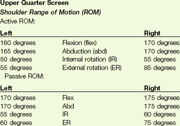
image


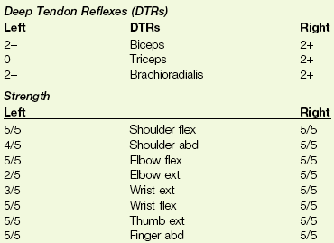
image


Special tests


Hawkins/Kennedy +


Neer +


Speed +


ER lag test −


IR lag test −


Cervical ROM  
Flexion 40 degrees  
Extension (ext) 20 degrees Report of left scapular pain
Left side bend 20 degrees Report of left scapular pain
Right side bend 25 degrees No report of pain
Left rotation 45 degrees Report of left scapular pain
Right rotation 70 degrees No report of pain
Quadrant position Right and left: Reproduced left posterior scapular pain with radicular pain to the thumb and second finger area


image


He did have intact sensation to light touch and proprioceptive sense. Strength testing on the Cybex weight-lifting machines showed he was able to do 10 triceps extensions on the right with four plates while on the left, he was only able to do one repetition with one plate.


Result: With the data obtained in the examination, the conclusion was made that he did have an impingement syndrome as described by Neer, with involvement of the bursa and rotator cuff tendons.72 Cyriax muscle testing revealed some musculotendon involvement with the strong/painful tests.61


The cervical findings required consultation with the referring physician. A provisional medical diagnosis was made of cervical radiculopathy with a C5-C6 herniated disk. The client was referred to a neurosurgeon for evaluation. An MRI confirmed the diagnosis and the client underwent an anterior cervical fusion with diskectomy.


Summary: This case example helps highlight the importance of a complete examination process, even if a physician specialist refers a client for physical therapy services. The therapist must “clear” or examine the joints above and below the region thought to be the cause of the dysfunction. The major reason for the symptoms or a secondary diagnosis may be missed if the screening step is left out because of a lack of time or assuming someone else checked out the entire client.


Voshell S: Case report presented in fulfillment of DPT 910, Institute for Physical Therapy Education, Widener University, Chester, PA, 2005. Used with permission.


In this chapter, we explore systemic and viscerogenic causes of shoulder and arm pain and take a look at each system that can refer pain or symptoms to the shoulder. This will include vascular, pulmonary, renal, gastrointestinal (GI), and gynecologic causes of shoulder and upper extremity pain and dysfunction. Primary or metastatic cancer as an underlying cause of shoulder pain also is included. The therapist must know how and what to look for to screen for cancer.


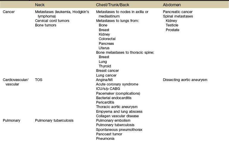
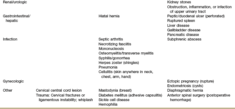
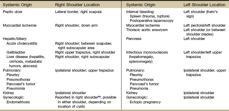
Systemic diseases and medical conditions affecting the neck, breast, and any organs in the chest or abdomen can present clinically as shoulder pain (Table 18-1).1 Peptic ulcers, heart disease, ectopic pregnancy, and myocardial ischemia are only a few examples of systemic diseases that can cause shoulder pain and movement dysfunction. Each disorder listed can present clinically as a shoulder problem before ever demonstrating systemic signs and symptoms.


Using the Screening Model to Evaluate Shoulder and Upper Extremity


Past Medical History


As you look over the various potential systemic causes of shoulder symptomatology listed in Table 18-1, think about the most common risk factors and red flag histories you might see with each of these conditions. For example, a history of any kind of cancer is always a red flag. Breast and lung cancer are the two most common types of cancer to metastasize to the shoulder.


Heart disease can cause shoulder pain, but it usually occurs in an age specific population.2,3 Anyone over 50 years old, postmenopausal women, and anyone with a positive first generation family history is at increased risk for symptomatic heart disease. Younger individuals may be more likely to demonstrate atypical symptoms such as shoulder pain without chest pain.4


Alternately, although atherosclerosis has been demonstrated in the blood vessels of children, teens, and young adults, they are rarely symptomatic unless some other heart anomaly is present.5,6


Hypertension, diabetes, and hyperlipidemia are other red flag histories associated with cardiac-related shoulder pain. Of course, a history of angina,7 heart attack, angiography, stent or pacemaker placement, coronary artery bypass graft (CABG), or other cardiac procedure is also a yellow (caution) flag to alert the therapist of the potential need for further screening.


Knowledge of risk factors associated with pathologic conditions, illnesses, and diseases helps the therapist navigate the screening process. For example, pulmonary tuberculosis (TB) is a possible cause of shoulder pain.810 Who is most likely to develop TB? Risk factors include:


In a case like tuberculosis, there will usually be other associated signs and symptoms such as fever, sweats, and cough. When completing a screening examination for a client with shoulder pain of unknown origin or an unusual clinical presentation, the therapist might look at vital signs, auscultate the client, and see what effect increased respiratory movements have on shoulder symptoms (Case Example 18-2).


Case Example 18-2   Homeless Man with Tuberculosis


Referral: A 36-year-old man was referred to physical therapy as an inpatient for a short-term hospitalization. He was a homeless man brought to the hospital by the police and admitted with an extensive medical problem list including:


There was no past medical history of cancer. The client was a smoker when he could get cigarettes. He would like to support a one-pack/day habit.


Medical service requested an evaluation of the client’s shoulder pain. X-rays were not taken because the man had full active ROM, no history of trauma, and no insurance to cover additional testing.


Clinical Presentation: The therapist was unable to reproduce the shoulder pain with palpation, position, or provocation testing. There was no sign of rotator cuff dysfunction, adhesive capsulitis, tendinitis, or trigger points in the upper quadrant. There was a noticeable stiffening of the neck with very limited cervical ROM in all planes and directions.


Vital signs were unremarkable, but the client was perspiring heavily despite being in threadbare clothing and at rest. He reported getting the “sweats” every day around this same time.


The therapist asked the client to take a deep breath and cough. He went into a paroxysm of coughing, which he said caused his shoulder to start aching. The cough was productive, but the client swallowed the sputum. Auscultation of lung sounds revealed rales (crackles) in the right upper lung lobe. Supraclavicular lymph nodes were palpable, tender, and moveable on both sides.


The therapist contacted the charge nurse and reported the following concerns:


Result: Consult with the physician on-call resulted in a medical evaluation and x-ray. Client was diagnosed with pulmonary tuberculosis, which was confirmed by a skin test. Shoulder and neck pain and dysfunction were attributed to a pulmonary source and not considered appropriate for physical therapy intervention.


The client was sent to a halfway house where he could receive adequate nutrition and medical services to treat his tuberculosis.


Clinical Presentation


Differential diagnosis of shoulder pain is sometimes especially difficult because any pain that is felt in the shoulder often affects the joint as though the pain were originating in the joint.3 Shoulder pain with any of the components listed in this chapter should be approached as a manifestation of systemic visceral illness, even if shoulder movements exacerbate the pain or if there are objective findings at the shoulder.


Many visceral diseases present as unilateral shoulder pain (Table 18-2). Esophageal, pericardial (or other myocardial diseases), aortic dissection, and diaphragmatic irritation from thoracic or abdominal diseases (e.g., upper GI, renal, hepatic/biliary) all can appear as unilateral pain.


Adhesive capsulitis, a condition in which both active and passive glenohumeral motions are restricted, can be associated with diabetes mellitus, hyperthyroidism,11,12 ischemic heart disease, infection, and lung diseases (tuberculosis, emphysema, chronic bronchitis, Pancoast’s tumors) (Case Example 18-3).9,10,1315


Case Example 18-3   Cardiac Cause of Shoulder Pain


A 65-year-old retired railroad engineer has come to you with a left “frozen shoulder.” During the course of the subjective examination, he tells you he is taking two cardiac medications.


What questions would you ask that might help you relate these two problems or rule out a cardiac condition as a possible cause? (shoulder/cardiac)


Try to organize your thoughts using these categories:


Physical Therapy Screening Interview


Onset/History


• What do you think is the cause of your shoulder problem?


• When did it occur, or how long have you had this problem (sudden or gradual onset)?


• Can you recall any specific incident when you injured your shoulder, for example, by falling, being hit by someone or something, automobile accident?


• Did you ever have a snapping or popping sensation just before your shoulder started to hurt? (Ligamentous or cartilaginous lesion)


• Did you injure your neck in any way before your shoulder developed these problems?


• Have you had a recent heart attack? Have you had nausea, fatigue, sweating, chest pain, or pressure? Any pain in your neck, jaw, left shoulder, or down your left arm?


• Has your left hand ever been stiff or swollen? (CRPS after myocardial infarction [MI])


• Do you think your shoulder pain is related to your heart problems?


• Shortly before you first noticed difficulty with your shoulder were you involved in any kind of activities that would require repetitive movements, such as painting, gardening, playing tennis or golf?


Medical Testing


Clinical Presentation


Pain/Symptoms


Follow the usual line of questioning regarding the pattern, frequency, intensity, and duration outlined in Fig. 3-6 to establish necessary information regarding pain.


Aggravating/Relieving Activities


Past Medical History


Evaluating subacute/acute/chronic musculoskeletal lesion versus systemic pain pattern (see Chapter 3 for specific meaning to the client’s answers to these questions):


• Can you lie on that side?


• Does the shoulder pain awaken you at night?


• Do you notice any chest pain, night sweats, fever, or heart palpitations when you wake up at night?


• Have you ever noticed these symptoms (e.g., chest pain, heart palpitations) with your shoulder pain during the day?


• Do these symptoms wake you up separately from your shoulder pain, or does your shoulder pain wake you up and you have these additional symptoms? (As always, when asking questions about sleep patterns, the person may be unsure of the answers to the questions. In such cases the physical therapist is advised to ask the client to pay attention to what happens related to sleep during the next few days up to 1 week and report back with more information.)


Other Clinical Tests: In addition to an orthopedic screening examination, the therapist should review potential side effects and interactions of cardiac medications, take vital signs, auscultate (including femoral bruits), and palpate for the aortic pulse (see Fig. 4-55).


Shoulder pain (unilateral or bilateral) progressing to adhesive capsulitis can occur 6 to 9 months after CABG. Similarly, anyone immobile in the intensive care unit (ICU) or coronary care unit (CCU) can experience loss of shoulder motion resulting in adhesive capsulitis (Case Example 18-4). Clients with pacemakers who have complications and revisions that result in prolonged shoulder immobilization can also develop complex regional pain syndrome (CRPS) and/or adhesive capsulitis.16


Case Example 18-4   Pleural Effusion with Fibrosis, Late Complication of Coronary Artery Bypass Graft


Referral: A 53-year-old man was referred to physical therapy by his primary care physician for left shoulder pain.


Past Medical History: The client had a recent (6 months ago) history of cardiac bypass surgery (also known as coronary artery bypass graft [CABG]) and had completed phase 1 and phase 2 cardiac rehab programs. He was continuing to follow an exercise program (phase 3 cardiac rehab) prescribed for him at the time of his physical therapy referral.


Clinical Presentation: The client looked in good health and demonstrated good posture and alignment. Shoulder range of motion (ROM) was equal and symmetric bilaterally, but the client reported pain when the left arm was raised over 90 degrees of flexion or abduction. His position of preference was left sidelying. The pain could be reduced in this position from a rated level of 6 to a 2 on a scale from 0 (no pain) to 10 (worst pain).


Scapulohumeral motion on the left was altered compared to the right. Medial and lateral rotations were within normal limits (WNL) with the upper arm against the chest. Lateral rotation reproduced painful symptoms when performed with the shoulder in 90 degrees of abduction. Physiologic motions were fully present in all directions on the left but seemed “sluggish” compared to the right.


Neurologic screen was negative.


Vital signs:  
Blood pressure: 122/68 mm Hg
Resting pulse: 60 bpm
Body temperature: 98.6° F


When asked if there were any symptoms of any kind anywhere else in the body, the client reported ongoing but intermittent chest pain and shortness of breath for the last 3 months. The client had not reported these “new” symptoms to the physician.


What are the red flags (if any)? Is an immediate medical referral indicated?


Red Flags


Medical Consultation: Shoulder problems are not uncommon following CABG, but the number and type of red flags present caught the therapist’s attention. The client was not in any apparent physiologic distress and vital signs were WNL (although he was on antihypertensive medications). Since he was referred by his primary care physician, the therapist made telephone contact with the physician’s office and faxed a summary of findings immediately.


A program of physical therapy intervention was determined, but the therapist insisted on speaking with the physician first before proceeding with the program. The physician approved the therapist’s treatment plan but requested immediate follow-up with the client who was seen the next day.


Result: The client was diagnosed with pleural effusion causing pleural fibrosis, a rare long-term complication of cardiac bypass surgery. The physician noted that the left lower lobe was adhered to the chest wall.


Pleural effusion is a common complication of cardiac surgery and is associated with other postoperative complications. It occurs more often in women and individuals with associated cardiac or vascular comorbidities and medications used to treat those conditions.7376


The client was treated medically but also continued in physical therapy to restore full and normal motion of the shoulder complex. The physician also asked the therapist to review the client’s cardiac rehab program and modify it accordingly due to the pulmonary complications.


The Shoulder Is Unique


It has been stressed throughout this text that the basic clues and approach to screening are similar, if not the same, from system to system and anatomic part to anatomic part.


So, for example, much of what was said about screening the neck and back (Chapter 14) applied to the sacrum, sacroiliac (SI), and pelvis (Chapter 15); buttock, hip, and groin (Chapter 16); and chest, breast, and rib (Chapter 17). Presenting the shoulder last in this text is by design. These principles do apply to the shoulder but beyond that:


It is not uncommon for the older adult to attribute “overdoing” it to the appearance of physical pain or neuromusculoskeletal (NMS) dysfunction. Any adult over age 65 presenting with shoulder pain and/or dysfunction must be screened for systemic or viscerogenic origin of symptoms, even when there is a known (or attributed) cause or injury.


In Chapter 2, it was stressed that clients who present with no known cause or insidious onset must be screened along with anyone who has a known or assumed cause of symptoms. Whether the client presents with an unknown etiology of injury or impairment or with an assigned cause, always ask yourself these questions:


The client may wrongly attribute onset of symptoms to an activity. The alert therapist may recognize a true causative factor.


Shoulder Pain Patterns


In Chapter 3, we presented three possible mechanisms for referred pain patterns from the viscera to the soma (embryologic development, multisegmental innervations, and direct pressure on the diaphragm). Multisegmental innervations (see Fig. 3-3) and direct pressure on the diaphragm (see Figs. 3-4 and 3-5) are two key mechanisms for referred shoulder pain.


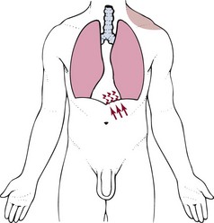
Diaphragmatic Irritation: Irritation of the peritoneal (outside) or pleural (inside) surface of the central diaphragm refers sharp pain to the ipsilateral upper trapezius, neck and/or supraclavicular fossa (Fig. 18-1). Shoulder pain from diaphragmatic irritation usually does not cause anterior shoulder pain. Pain is confined to the suprascapular, upper trapezius, and posterior portions of the shoulder.


If the irritation crosses the midline of the diaphragm, then it is possible to have bilateral shoulder pain. This does not happen very often and is most common with cardiac ischemia or pulmonary pathology affecting the lower lobes of the lungs on both sides. Irritation of the peripheral portion of the diaphragm is more likely to refer pain to the costal margins and lumbar region on the same side.


As you review Fig. 3-4, note how the heart, spleen, kidneys, pancreas (both the body and the tail), and the lungs can put pressure on the diaphragm. This illustration is key to remembering which shoulder can be involved based on organ pathology. For example, the spleen is on the left side of the body so pain from spleen rupture or injury is referred to the left shoulder (called Kehr’s sign) (Case Example 18-5).18


Case Example 18-5   Rugby Injury


Kehr’s Sign


Referral: A 27-year-old male accountant who has an office in the same complex with a physical therapy practice stopped by early Monday morning complaining of left shoulder pain.


When asked about repetitive motions or recent trauma or injuries, he reported playing in a rugby tournament over the weekend. “I got banged up quite a few times, but I had so much beer in me, I didn’t feel a thing.”


Clinical Presentation: Pain was described as a deep, sharp aching over the upper trapezius and shoulder area on the left side. There were no visual bruises or signs of bleeding in the upper left quadrant.


Vital signs:  
Pulse: 89 bpm
Respirations: 12 per minute
Blood pressure: 90/48 mm Hg (recorded sitting, left arm)
Temperature: 97° F (reported as the client’s “normal” morning temperature)
Pain: Rated as a 5 on a scale from 0 to 10

Range of motion was full in all planes and movements. No particular movement increased or decreased the pain. Gross manual muscle test of the upper extremities was normal (5/5 for flexion, abduction, extension, rotations).


Neurologic screen was negative. All special shoulder tests (e.g., impingement, anterior and posterior instability, quadrant position) were unremarkable.


What are the red flags here? What are your next questions, steps, or screening tests?


Red Flags


What are your next questions, steps, or screening tests?


Repeat blood pressure measurements, bilaterally. Perform percussive tests for the spleen (see Fig. 4-53).


Depending on the results of these clinical tests, referral might be needed immediately. In this case, the percussive test for enlarged spleen was inconclusive, but there was an observable and palpable “fullness” in the left flank compared to the right.


Result: This client was told:


“Mr. Smith, your exam does not look like what I would expect from a typical shoulder injury. Since I cannot find any way to make your pain better or worse and I cannot palpate or feel any areas of tenderness, there may be some other cause for your symptoms.


Given your history of playing rugby over the weekend, it is possible you have some internal injuries. I am not comfortable treating you until a medical doctor examines you first. Bleeding from the spleen can cause left shoulder pain. When I tapped over the area of your spleen, it did not sound quite like I expected it to, and it seems like there is some fullness along your left side that I am not seeing or feeling on the right.


I do not want to alarm you, but it may be best to go over to the emergency department of the hospital and see what they have to say. You can also call your regular doctor and see if you can get in right away. You can do that right from our clinic phone.”


Final Result: This accountant had clients already scheduled starting in 10 minutes. He did not feel he had the time to go check this out until his lunch hour. About 45 minutes later an ambulance was called to the building. Mr. Smith had collapsed, and his coworkers called 9-1-1.


He was rushed to the hospital and diagnosed with a torn and bleeding spleen, which the doctor called a “slow leak.” It eventually ruptured, leaving him unconscious from blood loss.


Either shoulder can be involved with renal colic or distention of the renal cap from any kidney disorder, but it is usually an ipsilateral referred pain pattern depending on which kidney is impaired (see Fig. 10-7; again, via pressure on the diaphragm). Bilateral shoulder pain from renal disease would only occur if and when both kidneys are compromised at the same time.


Look for history of a recent surgery as part of the past medical history and the presence of accompanying urologic symptoms.


The body of the pancreas lies along the midline of the diaphragm. When the body of the pancreas is enlarged, inflamed, obstructed, or otherwise impinging on the diaphragm, back pain is a possible referred pain pattern. Pain felt in the left shoulder may result from activation of pain fibers in the left diaphragm by an adjacent inflammatory process in the tail of the pancreas.


Postlaparoscopic shoulder pain (PLSP) frequently occurs after various laparoscopic surgical procedures. During the procedure air is introduced into the peritoneum to expand the area and move the abdominal contents out of the way. The mechanism of PLSP is commonly assumed to be overstretching of the diaphragmatic muscle fibers due to the pressure of a pneumoperitoneum (residual carbon dioxide [CO2] gas after surgery).19 Pressure from distention causes phrenic nerve–mediated referred pain to the shoulder.20


Keep in mind that shoulder pain also can occur from diaphragmatic dysfunction. For anyone with shoulder pain of an unknown origin or which does not improve with intervention, palpate the diaphragm and assess its excursion and timing during respiration. Reproduction of shoulder symptoms with direct palpation of the diaphragm and the presence of altered diaphragmatic movement with breathing offer clues to the possibility of diaphragmatic (muscular) involvement.


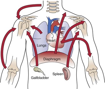
Fig. 18-2 reminds us that shoulder pain can be referred from the neck, back, chest, abdomen, and elbow. During orthopedic assessment, the therapist always checks “above and below” the impaired level for a possible source of referred pain. With this guideline in mind, we know to look for potential musculoskeletal or neuromuscular causes from the cervical and thoracic spine21 and elbow.


Associated Signs and Symptoms


One of the most basic clues in screening for a viscerogenic or systemic cause of shoulder pain is to look for shoulder pain accompanied by any of the following features:


Shoulder pain with any of these present should be approached as a manifestation of systemic visceral illness. This is true even if the pain is exacerbated by shoulder movement or if there are objective findings at the shoulder.24


Using the past medical history and assessing for the presence of associated signs and symptoms will alert the therapist to any red flags suggesting a systemic origin of shoulder symptoms. For example, a ruptured ectopic pregnancy with abdominal hemorrhage can produce left shoulder pain (with or without chest pain) in a woman of childbearing age.2527 The woman is sexually active, and there is usually a history of missed menses or recent unexplained/unexpected bleeding.


The client may not recognize the connection between painful urination and shoulder pain or the link between gallbladder removal by laparoscopy and subsequent shoulder pain. It is the therapist’s responsibility to assess musculoskeletal symptoms, making a diagnosis that includes ruling out the possibility of systemic disease.


Screening for Pulmonary Causes of Shoulder Pain


Extensive disease may occur in the periphery of the lung without pain until the process extends to the parietal pleura. Pleural irritation then results in sharp, localized pain that is aggravated by any respiratory movement.


Clients usually note that the pain is alleviated by lying on the affected side, which diminishes the movement of that side of the chest (called “autosplinting”) whereas shoulder pain of musculoskeletal origin is usually aggravated by lying on the symptomatic shoulder.


Shoulder symptoms made worse by recumbence are a yellow flag for pulmonary involvement. Lying down increases the venous return from the lower extremities. A compromised cardiopulmonary system may not be able to accommodate the increase in fluid volume. Referred shoulder pain from the taxed and overworked pulmonary system may result.


At the same time, recumbency or the supine position causes a slight shift of the abdominal contents in the cephalic direction. This shift may put pressure on the diaphragm, which in turn presses up against the lower lung lobes. The combination of increased venous return and diaphragmatic pressure may be enough to reproduce the musculoskeletal symptoms.


Pneumonia in the older adult may appear as shoulder pain when the affected lung presses on the diaphragm; usually there are accompanying pulmonary symptoms, but in older adults, confusion (or increased confusion) may be the only other associated sign.


The therapist should look for the presence of a pleuritic component such as a persistent or productive cough and/or chest pain. Look for tachypnea, dyspnea, wheezing, hyperventilation, or other noticeable changes. Chest auscultation is a valuable tool when screening for pulmonary involvement.


Screening for Cardiovascular Causes of Shoulder Pain


Pain of cardiac and diaphragmatic origin is often experienced in the shoulder because the heart and diaphragm are supplied by the C5 to C6 spinal segment, and the visceral pain is referred to the corresponding somatic area (see Fig. 3-3).


Exacerbation of the shoulder symptoms from a cardiac cause occurs when the client increases activity that does not necessarily involve the arm or shoulder. For example, walking up stairs or riding a stationary bicycle can bring on cardiac-induced shoulder pain.


In cases like this, the therapist should ask about the presence of nausea, unexplained sweating, jaw pain or toothache, back pain, or chest discomfort or pressure. For the client with known heart disease, ask about the effect of taking nitroglycerin (men) or antacids/acid-relieving drugs (women) on their shoulder symptoms.


Vital sign and physical assessment including chest auscultation are important screening tools. See Chapter 4 for details.


Angina or Myocardial Infarction


Angina and/or myocardial infarction (MI) can appear as arm and shoulder pain that can be misdiagnosed as arthritis or other musculoskeletal pathologic conditions (see complete discussion in Chapter 6 and see Figs. 6-8 and 6-9).


Look for shoulder pain that starts 3 to 5 minutes after the start of activity, including shoulder pain with isolated lower extremity motion (e.g., shoulder pain starts after the client climbs a flight of stairs or rides a stationary bicycle). If the client has known angina and takes nitroglycerin, ask about the influence of the nitroglycerin on shoulder pain.


Shoulder pain associated with MI is unaffected by position, breathing, or movement. Because of the well-known association between shoulder pain and angina, cardiac-related shoulder pain may be medically diagnosed without ruling out other causes, such as adhesive capsulitis or supraspinatus tendinitis, when, in fact, the client may have both a cardiac and a musculoskeletal problem (Case Example 18-6).


Case Example 18-6   Strange Case of the Flu


Referral: A 53-year-old butcher at the local grocery store stopped by the physical therapy clinic located in the same shopping complex with a complaint of unusual shoulder pain. He had been seen at this same clinic several years ago for shoulder bursitis and tendinitis from repetitive overuse (cutting and wrapping meat).


Clinical Presentation: His clinical presentation for this new episode of care was exactly as it had been during the last episode of shoulder impairment. The therapist reinstituted a program of soft tissue mobilization and stretching, joint mobilization, and postural alignment. Modalities were used during the first two sessions to help gain pain control.


At the third appointment, the client mentioned feeling “dizzy and sweaty” all day. His shoulder pain was described as a constant, deep ache that had increased in intensity from a 6 to a 10 on a scale from 0 to 10. He attributed these symptoms to having the flu.


It was not until this point that the therapist conducted a screening exam and found the following red flags:


Result: The therapist suggested the client get a medical checkup before continuing with physical therapy. Even though the clinical presentation supported shoulder impairment, there were enough red flags and soft signs of systemic distress to warrant further evaluation.


Taking vital signs would have been a good idea.


It turns out the client was having myocardial ischemia masquerading as shoulder pain, the flu, and an ear infection. He had an angioplasty with complete resolution of all his symptoms and even reported feeling energetic for the first time in years.


This is a good example of how shoulder pain and dysfunction can exactly mimic a true musculoskeletal problem—even to the extent of reproducing symptoms from a previous condition.


This case highlights the fact that we must be careful to fully assess our clients with each episode of care.


Using a review of symptoms approach and a specific musculoskeletal shoulder examination, the physical therapist can screen to differentiate between a medical pathologic condition and mechanical dysfunction28 (Case Example 18-7).


Case Example 18-7


Angina Versus Shoulder Pathology


Referral: A 54-year-old man was referred to physical therapy for pre-prosthetic training after a left transtibial (TT) amputation.


Past Medical History


Clinical Presentation: At the time of the initial evaluation for the left TT amputation, the client reported substernal chest pain and left upper extremity pain with activity. Typical anginal pain pattern was described as substernal chest pain. The pain occurs with exertion and is relieved by rest.


Arm pain has never been a part of his usual anginal pain pattern. He reports his arm pain began 10 months ago with intermittent pain starting in the left shoulder and radiating down the anterior-medial aspect of the arm, halfway between the shoulder and the elbow.


The pain is made worse by raising his left arm overhead, pushing his own wheelchair, and using a walker. He was not sure if the shoulder pain was caused by repetitive motions needed for mobility or by his angina. The shoulder pain is relieved by avoiding painful motions. He has not received any treatment for the shoulder problem.


Neurologic screen was negative.


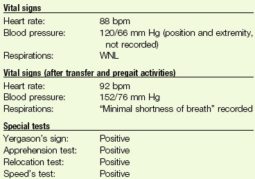
image


Palpation of the biceps and supraspinatus tendons increased the client’s shoulder pain.


image


There is a capsular pattern in the left glenohumeral joint with limitations in rotation and adduction. Significant capsular tightness is demonstrated with passive or physiologic motions (joint play) of the humerus on the glenoid.


image


Review of Systems: Dyspnea, fatigue, sweats with pain; when grouped together, these three symptoms fall under the Cardiovascular category; these do not occur at the same time as the shoulder pain.


• How can you differentiate between medical pathology and mechanical dysfunction as the cause of this client’s shoulder pain?


• Is a medical referral advised?


Result: Test results point to an untreated biceps and supraspinatus tendinitis. This tendinitis combined with adhesive capsulitis most likely accounted for the left shoulder pain. This assessment was based on the decreased left glenohumeral AROM and decreased joint mobility.


With objective clinical findings to support a musculoskeletal dysfunction, medical referral was not required. There were no indications that the shoulder pain was a signal of a change in the client’s anginal pattern.


Left shoulder impairments were limiting factors in his mobility and rehabilitation process. Shoulder intervention to alleviate pain and to improve upper extremity strength were included in the plan of care. The desired outcome was to improve transfer and gait activities.


Left shoulder pain resolved within the first week of physical therapy intervention. This gain made it possible to improve ambulation from 3 feet to 50 feet with a walker while wearing a right lower extremity prosthesis.


The client gained independence with bed mobility and supine-to-sit transfers. The client continued to make improvements in ambulation, range of motion, and functional mobility.


Physical therapy intervention for the shoulder impairments had a significant impact on the outcomes of this client’s rehab program. By differentiating and treating the shoulder movement dysfunction, the intervention enabled the client to progress faster in the transfer and gait training program than he would have had his left shoulder pain been attributed to angina.28


Data from Smith ML: Differentiating angina and shoulder pathology pain, Phys Ther Case Rep 1(4):210–212, 1998.

Only gold members can continue reading. Log In or Register to continue

Stay updated, free articles. Join our Telegram channel

Mar 20, 2017 | Posted by in MANUAL THERAPIST | Comments Off on Screening the Shoulder and Upper Extremity

Full access? Get Clinical Tree

Get Clinical Tree app for offline access