Screening for Pulmonary Disease

Chapter 7


Screening for Pulmonary Disease


For the client with neck, shoulder, or back pain at presentation, it may be necessary to consider the possibility of a pulmonary cause requiring medical referral. The most common pulmonary conditions to mimic those of the musculoskeletal system include pneumonia, pulmonary embolism, pleurisy, pneumothorax, and pulmonary arterial hypertension.


As always, using the past medical history, risk factor assessment, and clinical presentation, as well as asking about the presence of any associated signs and symptoms guide the screening process. In the case of pleuropulmonary disorders, the client’s recent personal medical history may include a previous or recurrent upper respiratory infection or pneumonia.


Pneumothorax may be preceded by trauma, overexertion, or recent scuba diving. Each pulmonary condition will have its own unique risk factors that can predispose clients to a specific respiratory disease or illness.


A previous history of cancer, especially primary lung cancer or cancers that metastasize to the lungs (e.g., breast, bone), is a red flag and a risk factor for cancer recurrence. Risk factor assessment also helps identify increased risk for other respiratory conditions or illnesses that can present as a primary musculoskeletal problem.


The material in this chapter will assist the therapist in treating both the client with a known pulmonary problem and the client with musculoskeletal signs and symptoms that may have an underlying systemic basis (Case Example 7-1).


Signs and Symptoms of Pulmonary Disorders


Signs and symptoms of pulmonary disorders can be many and varied; the most common symptoms associated with pulmonary disorders are cough and dyspnea. Other manifestations include chest pain, abnormal sputum, hemoptysis, cyanosis, digital clubbing, altered breathing patterns, and chest pain.


Dyspnea


Shortness of breath (SOB), or dyspnea, usually indicates hypoxemia but can be associated with emotional states, particularly fear and anxiety. Dyspnea is usually caused by diffuse and extensive rather than focal pulmonary disease; pulmonary embolism is the exception. Factors contributing to the sensation of dyspnea include increased work of breathing (WOB), respiratory muscle fatigue, increased systemic metabolic demands, and decreased respiratory reserve capacity. Dyspnea when the person is lying down is called orthopnea and is caused by redistribution of body water. Fluid shift leads to increased fluid in the lung, which interferes with gas exchange and leads to orthopnea. In supine and prone, the abdominal contents also exert pressure on the diaphragm, increasing the WOB and often limiting vital capacity.


The therapist must be careful when screening for dyspnea or SOB, either with exertion or while at rest. If a client denies compromised breathing, look for functional changes as the client accommodates for difficulty breathing by reducing activity or exertion.


Clubbing (see Chapter 4)


Thickening and widening of the terminal phalanges of the fingers and toes result in a painless clublike appearance recognized by the loss of the angle between the nail and the nail bed (see Figs. 4-36 and 4-37). Conditions that chronically interfere with tissue perfusion and nutrition may cause clubbing, including cystic fibrosis (CF), chronic obstructive pulmonary disease (COPD), lung cancer, bronchiectasis, pulmonary fibrosis, congenital heart disease, and lung abscess. Most of the time, clubbing is due to pulmonary disease and resultant hypoxia (diminished availability of blood to the body tissues), but clubbing can be a sign of heart disease, peripheral vascular disease, and disorders of the liver and gastrointestinal tract.


Altered Breathing Patterns


Changes in the rate, depth, regularity, and effort of breathing occur in response to any condition affecting the pulmonary system. Breathing patterns can vary, depending on the neuromuscular or neurologic disease or trauma (Box 7-1). Breathing pattern abnormalities seen with head trauma, brain abscess, diaphragmatic paralysis of chest wall muscles and thorax (e.g., generalized myopathy or neuropathy), heat stroke, spinal meningitis, and encephalitis can include apneustic breathing, ataxic breathing, or Cheyne-Stokes respiration (CSR).


Apneustic breathing (gasping inspiration with short expiration) localizes damage to the midpons and is most commonly a result of a basilar artery infarct. Ataxic, or Biot’s, breathing (irregular pattern of deep and shallow breaths with abrupt pauses) is caused by disruption of the respiratory rhythm generator in the medulla.


CSR may be evident in the well older adult, as well as in compromised clients. The most common cause of CSR is severe congestive heart failure (CHF), but it can also occur with renal failure, meningitis, drug overdose, and increased intracranial pressure. It may be a normal breathing pattern in infants and older persons during sleep.


Exercise may induce pleural pain, coughing, hemoptysis, SOB, and/or other abnormal changes in breathing patterns. When asked if the client is ever short of breath, the individual may say “no” because he or she has reduced activity levels to avoid dyspnea (see Appendix B-12).


Pulmonary Pain Patterns


The most common sites for referred pain from the pulmonary system are the chest, ribs, upper trapezius, shoulder, and thoracic spine. The first symptoms may not appear until the client’s respiratory system is stressed by the addition of exercise during physical therapy. On the other hand, the client may present with what appears to be primary musculoskeletal pain in any one of those areas. Auscultation may reveal the first signs of pulmonary distress (see Chapter 4 for screening examination by auscultation).


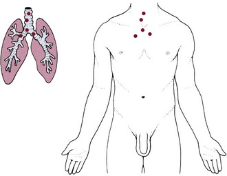
Pulmonary pain patterns are usually localized in the substernal or chest region over involved lung fields that may include the anterior chest, side, or back (Fig. 7-1). However, pulmonary pain can radiate to the neck, upper trapezius muscle, costal margins, thoracic back, scapulae, or shoulder. Shoulder pain may radiate along the medial aspect of the arm, mimicking other neuromuscular causes of neck or shoulder pain (see Fig. 7-10).


Pulmonary pain usually increases with inspiratory movements, such as laughing, coughing, sneezing, or deep breathing, and the client notes the presence of associated symptoms, such as dyspnea (exertional or at rest), persistent cough, fever, and chills. Palpation and resisted movements will not reproduce the symptoms, which may get worse with recumbency, especially at night or while sleeping.


The thoracic cavity is lined with pleura, or serous membrane. One surface of the pleura lines the inside of the rib cage (parietal) and the other surface covers the lungs (visceral). The parietal pleura is sensitive to painful stimulation, but the visceral pleura is insensitive to pain. Extensive disease may occur in the lung without occurrence of pain until the process extends to the parietal pleura. This explains why pathology of the lungs may be painless until obstruction or inflammation is enough to press on the parietal pleura.


Pleural irritation then results in sharp, localized pain that is aggravated by any respiratory movement. Clients usually note that the pain is alleviated by autosplinting, that is, lying on the affected side, which diminishes the movement of that side of the chest (see further discussion of pleural pain in this chapter).1


Pleural Pain


When the disease progresses enough to extend to the parietal pleura, pleural irritation occurs and results in sharp, localized pain that is aggravated by any respiratory movement. Clients usually note that the pain is alleviated by lying on the affected side, which diminishes the movement of that side of the chest called autosplinting.


Debate continues concerning the mechanism by which pain occurs in the parietal membrane. It has been long thought that friction between the two pleural surfaces (when the membranes are irritated and covered with fibrinous exudate) causes sharp pain. Other theories suggest that intercostal muscle spasm resulting from pleurisy or stretching of the parietal pleura causes this pain.


Pleural pain is present in pulmonary diseases such as pleurisy, pneumonia, pulmonary infarct (when it extends to the pleural surface, thus causing pleurisy), tumor (when it invades the parietal pleura), and pneumothorax. Tumor, especially bronchogenic carcinoma, may be accompanied by severe, continuous pain when the tumor tissue, extending to the parietal pleura through the lung, constantly irritates the pain nerve endings in the pleura.


Diaphragmatic Pleural Pain


The diaphragmatic pleura receives dual pain innervation through the phrenic and intercostal nerves. Damage to the phrenic nerve produces paralysis of the corresponding half of the diaphragm. The phrenic nerves are sensory and motor from both surfaces of the diaphragm.


Stimulation of the peripheral portions of the diaphragmatic pleura results in sharp pain felt along the costal margins, which can be referred to the lumbar region by the lower thoracic somatic nerves. Stimulation of the central portion of the diaphragmatic pleura results in sharp pain referred to the upper trapezius muscle and shoulder on the ipsilateral side of the stimulation (see Figs. 3-4 and 3-5).


Pain of cardiac and diaphragmatic origin is often experienced in the shoulder because the heart and diaphragm are supplied by the C5-6 spinal segment, and the visceral pain is referred to the corresponding somatic area.


Diaphragmatic pleurisy secondary to pneumonia is common and refers sharp pain along the costal margins or upper trapezius, which is aggravated by any diaphragmatic motion, such as coughing, laughing, or deep breathing.


There may be tenderness to palpation along the costal margins, and sharp pain occurs when the client is asked to take a deep breath. A change in position (sidebending or rotation of the trunk) does not reproduce the symptoms, which would be the case with a true intercostal lesion or tear.


Forceful, repeated coughing can result in an intercostal lesion in the presence of referred intercostal pain from diaphragmatic pleurisy, which can make differentiation between these two entities impossible without a medical referral and further diagnostic testing.


Pulmonary Physiology


The primary function of the respiratory system is to provide oxygen to and to remove carbon dioxide from cells in the body. The act of breathing, in which the oxygen and carbon dioxide exchange occurs, involves the two interrelated processes of ventilation and respiration.


Ventilation is the movement of air from outside the body to the alveoli of the lungs. Respiration is the process of oxygen uptake and carbon dioxide elimination between the body and the outside environment.


Breathing is an automatic process by which sensors detect changes in the levels of carbon dioxide and continuously direct data to the medulla. The medulla then directs respiratory muscles that adjust ventilation. Breathing patterns can be altered voluntarily when this automatic response is overridden by conscious thought.


The major sensors mentioned here are the central chemoreceptors (located near the medulla) and the peripheral sensors (located in the carotid body and aortic arch). The central chemoreceptors respond to increases in carbon dioxide and decreases in pH in cerebrospinal fluid.


As carbon dioxide increases, the medulla signals a response to increase respiration. The peripheral chemoreceptor system responds to low arterial blood oxygen and is believed to function only in pathologic situations such as when there are chronically elevated carbon dioxide levels (e.g., COPD).


Acid-Base Regulation


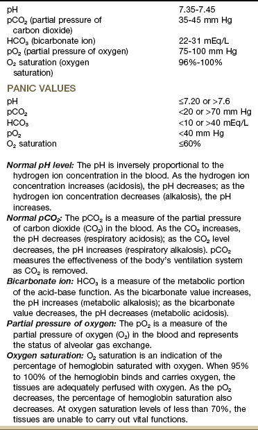
The proper balance of acids and bases in the body is essential to life. This balance is very complex and must be kept within the narrow parameters of a pH of 7.35 to 7.45 in the extracellular fluid. This number (or pH value) represents the hydrogen ion concentration in body fluid.


A reading of less than 7.35 is considered acidosis, and a reading greater than 7.45 is called alkalosis. Life cannot be sustained if the pH values are less than 7 or greater than 7.8.


Living human cells are extremely sensitive to alterations in body fluid pH (hydrogen ion concentration); thus various mechanisms are in operation to keep the pH at a relatively constant level.


Acid-base regulatory mechanisms include chemical buffer systems, the respiratory system, and the renal system. These systems interact very closely to maintain a normal acid-base ratio of 20 parts of bicarbonate to 1 part of carbonic acid and thus to maintain normal body fluid pH.


The blood test used most often to measure the effectiveness of ventilation and oxygen transport is the arterial blood gas (ABG) test (Table 7-1). The measurement of arterial blood gases is important in the diagnosis and treatment of ventilation, oxygen transport, and acid-base problems.


The ABG test measures the amount of dissolved oxygen and carbon dioxide in arterial blood and indicates acid-base status by measurement of the arterial blood pH. In simple terms a low pH reflects increased acid buildup, and a high pH reflects an increased base buildup.


Acid buildup occurs when there is an ineffective removal of carbon dioxide from the lungs or when there is excess acid production from the tissues of the body. These problems are corrected by adjusting the ventilation or buffering the acid with bicarbonate.


Pulmonary Pathophysiology


Respiratory Acidosis


Any condition that decreases pulmonary ventilation increases the retention and concentration of carbon dioxide (CO2), hydrogen, and carbonic acid; this results in an increase in the amount of circulating hydrogen and is called respiratory acidosis.


If ventilation is severely compromised, CO2 levels become extremely high and respiration is depressed even further, causing hypoxia as well.


During respiratory acidosis, potassium moves out of cells into the extracellular fluid to exchange with circulating hydrogen. This results in hyperkalemia (abnormally high potassium concentration in the blood) and cardiac changes that can cause cardiac arrest.


Respiratory acidosis can result from pathologic conditions that decrease the efficiency of the respiratory system. These pathologies can include damage to the medulla, which controls respiration, obstruction of airways (e.g., neoplasm, foreign bodies, pulmonary disease such as COPD, pneumonia), loss of lung surface ventilation (e.g., pneumothorax, pulmonary fibrosis), weakness of respiratory muscles (e.g., poliomyelitis, spinal cord injury, Guillain-Barré syndrome), or overdose of respiratory depressant drugs.


As hypoxia becomes more severe, diaphoresis, shallow rapid breathing, restlessness, and cyanosis may appear. Cardiac arrhythmias may also be present as the potassium level in the blood serum rises.


Treatment is directed at restoration of efficient ventilation. If the respiratory depression and acidosis are severe, injection of intravenous sodium bicarbonate and use of a mechanical ventilator may be necessary. Any client with symptoms of inadequate ventilation or CO2 retention needs immediate medical referral.


Respiratory Alkalosis


Increased respiratory rate and depth decrease the amount of available CO2 and hydrogen and create a condition of increased pH, or alkalosis. When pulmonary ventilation is increased, CO2 and hydrogen are eliminated from the body too quickly and are not available to buffer the increasingly alkaline environment.


Respiratory alkalosis is usually due to hyperventilation. Rapid, deep respirations are often caused by neurogenic or psychogenic problems, including anxiety, pain, and cerebral trauma or lesions. Other causes can be related to conditions that greatly increase metabolism (e.g., hyperthyroidism) or overventilation of clients who are using a mechanical ventilator.


If the alkalosis becomes more severe, muscular tetany and convulsions can occur. Cardiac arrhythmias caused by serum potassium loss through the kidneys may also occur. The kidneys keep hydrogen in exchange for potassium.


Treatment of respiratory alkalosis includes reassurance, assistance in slowing breathing and facilitating relaxation, sedation, pain control, CO2 administration, and use of a rebreathing device such as a rebreathing mask or paper bag. A rebreathing device allows the client to inhale and “rebreathe” the exhaled CO2.


Respiratory alkalosis related to hyperventilation is a relatively common condition and might be present more often in the physical therapy setting than respiratory acidosis. Pain and anxiety are common causes of hyperventilation, and treatment needs to be focused toward reduction of both of these interrelated elements. If hyperventilation continues in the absence of pain or anxiety, serious systemic problems may be the cause, and immediate physician referral is necessary.


If either respiratory acidosis or alkalosis persists for hours to days in a chronic and not life-threatening manner, the kidneys then begin to assist in the restoration of normal body fluid pH by selective excretion or retention of hydrogen ions or bicarbonate. This process is called renal compensation. When the kidneys compensate effectively, blood pH values are within normal limits (7.35 to 7.45) even though the underlying problem may still cause the respiratory imbalance.


Chronic Obstructive Pulmonary Disease


COPD, also called chronic obstructive lung disease (COLD), refers to a number of disorders that have in common abnormal airway structures resulting in obstruction of air in and out of the lungs. The most important of these disorders are obstructive bronchitis, emphysema, and asthma.


Although bronchitis, emphysema, and asthma may occur in a “pure form,” they most commonly coexist. For example, adult subjects with active asthma are as much as 12 times more likely to acquire COPD over time than subjects with no active asthma.25


COPD is a leading cause of morbidity and mortality among cigarette smokers. Other factors predisposing to COPD include air pollution; occupational exposure to aerosol pesticides, irritating dusts or gases, or art materials (e.g., paint, glass, ceramics, sculpture); hereditary factors; infection; allergies; aging; and potentially harmful drugs and chemicals.6


COPD rarely occurs in nonsmokers; however, only a minority of cigarette smokers develop symptomatic disease, suggesting that genetic factors or other underlying predisposition may contribute to the development of COPD.7


In all forms of COPD, narrowing of the airways obstructs airflow to and from the lungs (Table 7-2). This narrowing impairs ventilation by trapping air in the bronchioles and alveoli. The obstruction increases the resistance to airflow. The severity of symptoms depends on how much of the lungs have been damaged or destroyed.


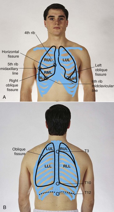
Trapped air hinders normal gas exchange and causes distention of the alveoli. Other mechanisms of COPD vary with each form of the disease. In the healthy adult the bottom margin of the respiratory diaphragm sits at T9 when the lungs are at rest. Taking a deep breath expands the diaphragm (and lungs) inferiorly to T11. For the client with COPD the lower lung lobes are already at T11 when the lungs are at rest from overexpansion as a result of alveoli distention and hyperinflation.


COPD develops earlier in life than is usually recognized, making it the most underdiagnosed and undertreated pulmonary disease. Smoking cessation is the only intervention shown to slow decline in lung function. Identifying risk factors and recognizing early signs and symptoms of COPD increases the affected individual’s chances of reduced morbidity through early intervention.6


Bronchitis:


Acute: Acute bronchitis is an inflammation of the trachea and bronchi (tracheobronchial tree) that is self-limiting and of short duration with few pulmonary signs. This condition may result from chemical irritation (e.g., smoke, fumes, gas) or may occur with viral infections such as influenza, measles, chickenpox, or whooping cough.


These predisposing conditions may become apparent during the subjective examination (i.e., Personal/Family History form or the Physical Therapy Interview). Although bronchitis is usually mild, it can become complicated in older clients and clients with chronic lung or heart disease. Pneumonia is a critical complication.


Chronic: Chronic bronchitis is a condition associated with prolonged exposure to nonspecific bronchial irritants and is accompanied by mucus hypersecretion and structural changes in the bronchi (large air passages leading into the lungs). This irritation of the tissue usually results from exposure to cigarette smoke or long-term inhalation of dust or air pollution and causes hypertrophy of mucus-producing cells in the bronchi.


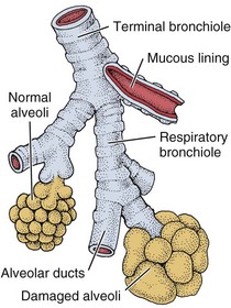
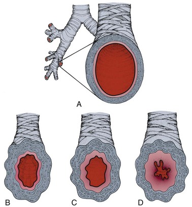
In bronchitis, partial or complete blockage of the airways from mucus secretions causes insufficient oxygenation in the alveoli (Fig. 7-3). The swollen mucous membrane and thick sputum obstruct the airways, causing wheezing, and the client develops a cough to clear the airways. The clinical definition of a person with chronic bronchitis is anyone who coughs for at least 3 months per year for 2 consecutive years without having had a precipitating disease.


To confirm that the condition is chronic bronchitis, tests are performed to determine whether the airways are obstructed and to exclude other diseases that may cause similar symptoms such as silicosis, tuberculosis, or a tumor in the upper airway. Sputum samples will be analyzed, and lung function tests may be performed.


Treatment is aimed at keeping the airways as clear as possible. Smokers are encouraged and helped to stop smoking. A combination of drugs may be prescribed to relieve the symptoms, including bronchodilators to open the obstructed airways and to thin the obstructive mucus so that it can be coughed up more easily.


Chronic bronchitis may develop slowly over a period of years, but it will not go away if untreated. Eventually, the bronchial walls thicken, and the number of mucous glands increases. The client is increasingly susceptible to respiratory infections, during which the bronchial tissue becomes inflamed and the mucus becomes even thicker and more profuse.


Chronic bronchitis can be incapacitating and lead to more serious and potentially fatal lung disease. Influenza and pneumococcal vaccines are recommended for these clients.


Bronchiectasis: Bronchiectasis is a form of obstructive lung disease that is actually a type of bronchitis. It is a progressive and chronic pulmonary condition that occurs after infections such as childhood pneumonia or CF.


Although bronchiectasis was once a common disease because of measles, pertussis, tuberculosis, and poorly treated bacterial pneumonias, the prevalence of bronchiectasis has diminished greatly since the introduction of antibiotics. It is characterized by abnormal and permanent dilatation of the large air passages leading into the lungs (bronchi) and by destruction of bronchial walls.


Bronchiectasis is caused by repeated damage to bronchial walls. The resultant destruction and bronchial dilatation reduce bronchial wall movement so that secretions cannot be removed effectively from the lungs, and the person is predisposed to frequent respiratory infections.


This vicious cycle of bacterial infection and inflammation of the bronchial wall leads to loss of ventilation and irreversible lung damage. Advanced bronchiectasis may cause pneumonia, cor pulmonale, or right-sided ventricular failure.


All pulmonary irritants, especially cigarette smoke, should be avoided. Postural drainage, adequate hydration, good nutrition, and bronchodilator therapy in bronchospasm are important components in treatment. Antibiotics are used during disease exacerbations (e.g., increased cough, purulent sputum, hemoptysis, malaise, and weight loss). The use of immunomodulatory therapy to alter the host response directly and thereby reduce tissue damage is under investigation.8,9


Emphysema: Emphysema may develop in a person after a long history of chronic bronchitis in which the alveolar walls are destroyed, leading to permanent overdistention of the air spaces and loss of normal elastic tension in the lung tissue.


Air passages are obstructed as a result of these changes (rather than as a result of mucus production, as in chronic bronchitis). Difficult expiration in emphysema is due to the destruction of the walls (septa) between the alveoli, partial airway collapse, and loss of elastic recoil.


As the alveoli and septa collapse, pockets of air form between the alveolar spaces (called blebs) and within the lung parenchyma (called bullae). This process leads to increased ventilatory “dead space,” or areas that do not participate in gas or blood exchange. The WOB is increased because there is less functional lung tissue to exchange oxygen and CO2. Emphysema also causes destruction of the pulmonary capillaries, further decreasing oxygen perfusion and ventilation.


In advanced emphysema, oxygen therapy is usually necessary to treat the progressive hypoxemia that occurs as the disease worsens. Oxygen therapy is carefully titrated and monitored to maintain venous oxygen saturation levels at or slightly above 90%. Too much oxygen can depress the respiratory drive of a person with emphysema.


The drive to breathe in a healthy person results from an increase in the arterial carbon dioxide level (pCO2). In the normal adult, increased CO2 levels stimulate chemoreceptors in the brainstem to increase the respiratory rate. With some chronic lung disorders these central chemoreceptors may become desensitized to pCO2 changes resulting in a dependence on the peripheral chemoreceptors to detect a fall in arterial oxygen levels (pO2) to stimulate the respiratory drive. Therefore too much oxygen delivered as a treatment can depress the respiratory drive in those individuals with COPD who have a dampening of the CO2 drive.


Monitoring respiratory rate, level of oxygen administered by nasal canula, and oxygen saturation levels is very important in this client population. Some pulmonologists agree that supplemental oxygen levels can be increased during activity without compromising the individual because they will “blow it (carbon dioxide) off” anyway. To our knowledge, there is no evidence yet to support this clinical practice.


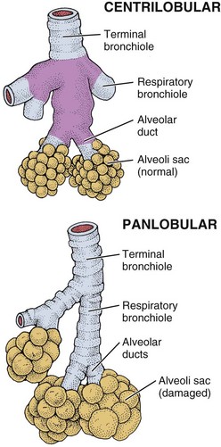
Types of Emphysema: There are three types of emphysema. Centrilobular emphysema (Fig. 7-4), the most common type, destroys the bronchioles, usually in the upper lung regions. Inflammation develops in the bronchioles, but usually the alveolar sac remains intact.


Panlobular emphysema destroys the more distal alveolar walls, most commonly involving the lower lung. This destruction of alveolar walls may occur secondary to infection or to irritants (most commonly, cigarette smoke). These two forms of emphysema, collectively called centriacinar emphysema, occur most often in smokers.


Paraseptal (or panacinar) emphysema destroys the alveoli in the lower lobes of the lungs, resulting in isolated blebs along the lung periphery. Paraseptal emphysema is believed to be the likely cause of spontaneous pneumothorax.


Clinical Signs and Symptoms: The irreversible destruction reduces elasticity of the lung and increases the effort to exhale trapped air, causing marked dyspnea on exertion later progressing to dyspnea at rest. Cough is uncommon.


The client is often thin, has tachypnea with prolonged expiration, and uses the accessory muscles for respiration. The client often leans forward with the arms braced on the knees to support the shoulders and chest for breathing. The combined effects of trapped air and alveolar distention change the size and shape of the client’s chest, causing a barrel chest and increased expiratory effort.


As the disease progresses, there is a loss of surface area available for gas exchange. In the final stages of emphysema, cardiac complications, especially enlargement and dilatation of the right ventricle, may develop. The overloaded heart reaches its limit of muscular compensation and begins to fail (cor pulmonale).


The most important factor in the treatment of emphysema is smoking cessation. The main goals for the client with emphysema are to improve oxygenation and decrease CO2 retention.


Pursed-lip breathing causes resistance to outflow at the lips, which in turn maintains intrabronchial pressure and improves the mixing of gases in the lungs. This type of breathing should be encouraged to help the client get rid of the stale air trapped in the lungs.


Exercise has not been shown to improve pulmonary function but is used to enhance cardiovascular fitness and train skeletal muscles to function more effectively. Routine progressive walking is the most common form of exercise.


Lung volume reduction surgery is available for some individuals and improves not only lung function and exercise performance, but also activities of daily function and quality of life.10,11


Inflammatory/Infectious Disease


Asthma


Asthma is a reversible obstructive lung disease caused by increased reaction of the airways to various stimuli. It is a chronic inflammatory condition with acute exacerbations that can be life-threatening if not properly managed. Our understanding of asthma has changed dramatically over the last decade.


Asthma was once viewed as a bronchoconstrictive disorder in which the airways narrowed, causing wheezing and breathing difficulties. Treatment with bronchodilators to open airways was the primary focus. Scientific evidence now supports the idea that asthma is primarily an inflammatory disorder in which the constriction of airways is a symptom of the underlying inflammation.


Asthma and other atopic disorders are the result of complex interactions between genetic predisposition and multiple environmental influences. The marked increase in asthma prevalence in the last 3 decades suggests environmental factors as a key contributor in the process of allergic sensitization.12


Fifteen million persons of all ages are affected by asthma in the United States. This represents a 61% increase over the last 15 years with a 45% increase in mortality during the last decade. Women are affected more than men, accounting for about 60% of the nearly 18 million cases of adult asthma. Hormones are thought to be a possible cause for this increase in incidence in women.13


Immune Sensitization and Inflammation


There are two major components to asthma. When the immune system becomes sensitized to an allergen, usually through heavy exposure in early life, an inflammatory cascade occurs, extending beyond the upper airways into the lungs.


The lungs become hyperreactive, responding to allergens and other irritants in an exaggerated way. This hyperresponsiveness causes the muscles of the airways to constrict, making breathing more difficult (Fig. 7-5). The second component is inflammation, which causes the air passages to swell and the cells lining the passages to produce excess mucus, further impairing breathing.


Asthma may be categorized as conventional asthma, occupational asthma, or exercise-induced asthma (EIA), but the underlying pathophysiologic complex remains the same. Since the triggers or allergens vary, each person reacts differently. SOB, wheezing, tightness in the chest, and cough are the most commonly reported symptoms, but other symptoms may also occur.


Clinical Signs and Symptoms


Anytime a client experiences SOB, wheezing, and cough and comments, “I’m more out of shape than I thought,” the therapist should ask about a past medical history of asthma and review the list of symptoms with the client. Therapists working with known asthmatic clients should encourage them to maintain hydration by drinking fluids to prevent mucous plugs from hardening and to take prescribed medications.


EIA or hyperventilation-induced asthma potentially can be prevented by exercising in a moist, humid environment and by grading exercise according to client tolerance using diaphragmatic breathing. Any type of sustained running or cycling or activity in the cold is more likely to precipitate EIA (Box 7-2).


Complications: Status asthmaticus is a severe, life-threatening complication of asthma. With severe bronchospasm the workload of breathing increases five to ten times, which can lead to acute cor pulmonale. When air is trapped, a severe paradoxic pulse develops as venous return is obstructed. This condition is seen as a blood pressure drop of more than 10 mm Hg during inspiration.


Pneumothorax can develop. If status asthmaticus continues, hypoxemia worsens and acidosis begins. If the condition is untreated or not reversed, respiratory or cardiac arrest will occur. An acute asthma episode may constitute a medical emergency.


Medical treatment for the underlying inflammation and resulting airway obstruction is with antiinflammatory agents and bronchodilators to prevent, interrupt, or terminate ongoing inflammatory reactions in the airways. A new class of antiinflammatory agents known as leukotriene modifiers works by blocking the activity of chemicals called leukotrienes, which are involved in airway inflammation. Reducing, eliminating, and avoiding allergens or triggers are important in self-care (see Box 7-2).


Pneumonia


Pneumonia is an inflammation of the lungs and can be caused by (1) aspiration of food, fluids, or vomitus; (2) inhalation of toxic or caustic chemicals, smoke, dust, or gases; or (3) a bacterial, viral, or mycoplasmal infection. It may be primary or secondary (a complication of another disease); it often follows influenza.


The common feature of all types of pneumonia is an inflammatory pulmonary response to the offending organism or agent. This response may involve one or both lungs at the level of the lobe (lobar pneumonia) or more distally beginning in the terminal bronchioles and alveoli (bronchopneumonia). Bronchopneumonia is seen more frequently than lobar pneumonia and is common in clients postoperatively and in clients with chronic bronchitis, particularly when these two situations coexist.


There are three main types of pneumonia: hospital-acquired pneumonia (HAP; also known as nosocomial pneumonia), ventilator-associated pneumonia (VAP), and community-acquired pneumonia (CAP). By definition, HAP occurs 48 hours or more after hospital admission; when HAP develops in a mechanically ventilated patient after endotracheal intubation, it becomes VAP.


Risk Factors


Infectious agents responsible for pneumonia are typically present in the upper respiratory tract and cause no harm unless resistance is lowered severely by some other factor such as smoking, a severe cold, disease, alcoholism, or generally poor health (e.g., poorly controlled diabetes, chronic renal problems, compromised immune function).


Older or bedridden clients are particularly at risk because of physical inactivity and immobility. Limited mobility causes normal secretions to pool in the airways and facilitates bacterial growth. Other risk factors predisposing a client to pneumonia are listed in Box 7-3.


Box 7-3   Risk Factors for Pneumonia


• Age: very young, very old


• Have not received a pneumococcal vaccination


• Smoking


• Air pollution


• Upper respiratory infection (URI)


• Altered consciousness: alcoholism, head injury, seizure disorder, drug overdose, general anesthesia


• Endotracheal intubation, nasogastric tube


• Recent chest surgery


• Prolonged immobility


• Immunosuppressive therapy: corticosteroids, cancer chemotherapy


• Nonfunctional immune system: acquired immunodeficiency syndrome (AIDS)


• Severe periodontal disease


• Prolonged exposure to virulent organisms


• Malnutrition, dehydration


• Chronic diseases: diabetes mellitus, heart disease, chronic lung disease, renal disease, cancer


• Prolonged debilitating disease


• Inhalation of noxious substances


• Aspiration of oral/gastric material (food or fluid), foreign materials (e.g., Petroleum products)


• Chronically ill, older clients who have poor immune systems, often residing in group-living situations; transfer from one health care facility to another (hospital-acquired or nosocomial pneumonia); hospitalization in the fall or winter

Stay updated, free articles. Join our Telegram channel

Mar 20, 2017 | Posted by in MANUAL THERAPIST | Comments Off on Screening for Pulmonary Disease

Full access? Get Clinical Tree

Get Clinical Tree app for offline access