Screening for Hematologic Disease

Chapter 5


Screening for Hematologic Disease


The blood consists of two major components: plasma, a pale yellow or gray-yellow fluid; and formed elements, erythrocytes (red blood cells [RBCs]), leukocytes (white blood cells [WBCs]), and platelets (thrombocytes). Blood is the circulating tissue of the body; the fluid and its formed elements circulate through the heart, arteries, capillaries, and veins.


The erythrocytes carry oxygen to tissues and remove carbon dioxide from them. Leukocytes act in inflammatory and immune responses. The plasma carries antibodies and nutrients to tissues and removes wastes from tissues. Platelets, together with coagulation factors in plasma, control the clotting of blood.


Primary hematologic diseases are uncommon, but hematologic manifestations secondary to other diseases are common. Cancers of the blood are discussed in Chapter 13.


In the physical therapist’s practice, symptoms of blood disorders are most common in relation to the use of nonsteroidal antiinflammatory drugs (NSAIDs) for inflammatory conditions, neurologic complications associated with pernicious anemia, and complications of chemotherapy or radiation.


Hematologic considerations in the orthopedic population fall into two main categories: bleeding and clotting. People with known abnormalities of hemostasis (either hypocoagulation or hypercoagulation problems) will require close observation.1


All surgical patients, neurologically compromised, or immobilized individuals must also be observed carefully for any signs or symptoms of venous thromboembolism.


Signs and Symptoms of Hematologic Disorders


There are many signs and symptoms that can be associated with hematologic disorders. Some of the most important indicators of dysfunction in this system include problems associated with exertion (often minimal exertion) such as dyspnea, chest pain, palpitations, severe weakness, and fatigue. Neurologic symptoms, such as headache, drowsiness, dizziness, syncope, or polyneuropathy, can also indicate a variety of possible problems in this system.2,3


Significant skin and fingernail bed changes that can occur with hematologic problems might include pallor of the face, hands, nail beds, and lips; cyanosis or clubbing of the fingernail beds; and wounds or easy bruising or bleeding in skin, gums, or mucous membranes, often with no reported trauma to the area. The presence of blood in the stool or emesis or severe pain and swelling in joints and muscles should also alert the physical therapist to the possibility of a hematologic-based systemic disorder and can sometimes be a critical indicator of bleeding disorders that can be life threatening.2,3


Many hematologic-induced signs and symptoms seen in the physical therapy practice occur as a result of medications. For example, chronic or long-term use of steroids and NSAIDs can lead to gastritis and peptic ulcer with gastrointestinal (GI) bleeding and subsequent iron deficiency anemia.4 Leukopenia, a common problem occurring during chemotherapy, or as a symptom of certain types of cancer, can produce symptoms of infections such as fever, chills, tissue inflammation; severe mouth, throat and esophageal pain; and mucous membrane ulcerations.5


Thrombocytopenia (decreased platelets) associated with easy bruising and spontaneous bleeding is a result of the pharmacologic treatment of common conditions seen in a physical therapy practice such as rheumatoid arthritis and cancer. More about this condition will be included later in this chapter.


Classification of Blood Disorders


Erythrocyte Disorders


Erythrocytes (red blood cells) consist mainly of hemoglobin and a supporting framework. Erythrocytes transport oxygen and carbon dioxide; they are important in maintaining normal acid-base balance. There are many more erythrocytes than leukocytes (600 to 1). The total number is determined by gender (women have fewer erythrocytes than men), altitude (less oxygen in the air requires more erythrocytes to carry sufficient amounts of oxygen to the tissues), and physical activity (sedentary people have fewer erythrocytes, athletes have more).


Disorders of erythrocytes are classified as follows (not all of these conditions are discussed in this text):


Anemia


Anemia is a reduction in the oxygen-carrying capacity of the blood as a result of an abnormality in the quantity or quality of erythrocytes. Anemia is not a disease but is a symptom of any number of different blood disorders. Excessive blood loss, increased destruction of erythrocytes, and decreased production of erythrocytes are the most common causes of anemia.6


In the physical therapy practice, anemia-related disorders usually occur in one of four broad categories:


Anemia with neoplasia may be a common complication of chemotherapy or develop as a consequence of bone marrow metastasis.7 Anemia can also occur as a symptom of leukemia. Adults with pernicious anemia have significantly higher risks for hip fracture even with vitamin B12 therapy. The hypothesized underlying factor is a lack of gastric acid (achlorhydria).8


Clinical Signs and Symptoms: Deficiency in the oxygen-carrying capacity of blood may result in disturbances in the function of many organs and tissues leading to various symptoms that differ from one person to another. Slowly developing anemia in young, otherwise healthy individuals is well tolerated, and there may be no symptoms until hemoglobin concentration and hematocrit fall below one half of normal (see values inside book cover).


However, rapid onset may result in symptoms of dyspnea, weakness and fatigue, and palpitations, reflecting the lack of oxygen transport to the lungs and muscles. Many people can have moderate-to-severe anemia without these symptoms. Although there is no difference in normal blood volume associated with severe anemia, there is a redistribution of blood so that organs most sensitive to oxygen deprivation (e.g., brain, heart, muscles) receive more blood than, for example, the hands and kidneys.


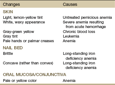
Changes in the hands and fingernail beds (Table 5-1) may be observed during the inspection/observation portion of the physical therapy evaluation (see Table 4-8 and Boxes 4-13 and 4-15). The physical therapist should look for pale palms with normal-colored creases (severe anemia causes pale creases as well). Observation of the hands should be done at the level of the client’s heart. In addition, the anemic client’s hands should be warm; if they are cold, the paleness is due to vasoconstriction.


Pallor in dark-skinned people may be observed by the absence of the underlying red tones that normally give brown or black skin its luster. The brown-skinned individual demonstrates pallor with a more yellowish-brown color, and the black-skinned person will appear ashen or gray.


Systolic blood pressure may not be affected, but diastolic pressure may be lower than normal, with an associated increase in the resting pulse rate. Resting cardiac output is usually normal in people with anemia, but cardiac output increases with exercise more than it does in people without anemia.9 As the anemia becomes more severe, resting cardiac output increases and exercise tolerance progressively decreases until dyspnea, tachycardia, and palpitations occur at rest.


Diminished exercise tolerance is expected in the client with anemia. Exercise testing and prescribed exercise(s) in clients with anemia must be instituted with extreme caution and should proceed very gradually to tolerance and/or perceived exertion levels.10,11 In addition, exercise for any anemic client should be first approved by his or her physician (Case Example 5-1).


Case Example 5-1   Anemia


A 72-year-old woman, status post hip fracture, was treated surgically with nails (used for the fixation of the ends of fractured bones) and was referred to physical therapy for follow-up treatment before hospital discharge. The physician’s preoperative examination and surgical report were unremarkable for physical therapy precautions or contraindications.


When the therapist met with the client for the first time, she had already been ambulating alone in her room from the bed to the bathroom and back using a hospital wheeled walker. She was wearing thigh length support hose, hospital gown, and open-heeled slippers from home. Although the nursing report indicated she was oriented to time and place, she seemed confused and required multiple verbal cues to follow the physical therapist’s directions.


After ambulating a distance of approximately 50 feet using her wheeled walker and standby assistance from the therapist, the client reported that she could not “catch her breath” and asked to sit down. She placed her hand over her heart and commented that her heart was “fluttering.” Blood pressure and pulse measurements were taken and recorded as 145/72 mm Hg (blood pressure) and 90 bpm (pulse rate).


The physical therapist consulted with nursing staff immediately regarding this episode and was given the “go ahead” to complete the therapy session. The physical therapist documented the episode in the medical record and left a note for the physician, briefly describing the incident and ending with the question: Are there any medical contraindications to continuing progressive therapy?


Result: A significant fall in hemoglobin (Hb) often occurs after hip fracture and surgical intervention secondary to the blood loss caused by the fracture and surgery. Other contributing factors may include blood transfusion and alcoholic liver cirrhosis.1214


In this case, although the physician did not offer a direct reply to the physical therapist, the physician’s notes indicated a suspected diagnosis of anemia. Follow-up blood work was ordered, and the diagnosis was confirmed. Nursing staff conferred with the physician, and the therapist was advised to work within the patient’s tolerance using perceived exertion as a guide while monitoring pulse and blood pressure.


Polycythemia


Polycythemia (also known as erythrocytosis) is characterized by increases in both the number of red blood cells and the concentration of hemoglobin. People with polycythemia have increased whole blood viscosity and increased blood volume.


The increased erythrocyte production results in this thickening of the blood and an increased tendency toward clotting. The viscosity of the blood limits its ability to flow easily, diminishing the supply of blood to the brain and to other vital tissues. Increased platelets in combination with the increased blood viscosity may contribute to the formation of intravascular thrombi.


There are two distinct forms of polycythemia: primary polycythemia (also known as polycythemia vera) and secondary polycythemia. Primary polycythemia is a relatively uncommon neoplastic disease of the bone marrow of unknown etiology. Secondary polycythemia is a physiologic condition resulting from a decreased oxygen supply to the tissues. It is associated with high altitudes, heavy tobacco smoking, radiation exposure, and chronic heart and lung disorders, especially congenital heart defects.


Clinical Signs and Symptoms: The symptoms of this disease are often insidious in onset with vague complaints. The most common first symptoms are shortness of breath and fatigue. The affected individual may be diagnosed only secondary to a sudden complication (e.g., stroke or thrombosis). Increased skin coloration and elevated blood pressure may develop as a result of the increased concentration of erythrocytes and increased blood viscosity.


Gout is sometimes a complication of primary polycythemia, and a typical attack of acute gout may be the first symptom of polycythemia. Gout is a metabolic disease marked by increased serum urate levels (hyperuricemia), which cause painfully arthritic joints. Uric acid level is an end product of purine metabolism. Purine metabolism is altered by excessive cellular proliferation and breakdown associated with increased red cells, granulocytes, and platelets. Hyperuricemia is uncommon in secondary polycythemia because the cellular proliferation is not as extensive as in primary polycythemia.


Blockage of the capillaries supplying the digits of either the hands or the feet may cause a peripheral vascular neuropathy with decreased sensation, burning, numbness, or tingling. This small blood vessel occlusion can also contribute to the development of cyanosis and clubbing. If the underlying disorder is not recognized and treated, the person may develop gangrene and have subsequent loss of tissue.


Watch for increase in blood pressure and elevated hematocrit levels.


Sickle Cell Anemia


Sickle cell disease is a generic term for a group of inherited, autosomal recessive disorders characterized by the presence of an abnormal form of hemoglobin, the oxygen-carrying constituent of erythrocytes. A genetic mutation resulting in a single amino acid substitution in hemoglobin causes the hemoglobin to aggregate into long chains, altering the shape of the cell. This sickled or curved shape causes the cell to lose its ability to deform and squeeze through tiny blood vessels, thereby depriving tissue of an adequate blood supply.3


The two features of sickle cell disorders, chronic hemolytic anemia and vasoocclusion, occur as a result of obstruction of blood flow to the tissues and early destruction of the abnormal cells. Anemia associated with this condition is merely a symptom of the disease and not the disease itself, despite the term sickle cell anemia.


Clinical Signs and Symptoms: A series of “crises,” or acute manifestations of symptoms, characterize sickle cell disease. Some people with this disease have only a few symptoms, whereas others are affected severely and have a short lifespan. Recurrent episodes of vasoocclusion and inflammation result in progressive damage to most organs, including the brain, kidneys, lungs, bones, and cardiovascular system, which becomes apparent with increasing age. Cerebrovascular accidents (CVAs) and cognitive impairment are a frequent and severe manifestation.16


Stress from viral or bacterial infection, hypoxia, dehydration, emotional disturbance, extreme temperatures, fever, strenuous physical exertion, or fatigue may precipitate a crisis. Pain caused by the blockage of sickled RBCs forming sickle cell clots is the most common symptom; it may be in any organ, bone, or joint of the body. Painful episodes of ischemic tissue damage may last 5 or 6 days and manifest in many different ways, depending on the location of the blood clot (Case Example 5-2).


Case Example 5-2


Sickle Cell Anemia


A 20-year-old African-American woman came to physical therapy with severe right knee joint pain. She could recall no traumatic injury but reported hiking 2 days previously in the Rocky Mountains with her brother, whom she was visiting (she was from New York City).


A general screen for systemic illness revealed frequent urination over the past 2 days. She also complained of stomach pain, but she thought this was related to the stress of visiting her family. Past medical history included one other similar episode when she had acute pneumonia at the age of 11 years. She stated that she usually felt fatigued but thought it was because of her active social life and busy professional career. She is a nonsmoker and a social drinker (1 to 3 drinks per week).


On examination, the right knee was enlarged and inflamed, with joint range of motion (ROM) limited by the local swelling. In fact, pain, swelling, and guarded motion in the joint prevented a complete evaluation. Given that restraint, there were no other physical findings, but not all special tests were completed. The neurologic screen was negative.


This woman was treated for local joint inflammation, but the combination of change in altitude, fatigue, increased urination, and stomach pains alerted the therapist to the possibility of a systemic process despite the client’s explanation for the fatigue and stomach upset. Because the client was from out of town and did not have a local physician, the therapist telephoned the hospital emergency department for a telephone consultation. It was suggested that a blood sample be obtained for preliminary screening while the client continued to receive physical therapy. Laboratory results included the following:


Based on these findings, the client was admitted to the hospital and diagnosed as having sickle cell anemia. It is likely that the change in altitude, the emotional stress of visiting family, and the physical exertion precipitated a “crisis” (now referred to as “episodes”). She received continued physical therapy treatment during her hospital stay and was discharged with further follow-up planned in her home city.


Adapted from Jennings B: Nursing role in management: hematological problems. In Lewis S, Collier I, editors: Medical-surgical nursing: assessment and management of clinical problems, St Louis, 1992, Mosby, pp 664-714. Used with permission.

Stay updated, free articles. Join our Telegram channel

Mar 20, 2017 | Posted by in MANUAL THERAPIST | Comments Off on Screening for Hematologic Disease

Full access? Get Clinical Tree

Get Clinical Tree app for offline access