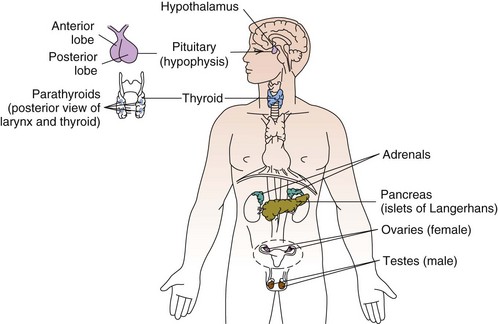
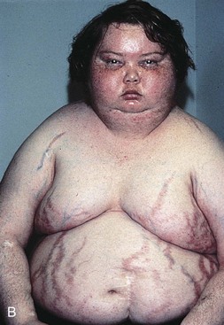
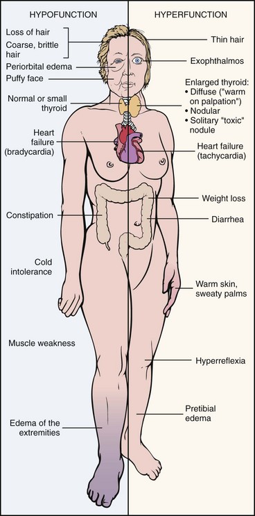
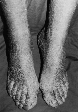
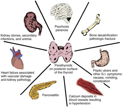
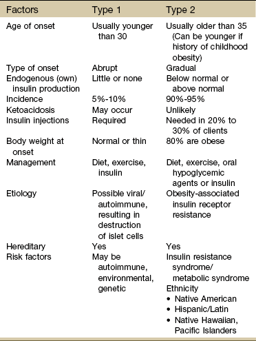
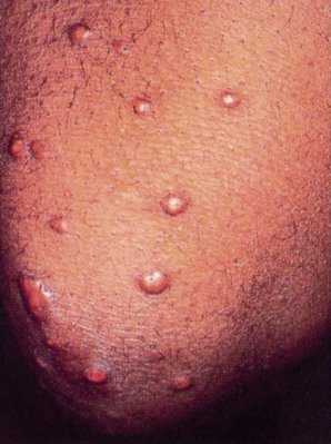
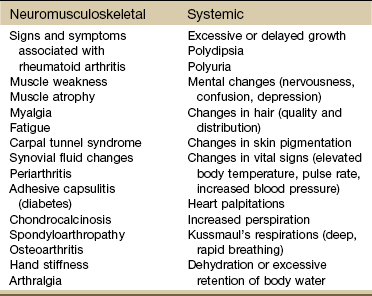
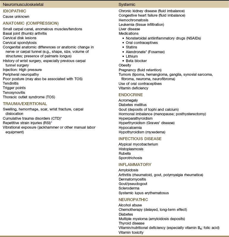
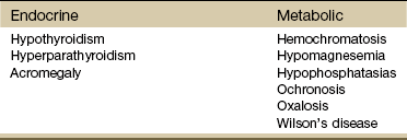
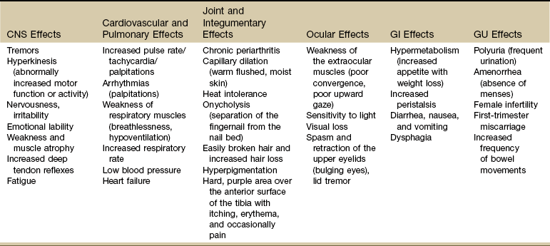
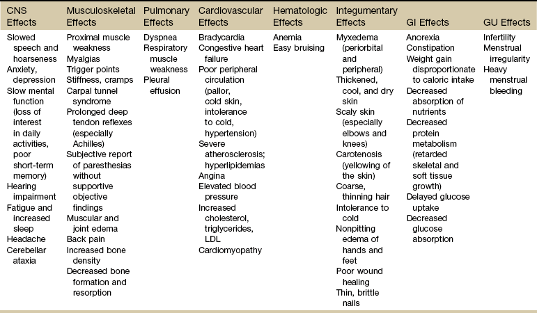
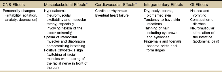
Screening for Endocrine and Metabolic Disease

Chapter 11


Screening for Endocrine and Metabolic Disease


Endocrinology is the study of ductless (endocrine) glands that produce hormones. A hormone acts as a chemical agent that is transported by the bloodstream to target tissues, where it regulates or modifies the activity of the target cell.


The endocrine system cannot be understood fully without consideration of the effects of the nervous system on the endocrine system. The endocrine system works with the nervous system to regulate metabolism, water and salt balance, blood pressure, response to stress, and sexual reproduction.


The endocrine system is slower in response and takes longer to act than the nervous system in transferring biochemical information. The pituitary (hypophysis), thyroid, parathyroids, adrenals, and pineal are glands of the endocrine system whose functions are solely endocrine related and have no other metabolic functions (Fig. 11-1). The hypothalamus controls pituitary function and thus has an important indirect influence on the other glands of the endocrine system. Feedback mechanisms exist to keep hormones at normal levels.


The endocrine system meets the nervous system in a complex series of interactions that link behavioral-neural-endocrine-immunologic responses. The hypothalamus and the pituitary form an integrated axis that maintains control over much of the endocrine system. The discovery and study of this complex interface axis is called psychoneuroimmunology (PNI) and has provided a new understanding of interactive biologic signaling.


The hypothalamus exerts direct control over both the anterior and posterior portions of the pituitary gland and can synthesize and release hormones from its axon terminals directly into the blood circulation. These neurosecretory cells are so-called because the neurons have a hormone-secreting function. Although neurons can have a hormone-secreting function, the opposite pathway is also present. Hormones that can stimulate the neural mechanism (e.g., acetylcholine) are called neurohormones. Acetylcholine is a neurotransmitter and a neurohormone. It is released at synapses to allow messages to pass along a nerve network, resulting in the release of both hormones and chemicals.


Associated Neuromuscular And Musculoskeletal Signs And Symptoms


The musculoskeletal system is composed of a variety of connective tissue structures in which normal growth and development are influenced strongly and sometimes controlled by various hormones and metabolic processes. Alterations in these control systems can result in structural changes and altered function of various connective tissues, producing systemic and musculoskeletal signs and symptoms (Table 11-1).


Bilateral Carpal Tunnel Syndrome


Bilateral carpal tunnel syndrome (CTS), resulting from median nerve compression at the wrist, is a common finding in a variety of systemic and neuromusculoskeletal conditions13 but especially with certain endocrine and metabolic disorders (Table 11-2).4 The fact that the majority of persons with CTS are women at or near menopause suggests that the soft tissues about the wrist could be affected in some way by hormones.58


Thickening of the transverse carpal ligament in certain systemic disorders (e.g., acromegaly, myxedema) may be sufficient to compress the median nerve. Any condition that increases the volume of the contents of the carpal tunnel (e.g., neoplasm, calcium, and gouty tophi deposits) can compress the median nerve.


The signs and symptoms often associated with CTS include paresthesia, tingling, and numbness and/or pain (or burning pain) with cutaneous distribution of the median nerve to the thumb, index, middle, and radial half of the ring finger. Nocturnal paresthesia is a common complaint, and this discomfort causes sleep disruption. It can be partially relieved by shaking of the hand or changing wrist and hand position. Pain may radiate into the palm and up the forearm and arm.9


It should be noted that bilateral tarsal syndrome affecting the feet also can occur either alone or in conjunction with CTS, although the incidence of tarsal tunnel syndrome is not high (see further discussion of tarsal tunnel syndrome in relation to carpal tunnel syndrome in Chapter 9). Bilateral median nerve neuritis can be characteristic of many systemic diseases, including rheumatoid arthritis, myxedema, localized amyloidosis, sarcoidosis, and infiltrative leukemia.10,11


Whenever a client presents with bilateral symptoms, it represents a red flag. With bilateral CTS the therapist can screen for medical disease by using the Special Questions to Ask: Bilateral Carpal Tunnel Syndrome section (see Appendix B-4).


Endocrine Pathophysiology


Disorders of the endocrine glands can be classified as primary (dysfunction of the gland itself) or secondary (dysfunction of an outside stimulus to the gland) and are a result of either an excess or an insufficiency of hormonal secretions.


Secondary dysfunction may also occur (iatrogenically) as a result of chemotherapy, surgical removal of the glands, therapy for a nonendocrine disorder (e.g., the use of large doses of corticosteroids resulting in Cushing’s syndrome), or excessive therapy for an endocrine disorder.


Pituitary Gland


Diabetes Insipidus


Diabetes insipidus (DI) is caused by a lack of secretion or action of vasopressin (antidiuretic hormone [ADH]). This hormone normally stimulates the distal tubules of the kidneys to reabsorb water. Without ADH, water moving through the kidney is not reabsorbed but is lost in the urine, resulting in severe water loss and dehydration through diuresis.


There are two main types of DI: central DI and nephrogenic DI. Central DI, which is the most common type, can be idiopathic (primary) or related to other causes (secondary), such as pituitary trauma, head injury (including neurosurgery), infections such as meningitis or encephalitis, pituitary neoplasm, anorexia, and vascular lesions such as aneurysms. Nephrogenic DI occurs as a result of some medications (e.g., lithium, phenytoin, corticosteroids, anticholinergics), alcohol, electrolyte imbalances such as hypercalcemia and hypokalemia, and diseases affecting the renal system (e.g., sarcoidosis, multiple myeloma, pyelonephritis, systemic lupus erythematosus).


If the person with DI is unconscious or confused and is unable to take in necessary fluids to replace those fluids lost, rapid dehydration, shock, and death can occur. Because sleep is interrupted by the persistent need to void (nocturia), fatigue and irritability result.


Syndrome of Inappropriate Secretion of Antidiuretic Hormone


Syndrome of inappropriate secretion of ADH (SIADH) is an excess or inappropriate secretion of vasopressin that results in marked retention of water in excess of sodium in the body. Urine output decreases dramatically as the body retains large amounts of water. Almost all the excess water is distributed within body cells, causing intracellular water gain and cellular swelling (water intoxication).


Acromegaly


Acromegaly is an abnormal enlargement of the extremities of the skeleton resulting from hypersecretion of growth hormone (GH) from the pituitary gland. This condition is relatively rare and occurs in adults, most often owing to a tumor of the pituitary gland. In children, overproduction of GH stimulates growth of long bones and results in gigantism, in which the child grows to exaggerated heights. With adults, growth of the long bones has already stopped, so the bones most affected are those of the face, jaw, hands, and feet. Other signs and symptoms include amenorrhea (in women), diabetes mellitus, profuse sweating, and hypertension.


Clinical Presentation: Degenerative arthropathy may be seen in the peripheral joints of a client with acromegaly, most frequently attacking the large joints. On x-ray studies, osteophyte formation may be seen, along with widening of the joint space because of increased cartilage thickness. In late-stage disease, joint spaces become narrowed, and occasionally chondrocalcinosis may be present.


Stiffness of the hand, typically of both hands, is associated with a broad enlargement of the fingers from bony overgrowth and with thickening of the soft tissue. Thickening and widening of the phalangeal tufts are typical x-ray findings in soft tissue. In clients with these x-ray findings, much of the pain and stiffness is believed to be due to premature osteoarthritis.


CTS is seen in up to 50% of people with acromegaly. The CTS that occurs with this growth disorder is thought to be caused by compression of the median nerve at the wrist from soft tissue hypertrophy or bony overgrowth or by hypertrophy of the median nerve itself.


Myopathy in people with acromegaly is commonly reported but poorly understood. Changes in muscle size and strength are associated with acromegaly and are probably multifactorial in origin. Screening individuals with acromegaly for muscle weakness and poor exercise tolerance is now recommended.12


About half the individuals with acromegaly have back pain. X-ray studies demonstrate increased intervertebral disk spaces and large osteophytes along the anterior longitudinal ligament (ALL), mimicking diffuse idiopathic skeletal hyperostosis (DISH).


DISH (also known as Forestier’s disease) is characterized by abnormal ossification of the ALL, resulting in an x-ray image of large osteophytes seemingly “flowing” along the anterior border of the spine. DISH is particularly common in the thoracic spine and has been reported to be more prevalent among persons with diabetes than among the nondiabetic population. DISH appears to be an age-related predisposition to ossification of tendon, joint capsule, and ligamentous attachments. Identification of the presence of DISH syndrome prior to surgery is important in the prevention of heterotropic bone formation.13


Adrenal Glands


The adrenals are two small glands located on the upper part of each kidney. Each adrenal gland consists of two relatively discrete parts: an outer cortex and an inner medulla. The outer cortex is responsible for the secretion of mineralocorticoids (steroid hormones that regulate fluid and mineral balance), glucocorticoids (steroid hormones responsible for controlling the metabolism of glucose), and androgens (sex hormones). The centrally located adrenal medulla is derived from neural tissue and secretes epinephrine and norepinephrine. Together, the adrenal cortex and medulla are major factors in the body’s response to stress.


Adrenal Insufficiency


Primary Adrenal Insufficiency: Chronic adrenocortical insufficiency (hyposecretion by the adrenal glands) may be primary or secondary. Primary adrenal insufficiency is also referred to as Addison’s disease (hypofunction), named after the physician who first studied and described the associated symptoms. It can be treated by the administration of exogenous cortisol (one of the adrenocortical hormones).


Primary adrenal insufficiency occurs when a disorder exists within the adrenal gland itself. This adrenal gland disorder results in decreased production of cortisol and aldosterone, two of the primary adrenocortical hormones. The most common cause of primary adrenal insufficiency is an autoimmune process that causes destruction of the adrenal cortex.


The most striking physical finding in the person with primary adrenal insufficiency is the increased pigmentation of the skin and mucous membranes. This discoloration may vary in the white population from a slight tan or a few black freckles to an intense generalized pigmentation, which has resulted in persons being mistakenly considered to be of a darker-skinned race. Members of darker-skinned races may develop a slate-gray color that may be obvious only to family members.


Melanin, the major product of the melanocyte, is largely responsible for the coloring of skin. In primary adrenal insufficiency, the increase in pigmentation is initiated by the excessive secretion of melanocyte-stimulating hormone (MSH) that occurs in association with increased secretion of adrenocorticotropic hormone (ACTH). ACTH is increased in an attempt to stimulate the diseased adrenal glands to produce and release more cortisol.


Most commonly, pigmentation is visible over extensor surfaces such as the backs of the hands; elbows; knees; and creases of the hands, lips, and mouth. Increased pigmentation of scars formed after the onset of the disease is common. However, it is possible for a person with primary adrenal insufficiency to demonstrate no significant increase in pigmentation.


Secondary Adrenal Insufficiency: Secondary adrenal insufficiency refers to a dysfunction of the gland because of insufficient stimulation of the cortex owing to a lack of pituitary ACTH. Causes of secondary disease include tumors of the hypothalamus or pituitary, removal of the pituitary, or rapid withdrawal of corticosteroid drugs. Clinical manifestations of secondary disease do not occur until the adrenals are almost completely nonfunctional and are primarily related to cortisol deficiency only.


Cushing’s Syndrome


Cushing’s syndrome (hyperfunction of the adrenal gland) is a general term for increased secretion of cortisol by the adrenal cortex. When corticosteroids are administered externally, a condition of hypercortisolism called iatrogenic Cushing’s syndrome occurs, producing a group of associated signs and symptoms. Hypercortisolism caused by excess secretion of ACTH (e.g., from pituitary stimulation) is called ACTH-dependent Cushing’s syndrome.14


Therapists often treat people who have developed Cushing’s syndrome after these clients have received large doses of cortisol (also known as hydrocortisone) or cortisol derivatives (e.g., dexamethasone) for a number of inflammatory disorders (Case Example 11-1).


Case Example 11-1   Cushing’s Syndrome


A 53-year-old woman with Cushing’s syndrome resulting from long-term use of cortisol for systemic lupus erythematosus reports the following problems:


Her primary complaint and reason for referral to physical therapy is for sacroiliac (SI) joint pain as a result of stepping down off an uneven curb.


You realize the signs and symptoms are of an endocrine origin, but you do not know whether they are part of the Cushing’s syndrome or a separate endocrine problem.


Should you send this client to a physician (or back to the referring physician)?


Not necessarily. This is more a case of need for additional information. Requesting a copy of the client’s most recent physician’s notes may answer all of your questions. Reading the physician’s systems review portion of the exam may reveal a record of these signs and symptoms with a corresponding medical problem list and plan.


If there is no mention of any of these associated signs and symptoms, a phone call to the physician’s office may be the next step. If you speak with the physician directly, identify yourself and your connection with the client by name. Briefly mention why you are seeing this client and make the following observation:


“Mrs. Jones reports muscle cramps and generalized weakness that do not seem consistent with her SI problem. She complains of temperature intolerance and hair and nail bed changes. These symptoms are outside the scope of my practice.


Can you help me understand this? Are they part of her lupus, Cushing’s syndrome, or something else?”


It is important to remember that whenever corticosteroids are administered externally, the increase in serum cortisol levels triggers a negative feedback signal to the anterior pituitary gland to stop adrenal stimulation. Adrenal atrophy occurs during this time, and adrenal insufficiency will result if external corticosteroids are abruptly withdrawn. Corticosteroid medications must be reduced gradually so that normal adrenal function can return.


Because cortisol suppresses the inflammatory response of the body, it can mask early signs of infection. Any unexplained fever without other symptoms should be a warning to the therapist of the need for medical follow-up.


Effects of Cortisol on Connective Tissue: Overproduction of cortisol or closely related glucocorticoids by abnormal adrenocortical tissue leads to a protein catabolic state. This overproduction causes liberation of amino acids from muscle tissue. The resultant weakened protein structures (muscle and elastic tissue) cause a protuberant abdomen, poor wound healing, generalized muscle weakness, and marked osteoporosis (demineralization of bone causing reduced bone mass), which is made worse by an excessive loss of calcium in the urine.


Excessive glucose resulting from this protein catabolic state is transformed mainly into fat and appears in characteristic sites, such as the abdomen, supraclavicular fat pads, and facial cheeks. The change in facial appearance may not be readily apparent to the client or to the therapist, but pictures of the client taken over a period of years may provide a visual record of those changes.


The effect of increased circulating levels of cortisol on the muscles of clients varies from slight to very marked. There may be so much muscle wasting that the condition simulates muscular dystrophy. Marked weakness of the quadriceps muscle often prevents affected clients from rising out of a chair unassisted. Those with Cushing’s syndrome of long duration almost always demonstrate demineralization of bone. In severe cases, this condition may lead to pathologic fractures, but it results more commonly in wedging of the vertebrae, kyphosis, bone pain, and back pain


Obesity, diabetes, polycystic ovarian syndrome, and other metabolic/endocrine problems can resemble Cushing’s syndrome. It is important to recognize critical indicators of this particular disorder, such as excessive hair growth, moonface, mood disorders, and increased muscle weakness, as indicators for further endocrine diagnostic testing.15


The poor wound healing that is characteristic of this syndrome becomes a problem when any surgical procedures are required. Inhibition of collagen formation with corticosteroid therapy is responsible for the frequency of wound breakdown in postsurgical clients.


Thyroid Gland


The thyroid gland is located in the anterior portion of the lower neck below the larynx, on both sides of and anterior to the trachea. The chief hormones produced by the thyroid are thyroxine (T4), triiodothyronine (T3), and calcitonin. Both T3 and T4 regulate the metabolic rate of the body and increase protein synthesis. Calcitonin has a weak physiologic effect on calcium and phosphorus balance in the body.


Genetics plays a role in thyroid disease. A family history of thyroid disease is a risk factor. Age and gender are also factors; most cases occur after age 50. Women are more likely than men to develop thyroid dysfunction.15 Data gathered on the medical history of the orthopedic physical therapy outpatient population indicate a 7% incidence of thyroid disease in the female population.16


Thyroid function is regulated by the hypothalamus and pituitary feedback controls, as well as by an intrinsic regulator mechanism within the gland itself. Basic thyroid disorders of significance to physical therapy practice include goiter, hyperthyroidism, hypothyroidism, and cancer. Alterations in thyroid function produce changes in hair, nails, skin, eyes, gastrointestinal (GI) tract, respiratory tract, heart and blood vessels, nervous tissue, bone, and muscle.


The risk of having thyroid diseases increases with age, but in people older than 60 years of age, it becomes more difficult to detect because it masquerades as other problems such as heart disease, depression, or dementia. Fatigue and weakness may be the first symptoms among older adults, often mistaken or attributed to normal aging. New-onset depression in the older adult population and anxiety syndromes are also symptoms that can indicate thyroid dysfunction.17


On the other hand, thyroid dysfunction can mimic signs and symptoms of aging such as hair loss, fatigue, and depression. The therapist may recognize problems early and make a medical referral, minimizing the client’s symptoms. A simple and inexpensive blood test called a thyroid-stimulating hormone (TSH) test is usually recommended to show whether the thyroid gland is hyperfunctioning or hypofunctioning.


Goiter


Goiter, an enlargement of the thyroid gland, occurs in areas of the world where iodine (necessary for the production of thyroid hormone) is deficient in the diet. It is believed that when factors (e.g., a lack of iodine) inhibit normal thyroid hormone production, hypersecretion of TSH occurs because of a lack of a negative feedback loop. The TSH increase results in an increase in thyroid mass.


Pressure on the trachea and esophagus causes difficulty in breathing, dysphagia, and hoarseness. With the use of iodized salt, this problem has almost been eliminated in the United States. Although the younger population in the United States may be goiter free, older adults may have developed goiter during their childhood or adolescent years and may still have clinical manifestations of this disorder.


Thyroiditis


Thyroiditis is an inflammation of the thyroid gland. Causes can include infection and autoimmune processes. The most common form of this problem is a chronic thyroiditis called Hashimoto’s thyroiditis. This condition affects women more frequently than men and is most often seen in the 30- to 50-year-old age group. Destruction of the thyroid gland from this condition can cause eventual hypothyroidism (Case Example 11-2).


Case Example 11-2   Hashimoto’s Thyroiditis


Referral: A 38-year-old woman with right-sided groin pain was referred to physical therapy by her physician. She says that the pain came on suddenly without injury. The pain is worse in the morning and hurts at night, waking her up when she changes position. The woman’s symptoms are especially acute when she tries to stand up after sitting, with weight bearing impossible for the first 5 to 10 minutes.


The woman, who looks athletic, reports that before the onset of this problem she was running 5 miles every other day without difficulty. The x-ray finding is reportedly within normal limits for structural abnormalities. Erythrocyte sedimentation rate (ESR) was 16 mm/hour.* The client has chronic sinusitis and has had two surgeries for that condition in the last 3 years. She is not a smoker and drinks only occasionally on a social basis.


This client was seen 6 weeks ago by another physical therapist, who tried ultrasound and stretching without improvement in symptoms or function.


Clinical Presentation: The physical therapy evaluation today revealed a positive Thomas test for right hip flexion contracture. However, it was difficult to assess whether there was a true muscle contracture or only loss of motion as a result of muscle splinting and guarding. Patrick’s test (FABER) for hip pathology and the iliopsoas test for intraabdominal infection were both negative. Joint accessory motions appeared to be within normal limits, given that the movements were tested in the presence of some residual muscle tension from protective splinting. A neurologic screen failed to demonstrate the presence of any neurologic involvement. Symptoms could be reproduced with deep palpation of the right groin area. There were no active or passive movements that could alter, provoke, change, or eliminate the pain. There were no trigger points in the abdomen or right lower quadrant that could account for the symptomatic presentation.


There was no apparent cause for her movement system impairment. Physical therapy intervention with soft tissue mobilization and proprioceptive neuromuscular facilitation techniques were initiated and used as a diagnostic tool. There was no change in the client’s symptoms or clinical presentation as the therapist continued trying a series of physical therapy techniques.


Result: In a young and otherwise healthy adult, a lack of measurable, reportable, or observable progress becomes a red flag for further medical follow-up. The results of the physical therapy examination and lack of response to treatment constitute a valuable medical diagnostic tool.


Further laboratory results revealed a medical diagnosis of Hashimoto’s thyroiditis. Treatment with thyroxine (T4) resulted in resolution of the musculoskeletal symptoms. The correlation between groin pain and loss of hip extension with Hashimoto’s remains unclear. Even so, response to the red flag (no change or improvement with intervention) resulted in a correct medical diagnosis.


*The ESR (an indication of possible infection or inflammation) was within normal limits for an adult woman.


Usually, both sides of the gland are enlarged, although one side may be larger than the other. Other symptoms are related to the functional state of the gland itself. Early involvement may cause mild symptoms of hyperthyroidism, whereas later symptoms cause hypothyroidism.


Hyperthyroidism


Hyperthyroidism (hyperfunction), or thyrotoxicosis, refers to those disorders in which the thyroid gland secretes excessive amounts of thyroid hormone. Graves’ disease is a common type of excessive thyroid activity characterized by a generalized enlargement of the gland (or goiter leading to a swollen neck) and often, protruding eyes caused by retraction of the eyelids and inflammation of the ocular muscles.


Clinical Presentation: Excessive thyroid hormone creates a generalized elevation in body metabolism. The effects of thyrotoxicosis occur gradually and are manifested in almost every system (Fig. 11-3 and Table 11-4).


In more than 50% of adults older than 70, three common signs are tachycardia, fatigue, and weight loss. In clients younger than 50, clinical signs and symptoms found most often include tachycardia, hyperactive reflexes, increased sweating, heat intolerance, fatigue, tremor, nervousness, polydipsia, weakness, increased appetite, dyspnea, and weight loss.18


Chronic periarthritis is also associated with hyperthyroidism. Inflammation that involves the periarticular structures, including the tendons, ligaments, and joint capsule, is termed periarthritis. The syndrome is associated with pain and reduced range of motion. Calcification, whether periarticular or tendinous, may be seen on x-ray studies. Both periarthritis and calcific tendinitis occur most often in the shoulder, and both are common findings in clients who have endocrine disease (Case Example 11-3).


Painful restriction of shoulder motion associated with periarthritis has been widely described among clients of all ages with hyperthyroidism. The involvement can be unilateral or bilateral and can worsen progressively to become adhesive capsulitis (frozen shoulder). Acute calcific tendinitis of the wrist also has been described in such clients. Although antiinflammatory agents may be needed for the acute symptoms, chronic periarthritis usually responds to treatment of the underlying hyperthyroidism.


Proximal muscle weakness (most marked in the pelvic girdle and thigh muscles), accompanied by muscle atrophy known as myopathy, occurs in up to 70% of people with hyperthyroidism. Muscle strength returns to normal in about 2 months after medical treatment, whereas muscle wasting resolves more slowly. In severe cases normal strength may not be restored for months.


The incidence of myasthenia gravis is increased in clients with hyperthyroidism, which in turn can aggravate muscle weakness. If the hyperthyroidism is corrected, improvement of myasthenia gravis follows in about two-thirds of clients.


Thyroid Storm: Life-threatening complications with hyperthyroidism are rare but still important for the therapist to recognize. Unrecognized disease, untreated disease, or incorrect treatment can result in a condition called thyroid storm. In addition, precipitating factors, such as trauma, infection, or surgery, can turn well-controlled hyperthyroidism into a thyroid storm.


Thyroid storm is characterized by signs and symptoms of hypermetabolism including severe tachycardia with heart failure, shock, and hyperthermia (up to 105.3° F [40.7° C]). Restlessness, agitation, chest pain, abdominal pain, nausea and vomiting, and coma can occur. Immediate medical referral is required to return the client to a normal thyroid state and prevent cardiovascular or hyperthermic collapse. Look for a recent history of the precipitating factors mentioned.


Hypothyroidism


Hypothyroidism (hypofunction) is more common than hyperthyroidism, results from insufficient thyroid hormone, and creates a generalized depression of body metabolism. Hypothyroidism in fetal development and infants is usually a result of absent thyroid tissue and hereditary defects in thyroid hormone synthesis. Untreated congenital hypothyroidism is referred to as cretinism.


The condition may be classified as either primary or secondary. Primary hypothyroidism results from reduced functional thyroid tissue mass or impaired hormonal synthesis or release (e.g., iodine deficiency, loss of thyroid tissue, autoimmune thyroiditis). Secondary hypothyroidism (which accounts for a small percentage of all cases of hypothyroidism) occurs as a result of inadequate stimulation of the gland because of anterior pituitary gland dysfunction.


Clinical Presentation: As with all disorders affecting the thyroid and parathyroid glands, clinical signs and symptoms affect many systems of the body (Table 11-5). Because the thyroid hormones play such an important role in the body’s metabolism, lack of these hormones seriously upsets the balance of body processes.


Among the primary symptoms associated with hypothyroidism are intolerance to cold, excessive fatigue and drowsiness, headaches, and weight gain. In women, menstrual bleeding may become irregular, and premenstrual syndrome (PMS) may worsen. Physical assessment often reveals dryness of the skin and increasing thinness and brittleness of the hair and nails. There may be nodules or other irregularities of the thyroid palpable during anterior neck examination.


Ichthyosis, or dry scaly skin (resembling fish scales; the word ichthyosis is derived from the Latin word ichthus, which means “fish”), may be an inherited dermatologic condition (Fig. 11-4). It may also be the result of a thyroid condition. It must not be assumed that clients who present with this condition are merely in need of better hydration or regular use of skin lotion. A medical referral is needed to rule out underlying pathology.


Myxedema: A characteristic sign of hypothyroidism and more rarely associated with hyperthyroidism (Graves’ disease) is myxedema (often used synonymously with hypothyroidism). Myxedema is a result of an alteration in the composition of the dermis and other tissues, causing connective tissues to be separated by increased amounts of mucopolysaccharides and proteins.


This mucopolysaccharide-protein complex binds with water, causing a nonpitting, boggy edema, especially around the eyes, hands, and feet and in the supraclavicular fossae (Case Example 11-4). The binding of this protein-mucopolysaccharide complex causes thickening of the tongue and the laryngeal and pharyngeal mucous membranes. This results in hoarseness and thick, slurred speech, which are also characteristic of untreated hypothyroidism.


Case Example 11-4   Myxedema


Referral: A 36-year-old African-American woman with a history of Graves’ disease came to an outpatient hand clinic as a self-referral with painless swelling in both hands and feet. She had seen her doctor 6 weeks ago and was told that she did not have rheumatoid arthritis and should see a physical therapist.


Past Medical History: The woman had a 3-year history of Graves’ disease, which was treated with thyroid supplementation. She had a family history of thyroid problems, maternal history of diabetes, and history of early death from heart attack (father). Aside from symptoms of hyperthyroidism, she did not have any health problems.


Clinical Presentation: There was a mild swelling apparent in the soft tissues of the fingers and toes. Presentation was painless and bilateral, although asymmetric (second and third digits of the right hand were affected; third and fourth digits of the left hand were symptomatic).


The therapist was alerted to the unusual clinical presentation by the following signs:


The client did not think these additional symptoms were present at the time she saw her physician 6 weeks ago, but she could not remember exactly.


Result: The therapist was unsure if the symptoms present were normal manifestations of Graves’ disease or an indication that the client’s thyroid levels were abnormal. The physician was contacted with information about the additional signs and questions about this client’s clinical presentation.


The physician requested a return visit from the client, at which time further testing was done. The skin changes and edema of the lower legs are called pretibial myxedema. Myxedema is more commonly associated with hypothyroidism. When accompanied by digital clubbing and new bone formation, the condition is called thyroid acropachy. This condition is seen most often in individuals who have been treated for hyperthyroidism.


Drug therapy for the thyroid function does not change the acropachy; treatment is palliative for relief of symptoms. Physical therapy intervention can be prescribed but has not been studied to prove effectiveness for this condition.


Clients who have myxedematous hypothyroidism may demonstrate synovial fluid that is highly distinctive. The fluid’s high viscosity results in a slow fluid wave that creates a sluggish “bulge” sign visible at the knee joint. Often, the fluid contains calcium pyrophosphate dihydrate (CPPD) crystal deposits that may be associated with chondrocalcinosis (deposit of calcium salts in joint cartilage). Thus a finding of a highly viscous, “noninflammatory” joint effusion containing CPPD crystals may suggest to the physician possible underlying hypothyroidism.


When such clients with hypothyroidism have been treated with thyroid replacement, some have experienced attacks of acute pseudogout caused by CPPD crystals remaining in the synovial fluid.


Neuromuscular Symptoms: Neuromuscular symptoms are among the most common manifestations of hypothyroidism. Flexor tenosynovitis with stiffness often accompanies CTS in people with hypothyroidism. CTS can develop before other signs of hypothyroidism become evident. It is thought that this CTS arises from deposition of myxedematous tissue in the carpal tunnel area. Acroparesthesias may occur as a result of median nerve compression at the wrist. The paresthesias are almost always located bilaterally in the hands. Most clients do not require surgical treatment because the symptoms respond to thyroid replacement.


Proximal muscle weakness sometimes accompanied by pain is common in clients who have hypothyroidism. As mentioned earlier, muscle weakness is not always related to either the severity or the duration of hypothyroidism and can be present several months before the diagnosis of hypothyroidism is made. Muscle bulk is usually normal; muscle hypertrophy is rare. Deep tendon reflexes are characterized by slowed muscle contraction and relaxation (prolonged reflex).


Characteristically, the muscular complaints of the client with hypothyroidism are aches and pains and cramps or stiffness. Involved muscles are particularly likely to develop persistent myofascial trigger points (TrPs). Of particular interest to the therapist is the concept that clinically any compromise of the energy metabolism of muscle aggravates and perpetuates TrPs. Treatment of the underlying hypothyroidism is essential in eliminating the TrPs,19 but new research also supports the need for soft tissue treatment to achieve full recovery.20


There appears to be an association between hypothyroidism and fibromyalgia syndrome (FMS). Individuals with FMS and clients with undiagnosed myofascial symptoms may benefit from a medical referral for evaluation of thyroid function.2124


Neoplasms


Cancer of the thyroid is a relatively uncommon, slow-growing neoplasm that rarely metastasizes. It is often the incidental finding in persons being treated for other disorders (e.g., musculoskeletal disorders involving the head and neck). Primary cancers of other endocrine organs are rare and are not encountered by the clinical therapist very often.


Risk factors for thyroid cancer include female gender, age over 40 years, Caucasian race, iodine deficiency, family history of thyroid cancer, and being exposed to radioactive iodine (I-131), especially as children. In addition, nuclear power plant fallout could expose large numbers of people to I-131 and subsequent thyroid cancer. The use of potassium iodide (KI) can protect the thyroid from the adverse effects of I-131 and is recommended to be made available in areas of the country near nuclear power plants in case of nuclear fallout.25 The initial manifestation in adults and especially in children is a palpable lymph node or nodule in the neck lateral to the sternocleidomastoid muscle in the lower portion of the posterior triangle overlying the scalene muscles26 (Fig. 11-5).


A physician must evaluate any client with a palpable nodule because a palpable nodule is often clinically indistinguishable from a mass associated with a benign condition. The presence of new-onset hoarseness, hemoptysis, or elevated blood pressure is a red-flag symptom for systemic disease.


Parathyroid Glands


Two parathyroid glands are located on the posterior surface of each lobe of the thyroid gland. These glands secrete parathyroid hormone (PTH), which regulates calcium and phosphorus metabolism. Parathyroid disorders include hyperparathyroidism and hypoparathyroidism.


The therapist may see clients with parathyroid disorders in acute care settings and postoperatively because these disorders can result from diseases and surgical procedures. If damage or removal of these glands occurs, the resulting hypoparathyroidism (temporary or permanent) causes hypocalcemia, which can result in cardiac arrhythmias and neuromuscular irritability (tetany).


Disorders of the parathyroid glands may produce periarthritis and tendinitis. Both types of inflammation may be crystal induced and can be associated with periarticular or tendinous calcification.


Hyperparathyroidism


Hyperparathyroidism (hyperfunction), or the excessive secretion of PTH, disrupts calcium, phosphate, and bone metabolism. The primary function of PTH is to maintain a normal serum calcium level. Elevated PTH causes release of calcium by the bone and accumulation of calcium in the bloodstream.


Symptoms of hyperparathyroidism are related to this release of bone calcium into the bloodstream. This causes demineralization of bone and subsequent loss of bone strength and density. At the same time, the increase of calcium in the bloodstream can cause many other problems within the body, such as renal stones. The incidence of hyperparathyroidism is highest in postmenopausal women.27


The major cause of primary hyperparathyroidism is a tumor of a parathyroid gland, which results in the autonomous secretion of PTH. Renal failure, another common cause of hyperparathyroidism, causes hypocalcemia and stimulates PTH production. Hyperplasia of the gland occurs as it attempts to raise the blood serum calcium levels. Thiazide diuretics (used for hypertension) and lithium carbonate (used for some psychiatric problems) can exacerbate or even cause hyperparathyroid disorders.28


Clinical Presentation: Many systems of the body are affected by hyperparathyroidism (Table 11-6). Proximal muscle weakness and fatigability are common findings and may be secondary to a peripheral neuropathic process. Myopathy of respiratory muscles with associated respiratory involvement often goes unnoticed. Striking reversal of muscle weakness and atrophy occur with successful treatment of the underlying hyperparathyroidism.


Other symptoms associated with hyperparathyroidism are muscle weakness, loss of appetite, weight loss, nausea and vomiting, depression, and increased thirst and urination (Case Example 11-5). Hyperparathyroidism can also cause GI problems, pancreatitis, bone decalcification, and psychotic paranoia (Fig. 11-6).


Case Example 11-5   Rheumatoid Arthritis and Hyperparathyroidism


Referral: A 58-year-old man was referred to physical therapy by his primary care physician with a diagnosis of new-onset rheumatoid arthritis. Chief complaint was bilateral sacroiliac (SI) joint pain and pain on palpation of the hands and wrists.


When asked if he had any symptoms of any kind anywhere else in the body, he mentioned constipation, nausea, and loss of appetite. The family took the therapist aside and expressed concerns about personality changes, including apathy, depression, and episodes of paranoia. These additional symptoms were first observed shortly after the hand pain developed.


Past Medical History: The client had a motorcycle accident 2 years ago but reported no major injuries and no apparent residual problems. He had a family history of heart disease and hypertension but was not hypertensive at the time of the physical therapy interview. There was no other contributory personal or family past medical history.


Clinical Presentation: The therapist was unable to account for the sacroiliac joint pain. There were no particular movements that made it better or worse, and no objective findings to suggest an underlying movement system impairment.


Other red flags included age, bilateral hand and SI symptoms, gastrointestinal (GI) distress, and psychologic/behavioral changes observed by the family.


Result: The therapist contacted the referring physician with the results of her evaluation. During the telephone conversation, the therapist mentioned the family’s concerns about the client’s personality change and the fact that the client had bilateral symptoms that could not be provoked or relieved. Additional GI symptoms were also discussed.


At the physician’s request, the client completed a short course of physical therapy intervention with an emphasis on posture, core training, and soft tissue mobilization. The client returned to the physician for a follow-up examination 4 weeks later. His symptoms were unchanged.


After additional testing, the client was eventually diagnosed with hyperparathyroidism and treated accordingly. Both his hand and SI pain went away, as well as most of the GI problems.


Bone erosion, bone resorption, and subsequent bone destruction from hypercalcemia associated with hyperparathyroidism occurs rarely today. In most cases, hypercalcemia is mild and detected before any significant skeletal disease develops. The classic bone disease osteitis fibrosa cystica affects persons with primary or renal hyperparathyroidism. Bone lesions called Brown tumors appear at the end stages of the cystic osteitis fibrosa. There are increasing reports of this condition in hyperparathyroidism secondary to renal failure because of the increasing survival rates of clients on hemodialysis.


Currently, skeletal manifestations of primary hyperparathyroidism are more likely to include bone pain secondary to osteopenia, especially diffuse osteopenia of the spine with possible vertebral fractures. In addition, a number of articular and periarticular disorders have been recognized in association with primary hyperparathyroidism. The therapist may encounter cases of ruptured tendons caused by bone resorption in clients with hyperparathyroidism.


Inflammatory erosive polyarthritis may be associated with chondrocalcinosis and CPPD deposits in the synovial fluid. This erosion is called osteogenic synovitis. Concurrent illness and surgery (most often parathyroidectomy) are recognized inducers of acute arthritic episodes.


Hypoparathyroidism


Hypoparathyroidism (hypofunction), or insufficient secretion of PTH, most commonly results from accidental removal or injury of the parathyroid gland during thyroid or anterior neck surgery. A less common form of the disease can occur from a genetic autoimmune destruction of the gland. Hypofunction of the parathyroid gland results in insufficient secretion of PTH and subsequent hypocalcemia, hyperphosphatemia, and pronounced neuromuscular and cardiac irritability.


Clinical Presentation: Hypocalcemia occurs when the parathyroids become inactive. The resultant deficiency of calcium in the blood alters the function of many tissues in the body. These altered functions are described by the systemic manifestations of signs and symptoms associated with hypoparathyroidism (Table 11-7).


The most significant clinical consequence of hypocalcemia is neuromuscular irritability. This irritability results in muscle spasms, paresthesias, tetany, and life-threatening cardiac arrhythmias. Muscle weakness and pain have been reported along with hypocalcemia in clients with hypoparathyroidism.


Hypoparathyroidism is primarily treated through pharmacologic management with intravenous calcium gluconate, oral calcium salts, and vitamin D. Acute hypoparathyroidism is a life-threatening emergency and is treated rapidly with calcium replacement, anticonvulsants, and prevention of airway obstruction.


Pancreas


The pancreas is a fish-shaped organ that lies behind the stomach. Its head and neck are located in the curve of the duodenum, and its body extends horizontally across the posterior abdominal wall.


The pancreas has dual functions. It acts as both an endocrine gland, secreting the hormones insulin and glucagon, and an exocrine gland, producing digestive enzymes. Disorders of endocrine function are included in this chapter, whereas disorders of exocrine function affecting digestion are included in Chapter 8.


Diabetes Mellitus


Diabetes mellitus (DM) is a chronic disorder caused by deficient insulin or defective insulin action in the body. It is characterized by hyperglycemia (excess glucose in the blood) and disruption of the metabolism of carbohydrates, fats, and proteins. Over time, it results in serious small vessel and large vessel vascular complications and neuropathies.


Diabetes is the leading cause of end-stage renal disease (ESRD) [kidney failure requiring dialysis or transplantation], nontraumatic lower extremity amputations, and new cases of blindness among adults in the United States, and a major cause of heart disease and stroke.29,30


Type 1 DM is a condition in which little or no insulin is produced. It occurs in about 10% of all cases and usually occurs in children or young adults. Type 2 DM commonly occurs after age 40 and is a condition of defective insulin and/or impaired cell receptor binding of insulin. Table 11-8 depicts the major differences between type 1 and type 2 in presentation and treatment. There has been some discussion as to whether Alzheimer’s disease is type 3 diabetes (“brain diabetes”), unique to the brain or if diabetes is just a risk factor for Alzheimer’s disease.31 A relationship between DM and dementia is undeniable, with numerous studies concluding that DM increases the risk of cognitive decline and dementia, including Alzheimer’s disease.32


Native Americans, Latino Americans, Native Hawaiians, and some Asian Americans and Pacific Islanders have been identified at particularly high risk for type 2 DM and its complications.33 Lack of exercise and obesity are two major risk factors for type 2 DM. As a result of these lifestyle factors (sedentary lifestyle, obesity), the overall number of persons in the United States with diabetes has increased from 10 million in 1977 to 24 million in 2007 and is projected to double or triple by 2050 if current trends in diabetes prevalence continue.34,35


Clinical Presentation: Specific physiologic changes occur when insulin is lacking or ineffective. Normally, the blood glucose level rises after a meal. A large amount of this glucose is taken up by the liver for storage or for use by other tissues such as skeletal muscle and fat. When insulin function is impaired, the glucose in the general circulation is not taken up or removed by these tissues; thus it continues to accumulate in the blood. Because new glucose has not been “deposited” into the liver, the liver synthesizes more glucose and releases it into the general circulation, which increases the already elevated blood glucose level.


Protein synthesis is also impaired because amino acid transport into cells requires insulin. The metabolism of fats and fatty acids is altered, and instead of fat formation, fat breakdown begins in an attempt to liberate more glucose. The oxidation of these fats causes the formation of ketone bodies. Because the formation of these ketones can be rapid, they can build quickly and reach very high levels in the bloodstream. When the renal threshold for ketones is exceeded, the ketones appear in the urine as acetone (ketonuria).


The accumulation of high levels of glucose in the blood creates a hyperosmotic condition in the blood serum. This highly concentrated blood serum then “pulls” fluid from the interstitial areas, and fluid is lost through the kidneys (osmotic diuresis). Because large quantities of urine are excreted (polyuria), serious fluid losses occur, and the conscious individual becomes extremely thirsty and drinks large amounts of water (polydipsia). In addition, the kidney is unable to resorb all the glucose, so glucose begins to be excreted in the urine (glycosuria).


Certain medications can cause or contribute to hyperglycemia. Corticosteroids taken orally have the greatest glucogenic effect. Any person with diabetes taking corticosteroid medications must be monitored for changes in blood glucose levels.


Other hormones produced by the body also affect blood glucose levels and can have a direct influence on the severity of diabetic symptoms. Epinephrine, glucocorticoids, and growth hormone can cause significant elevations in blood glucose levels by mobilizing stored glucose to blood glucose during times of physical or psychologic stress.


When persons with DM are under stress, such as during surgery, trauma, pregnancy, puberty, or infectious states, blood glucose levels can rise and result in the need for increased amounts of insulin. If these insulin needs cannot be met, a hyperglycemic emergency such as diabetic ketoacidosis can result.


It is essential to remember that clients with DM who are under stress will have increased insulin requirements and may become symptomatic even though their disease is usually well controlled in normal circumstances.


Diagnosis: To be diagnosed with diabetes, a person must have fasting plasma glucose (FPG) readings of 126 mg/dL or higher on two different days. The previous cutoff, set in 1979, was 140 mg/dL. This change occurred as a result of research showing that individuals with readings as low as the mid-120s have already started developing tissue damage from diabetes. A value greater than 100 mg/dL is a risk factor for future diabetes and cardiovascular disease. It has been suggested that the term “prediabetes” is no longer used: the person either has diabetes or does not.36


The American Diabetes Association offers consumers a risk test for diabetes (http://www.diabetes.org/risk-test.jsp). All adults should take this risk test; anyone 45 or older should be tested for diabetes every 3 years. Individuals with elevated FPG values as described should be tested every 1 to 2 years. The therapist can offer at-risk clients information on increased activity and exercise as a means of lowering their risk of developing diabetes.37


Physical Complications: At presentation, the client with DM may have a variety of serious physical problems. Infection and atherosclerosis are the two primary long-term complications of this disease and are the usual causes of severe illness and death in the person with diabetes.


Blood vessels and nerves sustain major pathologic changes in the person affected by DM. Atherosclerosis in both large vessels (macrovascular changes) and small vessels (microvascular changes) develops at a much earlier age and progresses much faster in the individual with DM. The blood vessel changes result in decreased blood vessel lumen size, compromised blood flow, and resultant tissue ischemia. The pathologic end-products are cerebrovascular disease (CVD), coronary artery disease (CAD), renal artery stenosis, and peripheral vascular disease (PVD).


Microvascular changes, characterized by the thickening of capillaries and damage to the basement membrane, result in diabetic nephropathy (kidney disease) and diabetic retinopathy (disease of the retina). Diabetes is the leading cause of kidney failure and new cases of blindness in the United States as of 2007.30,38


Poorly controlled DM can lead to various tissue changes that result in impaired wound healing. Decreased circulation to the skin can further delay or diminish healing. Skin eruptions called xanthomas (Fig. 11-7) may appear when high lipid levels (e.g., cholesterol and triglycerides) in the blood cause fat deposits in the skin over extensor surfaces such as the elbows, knees, back of the head and neck, and heels. Yellow patches on the eyelids are another sign of hyperlipidemia. Medical referral is required to normalize lipid levels.


Stay updated, free articles. Join our Telegram channel

Mar 20, 2017 | Posted by in MANUAL THERAPIST | Comments Off on Screening for Endocrine and Metabolic Disease

Full access? Get Clinical Tree

Get Clinical Tree app for offline access