Scapulothoracic Fusion

Chapter 32


Scapulothoracic Fusion


The scapulothoracic articulation is one of four joints that work in concert to allow the shoulder to have the greatest range of motion of any joint in the body. Scapulothoracic motion is a significant contributor because it helps account for one third of shoulder elevation. Causes of dysfunction of the scapulothoracic joint can essentially be broken into two categories: dystrophic and nondystrophic. The primary dystrophic cause is facioscapulohumeral dystrophy (FSHD). Nondystrophic causes include peripheral nerve injury, failed tendon transfers for nerve injury, brachial plexus injuries, and stroke. These conditions ultimately alter the stability of the scapular platform and affect glenohumeral motion. Commonly this manifests as scapular winging and loss of shoulder motion that lead to pain. Scapulothoracic arthrodesis has been described as a viable salvage operation.1,2 The goal of this procedure is to create a solid union between the anterior surface of the scapula and the posterior thorax to stabilize this articulation, restore some level of function, and alleviate pain.3


Preoperative Considerations


Physical Examination


Primarily patients have pain combined with loss of forward elevation and abduction. Depending on the cause, various forms of scapular winging may be present. Medial winging of the scapula caused by serratus anterior or long thoracic nerve injury typically occurs with superior migration and medial rotation of the inferior border of the scapula. Lateral winging of the scapula caused by trapezius or spinal accessory nerve injury is associated with inferior migration and lateral rotation of the inferior border of the scapula. More subtle winging resulting from rhomboid or dorsal scapular nerve injury will manifest similarly to lateral winging with inferior migration and lateral rotation of the inferior border. In most patients the deltoid and rotator cuff function is preserved. Range of motion is usually less than 90 degrees of forward elevation and abduction. Atrophy of the shoulder girdle should also be noted. These visual cues will help lead the examiner to determine if any nerve injury is present and which nerves are involved. Another test that is helpful is to stabilize the scapula with one’s hand and determine if the patient’s range of motion improves. This will provide insight to the possible motion to be gained from the procedure by providing a stable platform for function.


Imaging


No imaging modalities will provide significant insight into this particular diagnosis. The pathology is a dynamic phenomenon and is not structural. Plain radiographs may demonstrate abnormal scapular positioning with significant elevation of the superomedial border or lateral translation depending on the underlying pathology. Nerve injury patterns might be identified on magnetic resonance imaging (MRI) but again are not diagnostic. Electromyography can be useful in determining nerve injury and the extent of injury. It is important to identify whether transient neurapraxic changes are present versus a more permanent injury. Neurapraxic injuries resulting from trauma often will spontaneously recover within 1 year. These patients often can be followed and conservative measures used to maintain range of motion and muscle strength in functioning groups.


Surgical Procedure


Anesthesia and Positioning


The patient is brought to the operating room and given preoperative antibiotics. After induction of general anesthesia and intubation, the patient is moved to a prone position. Care must be taken to ensure that the patient’s abdomen is free to maximize ventilation and minimize intra-abdominal pressure. Bony prominences should be padded and the arms should be in a position to avoid awkward shoulder positioning and nerve compression. The operative arm is placed in 90 degrees of abduction and external rotation with 30 degrees of horizontal adduction. The upper arm, neck, and spine all the way to the posterior superior iliac spine should be prepped into the surgical field. This allows for some movement of the extremity and harvesting of bone graft during the procedure.


Surgical Landmarks


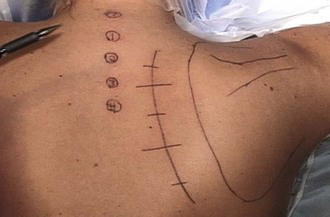
Surgical landmarks that should be marked before the procedure include the spinous processes of C7 to T4, the associated thoracic ribs, and the superior, medial, and lateral borders of the scapula (Fig. 32-1). Typically with the arm positioned as described earlier, the spine of the scapula overlies the fourth rib and the scapula is rotated approximately 30 degrees in relation to the spinous processes.


Stay updated, free articles. Join our Telegram channel

Sep 11, 2016 | Posted by in SPORT MEDICINE | Comments Off on Scapulothoracic Fusion

Full access? Get Clinical Tree

Get Clinical Tree app for offline access