Sacroiliac Orthopaedic Tests
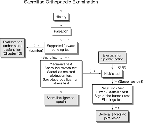
Palpation
Descriptive Anatomy
The iliac crest and posterior superior iliac spine are important bony landmarks used to assess postural deviations and leg length deficiencies. The iliac crest extends through the inferior margin of the flank and is easily palpable. The posterior superior iliac spine is inferior to the iliac crest and lateral to the S2 sacral segment (Fig. 12-1).
Procedure
With the patient standing, palpate the iliac crest for tenderness (Fig. 12-2). Tenderness may be caused by contusions, periosteitis, or avulsion fractures. Next, place your forefingers on each iliac crest and your thumbs on the posterior superior iliac spines of each ilium (Fig. 12-3). Note any difference in longitudinal position, which may indicate a leg length discrepancy, scoliosis, sacroiliac joint subluxation, or hip joint dislocation.
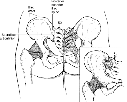 Figure 12-1 |
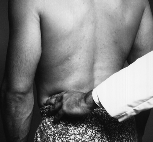 Figure 12-2 |
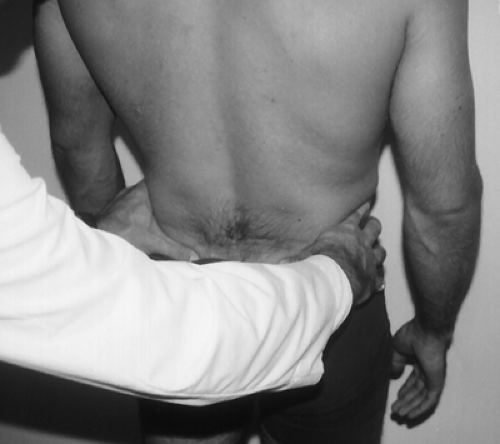 Figure 12-3 |
Descriptive Anatomy
The sacroiliac articulations are inferior and medial to the posterior superior iliac spine (Fig. 12-1). The articulations are synovial and are held together by interosseous ligaments and ventral and dorsal sacroiliac ligaments. The movement of the sacroiliac joints is limited to slight gliding and rotation. These joints are primarily weight bearing; they transfer the weight of the trunk to the hip joints.
Procedure
With the patient prone, flex the patient’s knee to 90° and externally rotate the leg. With your opposite hand, palpate the sacroiliac joint from just below the posterior superior iliac spine to the sacral notch (Fig. 12-4). Note any pain or tenderness that may indicate an inflammatory process in the sacroiliac joint.
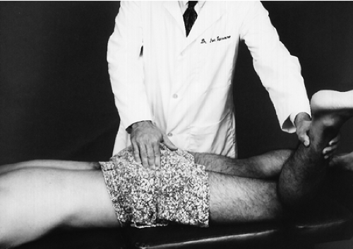 Figure 12-4 |
Descriptive Anatomy
The ischial tuberosity is just below the gluteal fold (Fig. 12-1) and can be palpated easily with the hip flexed. The hamstring muscles originate from the tuberosity. The tuberosity may be injured by direct trauma or by trauma to the hamstring muscles.
Procedure
Have the patient lie on one side and flex the hip by bringing the knee to the chest. Palpate the ischial tuberosity, noting any pain or tenderness (Fig. 12-5). Pain may indicate a contusion secondary to trauma, an avulsion fracture caused by severe hamstring pull, or ischial tuberosity bursitis.
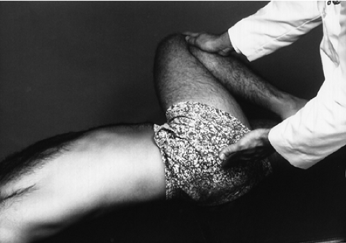 Figure 12-5 |
Sacroiliac Sprain
Clinical Description
The sacroiliac joint is a very strong cartilaginous joint that produces little movement. The sacrum is suspended between the two iliac bones and held in place by very strong interosseous dorsal sacroiliac ligaments. The joint is under great stress that predisposes it to sprains. The joint movement is limited by the tension of the sacrotuberous, sacrospinous, and sacroiliac ligaments (Fig. 12-6). These ligaments may be stretched, causing ligamentous sprain by some of the following movements:
Sudden contracture of the hamstrings or abdominal muscles, which exerts a rotating force on the ilium
Sudden unexpected twisting motions of the trunk, which may occur in sports such as football and basketball
Vigorous pulling while bending forward
A fall on one or both buttocks
The patient may have acute lower back pain and difficulty in bending. In unilateral cases it may cause the patient to elevate the hip when standing on the painful side to avoid weight bearing over the sacroiliac joint. Local tenderness is a common finding.
Clinical Signs and Symptoms
Sacroiliac joint pain
Abnormal gait
Tender sacroiliac joint on palpation
Pain on forward flexion
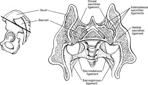 Figure 12-6 |
Gaenslen’s Test (4)
 |
Procedure
Rationale
Extension of the leg stresses the sacroiliac joint and anterior sacroiliac joint ligaments on the side of leg extension. Pain on that side indicates a general sacroiliac lesion, that is, anterior sacroiliac ligament sprain (iliofemoral, ischiofemoral) or inflammatory process in the sacroiliac joint.
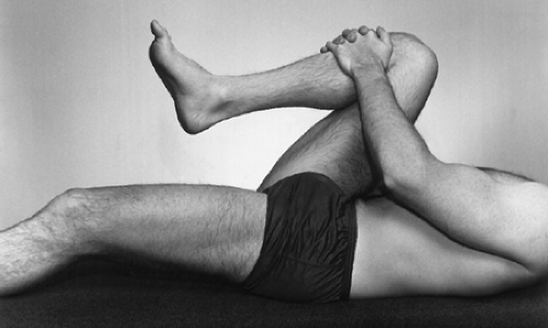 Figure 12-7 |
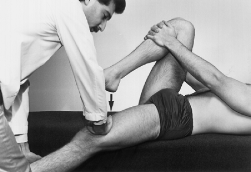 Figure 12-8 |
Lewin-Gaenslen Test
 |
Procedure
Instruct the patient to lie on the unaffected side and flex the inferior leg (Fig. 12-9). Take the superior leg and extend it while you stabilize the lumbosacral joint (Fig. 12-10).
Rationale
Extension of the leg stresses the sacroiliac joint and anterior sacroiliac joint ligaments on the side of leg extension. Pain on that side indicates a general sacroiliac joint lesion, that is, anterior sacroiliac ligament sprain (iliofemoral, ischiofemoral ligaments) or inflammatory process in the sacroiliac joint.
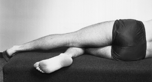 Figure 12-9 |
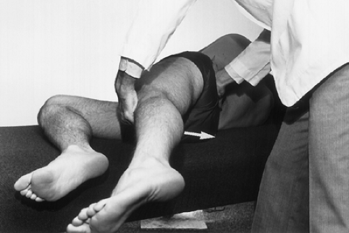 Figure 12-10 |
 |
Procedure
Related posts:

Stay updated, free articles. Join our Telegram channel

Full access? Get Clinical Tree


