Rotator Cuff Cases
Followin’ the crowd is the quickest way to get nowhere.
CASE 1: MASSIVE TRAUMATIC ROTATOR CUFF TEAR WITH INTRATENDINOUS DISRUPTION NEAR THE MUSCULOTENDINOUS JUNCTION
History:
- 43-year-old contractor who fell through a hole in the attic of a home he was building
- Tried to break his fall by grabbing a support beam
- Hyperabduction injury to left shoulder
Exam:
- Pain with elevation above 90°
- Very weak resisted external rotation
Imaging:
- X-rays are normal.
- MRI shows complete retracted tears of supraspinatus and infraspinatus, with tendon disruption near the musculotendinous junction, and a large tendon stump attached to the greater tuberosity.
Arthroscopic Findings:
- Large retracted traumatic disruption of supraspinatus (SS) and infraspinatus (IS) tendons
- Short tendon leader still attached to SS and IS muscles
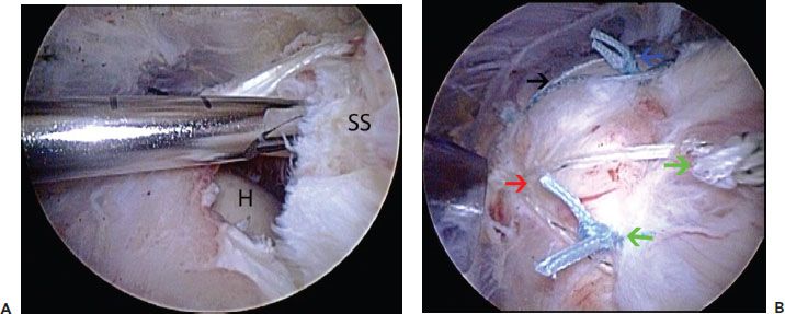
- Large viable tendon stump still attached to greater tuberosity (Fig. 26.1A)
Procedures Performed:
- Arthroscopic repair with suture anchors reinforced by a FiberTape rip-stop load-sharing tendon-to-bone construct (Fig. 26.1B)
Key Points:
- With a large distal tendon stump, simple sutures between tendon ends will not provide good fixation and will not hold.
- If the distal stump were removed and the proximal musculotendinous segment were advanced to the bone bed, the muscle-tendon unit would likely be overtensioned.
- In order to accomplish the two goals of strong fixation and normal length–tension relationship of the muscle-tendon unit, we perform an augmented suture anchor repair using a FiberTape rip-stop load-sharing tendon-to-bone construct. Multiple knotted sutures around the FiberTape rip-stop enhance the tendon fixation.
CASE 2: “RESCUE ANCHOR” TECHNIQUE FOR AUGMENTING SUTURE ANCHOR REPAIR IN SOFT BONE
History:
- 67-year-old female
- Chronic pain and weakness in dominant shoulder for 4 years
- Temporary relief from injections
Exam:
- Very weak resisted external rotation
- Normal bear-hug and belly-press tests
- Active elevation 30° and passive elevation 180° (pseudoparalysis)

Figure 26.1 A: Left shoulder, posterior subacromial viewing portal, demonstrates a medial rotator cuff tear with a lateral tendon stump. B: Same shoulder. Two rip-stop sutures have been placed; the anterior FiberTape (Arthrex, Inc., Naples, FL) rip-stop (blackarrows) encircles simple stitches (bluearrows) based from the anteromedial anchor, and the posterior rip-stop (redarrows) encircles simples stitches (greenarrows) based from the posteromedial anchor. H, humeral head; SS, supraspinatus tendon.
Imaging:
- X-rays show marked osteopenia of proximal humerus.
- MRI shows retracted tear of supraspinatus and infraspinatus, with good quality muscle on T-1 parasagittal sections.
Arthroscopic Findings:
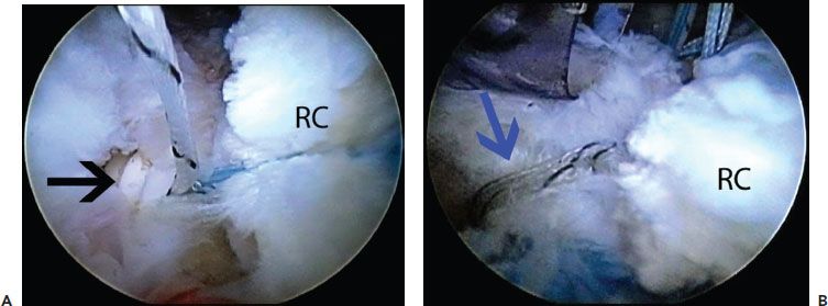
- Large crescent-shaped tear of SS and IS, easily reducible to bone bed with relatively small amount of tension (Fig. 26.2A)
Procedures Performed:
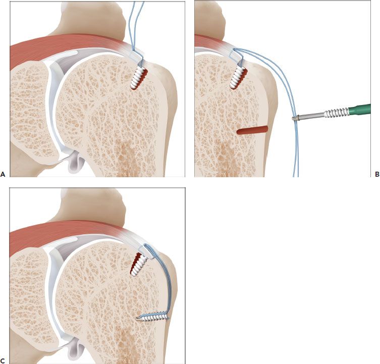
- Arthroscopic suture anchor repair of the rotator cuff, augmented by “rescue anchor” technique (Figs. 26.2B and 26.3)
Key Points:
- In osteopenic bone, a suture anchor may begin to tip medially, even under low loads, resulting in a loose anchor with a “reverse deadman angle.”
- A laterally placed “rescue anchor” (SwiveLock) can restore an anatomic footprint reduction by anchoring FiberWire suture into the relatively stronger metaphyseal cortex (lateral to the corner of the greater tuberosity).

Figure 26.2 A: Left shoulder, posterior subacromial viewing portal. After knot tying, an anchor placed for a rotator cuff repair has tilted medially (blackarrow). B: Same shoulder demonstrates a rescue anchor (blue arrow) that has been used to reinforce the medial anchor. RC, rotator cuff.

Figure 26.3 Schematic of the “rescue anchor” technique for a loose anchor. A: An anchor has been placed in the greater tuberosity for rotator cuff repair. However, while securing the rotator cuff the anchor has tilted medially under tension in poor quality bone. B: Although the tear is not amenable to double-row fixation because of limited mobility, a lateral anchor can be used to salvage the construct. The suture tails passed through the rotator cuff are left long and secured in the lateral greater tuberosity with a knotless SwiveLock anchor (Arthrex, Inc., Naples, FL). C: The lateral anchor effectively shares the load of the medial anchor.
Stay updated, free articles. Join our Telegram channel

Full access? Get Clinical Tree


